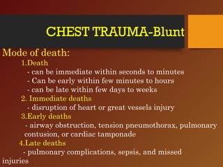
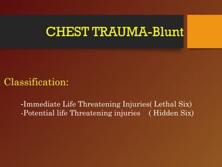
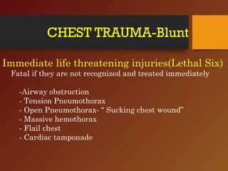
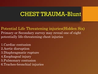
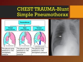
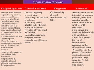
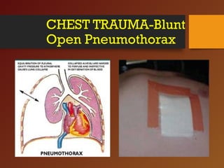
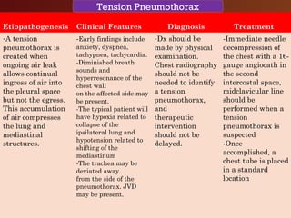
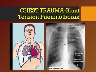
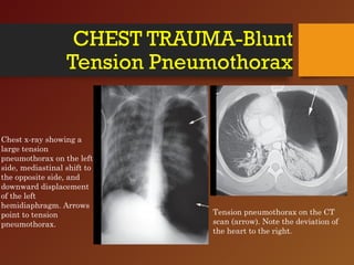
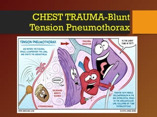
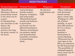
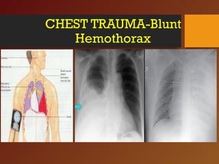
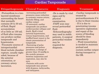
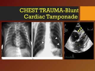
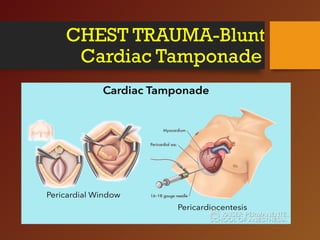
Chest trauma, especially blunt chest trauma, can cause many serious injuries from rib fractures to life-threatening conditions like tension pneumothorax. It is the second leading cause of trauma deaths. Immediate life-threatening injuries include tension pneumothorax, massive hemothorax, flail chest, and cardiac tamponade which must be quickly diagnosed and treated to prevent death. Other potential injuries include pulmonary contusions, pneumothorax, and aortic disruption which require close monitoring and treatment.
there is the introduction part of the torso trauma,
check out my next ppts for further more about torso trauma.
contents are in following order...
introduction
mechanism of injury
junctional zones of torso
tension pneumothorax
cardiac temponade
massive hemothorax
etc.
check out all slides
there is the introduction part of the torso trauma,
check out my next ppts for further more about torso trauma.
contents are in following order...
introduction
mechanism of injury
junctional zones of torso
tension pneumothorax
cardiac temponade
massive hemothorax
etc.
check out all slides
there is the introduction part of the torso trauma,
check out my next ppts for further more about torso trauma.
contents are in following order...
introduction
mechanism of injury
junctional zones of torso
tension pneumothorax
cardiac temponade
massive hemothorax
etc.
check out all slides
chest trauma is one of the leading cause of death in poly trauma patients. ER doctor should be aware of how to suspect and how to deal with life threatening conditions resulting from chest trauma
Mediastinal injury includes cardiac, thoracic aorta, pulmonary vessels and SVC injury. Pelvic includes pelvic fracture. GI injury includes upper and lower GI injuries. Abdominal vascular includes abdominal aorta, IVC and other named vascular injury.
In this presentation I am talking about the overview of So-Hum meditation- the universal mantra.
I have discussed the meaning, how to do it, it's advantages and an advanced visualisation technique.
This is a small handbook on individual surgical disease and its management . I have discussed about Acute Appendicitis and then step by step I explain both open and laparoscopic appendicectomy in this book.
The operative surgery part is very useful for surgical trainees.
POWER OF YOUTUBE IN MEDICAL EDUCATION- Surgical Educator Channel
#powerofyoutube #surgicaleducator #babysurgeon #usmle
Website Link: www.surgicaleducator.com
Dear viewers,
• Greetings from “Surgical Educator’
• In this episode, I am talking about the Power of YouTube in medical education
• I will be discussing the various benefits of using YouTube in medical education. YouTube is definitely revolutionize the way in which we are teaching our students.
• You can enjoy all my videos in the following links:
•
/ surgicaleducator surgicaleducator.com
• Thank you for watching the video.
All my videos are problem-based, because patients are coming to us with problems and not with a diagnosis.
• I have made modules for each surgical problem which consists of
many of my YouTube videos and my PPT slides
• I request you all to watch all the videos in a playlist together, so
that you will become confident in dealing with these problems.
• Links to the Playlists based on the Surgical Problems:
• Module 1: Scrotal Swellings:
https://www.youtube.com/playlist?list...
uXwt0JH0YG8m4JmzgAli9jj
https://www.slideshare.net/babysurgeo...
• Module 2: Groin Swellings:
https://www.youtube.com/playlist?list...
uVaDboG_ddw2S6xInNnB80D
https://www.slideshare.net/babysurgeo...
• Module 3: Abdominal Pain:
https://www.youtube.com/playlist?list...
uUcXb96A3tFpTrWOVa2F7j1
https://www.slideshare.net/babysurgeo...
case-based-learning-82091549
• Module 4: Abdominal Lumps:
https://youtube.com/playlist?list=PLx...
uWBKVnBkhdE4XkW-xEoiIwB
• Module 5: Obstructive Jaundice:
https://www.youtube.com/playlist?list...
uX6MsQnsCTGl8YDFN1TYiQm
https://www.slideshare.net/babysurgeo...
127314632
• Module 6: Upper GI Hemorrhage:
https://www.youtube.com/playlist?list...
uUtV67AdUQYEUKdhX9vL576
https://www.slideshare.net/babysurgeo...
227888333
• Module 7: Lower GI Hemorrhage:
https://www.youtube.com/playlist?list...
https://www.slideshare.net/babysurgeo...
• Module 8: Thyroid Pathologies:
https://www.youtube.com/playlist?list...
uWg55odQfB_7JT0NYIP8ELp
https://www.slideshare.net/babysurgeo...
benign-diseases-and-carcinoma-thyroid
• Module 9: Breast Pathologies:
https://www.youtube.com/playlist?list...
uVTLcGtam1kFBzjY4NAf7MZ
https://www.slideshare.net/babysurgeo...
diseases-and-carcinoma-breast
• Module 10: Peripheral Arterial Diseases:
https://www.youtube.com/playlist?list...
6VIbQR4g8MdOi0z
https://www.slideshare.net/babysurgeo...
106254612
• Module 11: Venous Diseases:
https://www.youtube.com/playlist?list...
uVf1aYodgILbxVpC-fkdqNo
https://www.slideshare.net/babysurgeo...
127314847
• Module 12: Dysphagia:
https://www.youtube.com/playlist?list...
4DlU1Lp
# Dear Viewers/Friends/Colleagues,
# Greetings from Surgical Educator YouTube channel
# I am sharing an E-book where you can find out the hyperlinks for all my surgery teaching videos and their PPTs
# In this E-book you will learn the purpose of my YouTube channel Surgical Educator, core clinical problems you should master, how to utilize the channel effectively, statistics and analytics for the channel, all the teaching modules with hyperlinks to all my teaching videos and their PPTs and other learning resources created by me like the android app for the channel and other E-books.
In this presentation, I discussed the various liver swellings- both cystic and solid swellings. Cystic lumps are Pyogenic liver abscess, Amebic liver abscess and hydatid cyst. Benign solid swellings are Hepatic adenoma, Focal nodular hyperplasia and Hemangioma. The malignant solid swelings are secondary carcinoma of the liver, primary Hepatocellular carcinoma and Hepatoblastoma.
In this presentation, I have shown how to do open anterior resection both high and low varieties in a step-by-step manner with clear pictures as if reading an atlas of operative surgery.
The surgical causes for jaundice in children- both in neonates and infants- are Biliary atresia, Choledochal cyst, Biliary hypoplasia, Inspissated bile syndrome, and spontaneous perforation of CBD. How to Diagnose & Treat all these causes.
I am sharing a 10 paged e-book that consists of the hyperlinks to all my surgery teaching videos and to all the PPTs used for these videos from SlideShare. You can watch these videos problem based and can become competent to deal with it. You can read this to cover the whole undergraduate curriculum.
In this presentation I discussed 5 scrotal swellings case scenarios with my MBBS students. I have shared these case scenarios prior to the PBL class and asked the students to come prepared to the class. In the class i tested the knowledge gaind by the students by watching my didactic YouTube videos on the subject by asking so many questions. So this online class was highly interactive based on flip class model.
Flu Vaccine Alert in Bangalore Karnatakaaddon Scans
As flu season approaches, health officials in Bangalore, Karnataka, are urging residents to get their flu vaccinations. The seasonal flu, while common, can lead to severe health complications, particularly for vulnerable populations such as young children, the elderly, and those with underlying health conditions.
Dr. Vidisha Kumari, a leading epidemiologist in Bangalore, emphasizes the importance of getting vaccinated. "The flu vaccine is our best defense against the influenza virus. It not only protects individuals but also helps prevent the spread of the virus in our communities," he says.
This year, the flu season is expected to coincide with a potential increase in other respiratory illnesses. The Karnataka Health Department has launched an awareness campaign highlighting the significance of flu vaccinations. They have set up multiple vaccination centers across Bangalore, making it convenient for residents to receive their shots.
To encourage widespread vaccination, the government is also collaborating with local schools, workplaces, and community centers to facilitate vaccination drives. Special attention is being given to ensuring that the vaccine is accessible to all, including marginalized communities who may have limited access to healthcare.
Residents are reminded that the flu vaccine is safe and effective. Common side effects are mild and may include soreness at the injection site, mild fever, or muscle aches. These side effects are generally short-lived and far less severe than the flu itself.
Healthcare providers are also stressing the importance of continuing COVID-19 precautions. Wearing masks, practicing good hand hygiene, and maintaining social distancing are still crucial, especially in crowded places.
Protect yourself and your loved ones by getting vaccinated. Together, we can help keep Bangalore healthy and safe this flu season. For more information on vaccination centers and schedules, residents can visit the Karnataka Health Department’s official website or follow their social media pages.
Stay informed, stay safe, and get your flu shot today!
Title: Sense of Taste
Presenter: Dr. Faiza, Assistant Professor of Physiology
Qualifications:
MBBS (Best Graduate, AIMC Lahore)
FCPS Physiology
ICMT, CHPE, DHPE (STMU)
MPH (GC University, Faisalabad)
MBA (Virtual University of Pakistan)
Learning Objectives:
Describe the structure and function of taste buds.
Describe the relationship between the taste threshold and taste index of common substances.
Explain the chemical basis and signal transduction of taste perception for each type of primary taste sensation.
Recognize different abnormalities of taste perception and their causes.
Key Topics:
Significance of Taste Sensation:
Differentiation between pleasant and harmful food
Influence on behavior
Selection of food based on metabolic needs
Receptors of Taste:
Taste buds on the tongue
Influence of sense of smell, texture of food, and pain stimulation (e.g., by pepper)
Primary and Secondary Taste Sensations:
Primary taste sensations: Sweet, Sour, Salty, Bitter, Umami
Chemical basis and signal transduction mechanisms for each taste
Taste Threshold and Index:
Taste threshold values for Sweet (sucrose), Salty (NaCl), Sour (HCl), and Bitter (Quinine)
Taste index relationship: Inversely proportional to taste threshold
Taste Blindness:
Inability to taste certain substances, particularly thiourea compounds
Example: Phenylthiocarbamide
Structure and Function of Taste Buds:
Composition: Epithelial cells, Sustentacular/Supporting cells, Taste cells, Basal cells
Features: Taste pores, Taste hairs/microvilli, and Taste nerve fibers
Location of Taste Buds:
Found in papillae of the tongue (Fungiform, Circumvallate, Foliate)
Also present on the palate, tonsillar pillars, epiglottis, and proximal esophagus
Mechanism of Taste Stimulation:
Interaction of taste substances with receptors on microvilli
Signal transduction pathways for Umami, Sweet, Bitter, Sour, and Salty tastes
Taste Sensitivity and Adaptation:
Decrease in sensitivity with age
Rapid adaptation of taste sensation
Role of Saliva in Taste:
Dissolution of tastants to reach receptors
Washing away the stimulus
Taste Preferences and Aversions:
Mechanisms behind taste preference and aversion
Influence of receptors and neural pathways
Impact of Sensory Nerve Damage:
Degeneration of taste buds if the sensory nerve fiber is cut
Abnormalities of Taste Detection:
Conditions: Ageusia, Hypogeusia, Dysgeusia (parageusia)
Causes: Nerve damage, neurological disorders, infections, poor oral hygiene, adverse drug effects, deficiencies, aging, tobacco use, altered neurotransmitter levels
Neurotransmitters and Taste Threshold:
Effects of serotonin (5-HT) and norepinephrine (NE) on taste sensitivity
Supertasters:
25% of the population with heightened sensitivity to taste, especially bitterness
Increased number of fungiform papillae
MANAGEMENT OF ATRIOVENTRICULAR CONDUCTION BLOCK.pdfJim Jacob Roy
Cardiac conduction defects can occur due to various causes.
Atrioventricular conduction blocks ( AV blocks ) are classified into 3 types.
This document describes the acute management of AV block.
Tom Selleck Health: A Comprehensive Look at the Iconic Actor’s Wellness Journeygreendigital
Tom Selleck, an enduring figure in Hollywood. has captivated audiences for decades with his rugged charm, iconic moustache. and memorable roles in television and film. From his breakout role as Thomas Magnum in Magnum P.I. to his current portrayal of Frank Reagan in Blue Bloods. Selleck's career has spanned over 50 years. But beyond his professional achievements. fans have often been curious about Tom Selleck Health. especially as he has aged in the public eye.
Follow us on: Pinterest
Introduction
Many have been interested in Tom Selleck health. not only because of his enduring presence on screen but also because of the challenges. and lifestyle choices he has faced and made over the years. This article delves into the various aspects of Tom Selleck health. exploring his fitness regimen, diet, mental health. and the challenges he has encountered as he ages. We'll look at how he maintains his well-being. the health issues he has faced, and his approach to ageing .
Early Life and Career
Childhood and Athletic Beginnings
Tom Selleck was born on January 29, 1945, in Detroit, Michigan, and grew up in Sherman Oaks, California. From an early age, he was involved in sports, particularly basketball. which played a significant role in his physical development. His athletic pursuits continued into college. where he attended the University of Southern California (USC) on a basketball scholarship. This early involvement in sports laid a strong foundation for his physical health and disciplined lifestyle.
Transition to Acting
Selleck's transition from an athlete to an actor came with its physical demands. His first significant role in "Magnum P.I." required him to perform various stunts and maintain a fit appearance. This role, which he played from 1980 to 1988. necessitated a rigorous fitness routine to meet the show's demands. setting the stage for his long-term commitment to health and wellness.
Fitness Regimen
Workout Routine
Tom Selleck health and fitness regimen has evolved. adapting to his changing roles and age. During his "Magnum, P.I." days. Selleck's workouts were intense and focused on building and maintaining muscle mass. His routine included weightlifting, cardiovascular exercises. and specific training for the stunts he performed on the show.
Selleck adjusted his fitness routine as he aged to suit his body's needs. Today, his workouts focus on maintaining flexibility, strength, and cardiovascular health. He incorporates low-impact exercises such as swimming, walking, and light weightlifting. This balanced approach helps him stay fit without putting undue strain on his joints and muscles.
Importance of Flexibility and Mobility
In recent years, Selleck has emphasized the importance of flexibility and mobility in his fitness regimen. Understanding the natural decline in muscle mass and joint flexibility with age. he includes stretching and yoga in his routine. These practices help prevent injuries, improve posture, and maintain mobilit
- Video recording of this lecture in English language: https://youtu.be/lK81BzxMqdo
- Video recording of this lecture in Arabic language: https://youtu.be/Ve4P0COk9OI
- Link to download the book free: https://nephrotube.blogspot.com/p/nephrotube-nephrology-books.html
- Link to NephroTube website: www.NephroTube.com
- Link to NephroTube social media accounts: https://nephrotube.blogspot.com/p/join-nephrotube-on-social-media.html
These simplified slides by Dr. Sidra Arshad present an overview of the non-respiratory functions of the respiratory tract.
Learning objectives:
1. Enlist the non-respiratory functions of the respiratory tract
2. Briefly explain how these functions are carried out
3. Discuss the significance of dead space
4. Differentiate between minute ventilation and alveolar ventilation
5. Describe the cough and sneeze reflexes
Study Resources:
1. Chapter 39, Guyton and Hall Textbook of Medical Physiology, 14th edition
2. Chapter 34, Ganong’s Review of Medical Physiology, 26th edition
3. Chapter 17, Human Physiology by Lauralee Sherwood, 9th edition
4. Non-respiratory functions of the lungs https://academic.oup.com/bjaed/article/13/3/98/278874
New Directions in Targeted Therapeutic Approaches for Older Adults With Mantl...i3 Health
i3 Health is pleased to make the speaker slides from this activity available for use as a non-accredited self-study or teaching resource.
This slide deck presented by Dr. Kami Maddocks, Professor-Clinical in the Division of Hematology and
Associate Division Director for Ambulatory Operations
The Ohio State University Comprehensive Cancer Center, will provide insight into new directions in targeted therapeutic approaches for older adults with mantle cell lymphoma.
STATEMENT OF NEED
Mantle cell lymphoma (MCL) is a rare, aggressive B-cell non-Hodgkin lymphoma (NHL) accounting for 5% to 7% of all lymphomas. Its prognosis ranges from indolent disease that does not require treatment for years to very aggressive disease, which is associated with poor survival (Silkenstedt et al, 2021). Typically, MCL is diagnosed at advanced stage and in older patients who cannot tolerate intensive therapy (NCCN, 2022). Although recent advances have slightly increased remission rates, recurrence and relapse remain very common, leading to a median overall survival between 3 and 6 years (LLS, 2021). Though there are several effective options, progress is still needed towards establishing an accepted frontline approach for MCL (Castellino et al, 2022). Treatment selection and management of MCL are complicated by the heterogeneity of prognosis, advanced age and comorbidities of patients, and lack of an established standard approach for treatment, making it vital that clinicians be familiar with the latest research and advances in this area. In this activity chaired by Michael Wang, MD, Professor in the Department of Lymphoma & Myeloma at MD Anderson Cancer Center, expert faculty will discuss prognostic factors informing treatment, the promising results of recent trials in new therapeutic approaches, and the implications of treatment resistance in therapeutic selection for MCL.
Target Audience
Hematology/oncology fellows, attending faculty, and other health care professionals involved in the treatment of patients with mantle cell lymphoma (MCL).
Learning Objectives
1.) Identify clinical and biological prognostic factors that can guide treatment decision making for older adults with MCL
2.) Evaluate emerging data on targeted therapeutic approaches for treatment-naive and relapsed/refractory MCL and their applicability to older adults
3.) Assess mechanisms of resistance to targeted therapies for MCL and their implications for treatment selection
Anti ulcer drugs and their Advance pharmacology ||
Anti-ulcer drugs are medications used to prevent and treat ulcers in the stomach and upper part of the small intestine (duodenal ulcers). These ulcers are often caused by an imbalance between stomach acid and the mucosal lining, which protects the stomach lining.
||Scope: Overview of various classes of anti-ulcer drugs, their mechanisms of action, indications, side effects, and clinical considerations.
Lung Cancer: Artificial Intelligence, Synergetics, Complex System Analysis, S...Oleg Kshivets
RESULTS: Overall life span (LS) was 2252.1±1742.5 days and cumulative 5-year survival (5YS) reached 73.2%, 10 years – 64.8%, 20 years – 42.5%. 513 LCP lived more than 5 years (LS=3124.6±1525.6 days), 148 LCP – more than 10 years (LS=5054.4±1504.1 days).199 LCP died because of LC (LS=562.7±374.5 days). 5YS of LCP after bi/lobectomies was significantly superior in comparison with LCP after pneumonectomies (78.1% vs.63.7%, P=0.00001 by log-rank test). AT significantly improved 5YS (66.3% vs. 34.8%) (P=0.00000 by log-rank test) only for LCP with N1-2. Cox modeling displayed that 5YS of LCP significantly depended on: phase transition (PT) early-invasive LC in terms of synergetics, PT N0—N12, cell ratio factors (ratio between cancer cells- CC and blood cells subpopulations), G1-3, histology, glucose, AT, blood cell circuit, prothrombin index, heparin tolerance, recalcification time (P=0.000-0.038). Neural networks, genetic algorithm selection and bootstrap simulation revealed relationships between 5YS and PT early-invasive LC (rank=1), PT N0—N12 (rank=2), thrombocytes/CC (3), erythrocytes/CC (4), eosinophils/CC (5), healthy cells/CC (6), lymphocytes/CC (7), segmented neutrophils/CC (8), stick neutrophils/CC (9), monocytes/CC (10); leucocytes/CC (11). Correct prediction of 5YS was 100% by neural networks computing (area under ROC curve=1.0; error=0.0).
CONCLUSIONS: 5YS of LCP after radical procedures significantly depended on: 1) PT early-invasive cancer; 2) PT N0--N12; 3) cell ratio factors; 4) blood cell circuit; 5) biochemical factors; 6) hemostasis system; 7) AT; 8) LC characteristics; 9) LC cell dynamics; 10) surgery type: lobectomy/pneumonectomy; 11) anthropometric data. Optimal diagnosis and treatment strategies for LC are: 1) screening and early detection of LC; 2) availability of experienced thoracic surgeons because of complexity of radical procedures; 3) aggressive en block surgery and adequate lymph node dissection for completeness; 4) precise prediction; 5) adjuvant chemoimmunoradiotherapy for LCP with unfavorable prognosis.
Lung Cancer: Artificial Intelligence, Synergetics, Complex System Analysis, S...
CHEST INJURY- BLUNT- Trauma Surgery
1. CHEST TRAUMA-
BLUNT
Dr.B.Selvaraj MS;Mch;FICS;
Professor of surgery
Melaka Manipal Medical College
Melaka 75150 Malaysia
AN OVERVIEW
Dr.B.Selvaraj MS;Mch;FICS;
Professor of Surgery
Melaka Manipal Medical College
Melaka 75150 Malaysia
3. CHEST TRAUMA-Blunt
INTRODUCTION:
1.Second leading cause of trauma deaths
2. 65-70% of Chest injuries are due to RTA
3. 25% of trauma deaths are due to chest injury
4. 50% of patients who die from multiple trauma have
significant chest injury
5. Jagged edges of the broken ribs due to blunt trauma
can cause penetrating injuries to underlying structures
4. CHEST TRAUMA-Blunt
Mechanism of chest injury:
1.Body acceleration and deceleration (organ inertia lags
behind skeletal acceleration or deceleration) Eg: RTA
2.Body compression (force > the strength of skeleton) Eg:
Crush injury and falls
3.Penetrating wounds (open pneumothorax and organ
injury) Eg: Assaults- Stab and Missile injuries
5. CHEST TRAUMA-Blunt
Types of Chest Injury:
1.Blunt Chest Injury (Closed Chest Injury)
-Eg. RTA, Fall, Crush injury
- Associated with multiple injuries such as
head, limb, abdomen
2. Penetrating chest injury (Open chest injury)
- mostly by assault
- Associated with chest wall damage, open
pneumothorax and organ injury
6. CHEST TRAUMA-Blunt
Mode of death:
1.Death
- can be immediate within seconds to minutes
- Can be early within few minutes to hours
- can be late within few days to weeks
2. Immediate deaths
- disruption of heart or great vessels injury
3.Early deaths
- airway obstruction, tension pneumothorax, pulmonary
contusion, or cardiac tamponade
4.Late deaths
- pulmonary complications, sepsis, and missed
injuries
8. CHEST TRAUMA-Blunt
Immediate life threatening injuries(Lethal Six)
Fatal if they are not recognized and treated immediately
-Airway obstruction
- Tension Pneumothorax
- Open Pneumothorax- “ Sucking chest wound”
- Massive hemothorax
- Flail chest
- Cardiac tamponade
9. CHEST TRAUMA-Blunt
Potential Life Threatening injuries(Hidden Six)
Primary or Secondary survey may reveal one of eight
potentially life-threatening chest injuries
1.Cardiac contusion
2.Aortic disruption
3.Diaphragmatic rupture
4.Esophageal injury
5.Pulmonary contusion
6.Tracheo-bronchial injuries
10. CHEST TRAUMA-Blunt
Clinical Approach
The most common injuries to the chest wall—fractures of the
ribs, sternum, and clavicle—are rarely life threatening
They may portend more significant underlying visceral or
neurovascular injury.
The primary survey (ABCs) of the ATLS algorithm will direct
you to evaluate for the six conditions that results from
immediate life threatening injuries- Lethal Six.
Only after assessment of hemodynamic stability and
stabilization of airway, breathing, and circulation, you have
to proceed to the secondary survey
11. CHEST TRAUMA-Blunt
Clinical Approach
Chest radiography assists in the dx of pneumothorax,
hemothorax, chest wall injuries, or pulmonary contusions.
Focused assessment sonography in trauma (FAST)
examination will rule out cardiac tamponade and will assist
in the dx of associated abdominal injury.
Once primary and secondary surveys are completed, obtain
chest, abdominal, and pelvic CT scans in stable patients.
Unstable patients may need urgent operative intervention,
but ED thoracotomy in blunt trauma is virtually never
successful.
12. CHEST TRAUMA-Blunt
History/Symptoms
What was the mechanism of injury?
If a motor vehicle collision, what details can be obtained from
paramedics? VS in the field and en route?
Patient’s complaints; localization of pain?
AMPLE (Allergies; Medications; Past medical hx; Last meal;
Events leading to presentation)
13. CHEST TRAUMA-Blunt
Physical Exam/Signs
Head-to-toe physical examination (secondary survey)
Evaluate and re-evaluate the VS and pulse oximetry. Look
for JVD and observe chest wall motion, flail segments, or
sucking wounds. Check for pulsus paradoxus.
Interrupt the physical examination if a life-saving procedure
such as airway or chest tube placement is needed.
Evaluate and re-evaluate areas of ongoing blood loss,
including open wounds and fractures. Are there associated
abdominal, pelvic, or extremity injuries that could account
for blood loss?
14. CHEST TRAUMA-Blunt
Investigations
CXR: Evaluate for pneumothorax, tension pneumothorax,
hemothorax, chest wall fractures, and pulmonary contusions.
FAST examination: Evaluate for cardiac tamponade.
CT of chest, abdomen, and pelvis (for stable patients only):
Perform with IV contrast. Rapid, reliable imaging of
lacerations and contusions.
CT angiography: Can be performed at the same time as
routine CT on many high-speed spiral scanners.
Catheter angiography: Remains the gold standard for
evaluation of aortic injury.
15. CHEST TRAUMA-Blunt
Rib Fractures
Etiopathogenesis Clinical Features Diagnosis Treatment
-With excessive
energy transfer
during blunt
trauma, any of the
thoracic bones may
potentially fracture
—ribs, sternum,
clavicle,
or scapula.
-Simple rib
fractures are the
most common
injuries and,
although rarely life
threatening, may
be indicators of
more
significant
underlying injury.
- Pain and tenderness
over the fractured area
and discomfort with
deep breathing
- Local tenderness,
swelling or ecchymosis
around the fracture.
-A palpable defect or
fracture-related
crepitus may be
present.
-Respiratory insuffi
ciency seen in more
severe cases and can be
identified by observing
for tachypnea, use of
accessory muscles, and
cyanosis.
-AP chest films
are used routinely
in the initial
assessment
-CT of
the chest may
provide more
specific
information
regarding
location and
extent of specific
injury.
-Pain control and
observation for
patients with simple
chest wall fractures
-Analgesic agents,
intercostal
nerve blocks, and
epidural analgesia all
have been used
-Aggressive
pulmonary toilet to
prevent
atelectasis and
pneumonia.
-Operation is rarely
needed for simple
fractures but required
for significant
comminution,
hemorrhage from
fractures that lacerate
vessels, or
chronic non-union.
CHEST WALL FRACTURES
16. CHEST TRAUMA-Blunt
Chest Wall Fractures
Chest x-ray with fractures of the upper
ribs. These cases should always be
evaluated for thoracic inlet vascular
injuries, especially the subclavian
vessels.
Plain radiographs of multiple mid rib fractures,
with underlying lung contusion . Adequate
analgesia with intercostal block, epidural or
patient-controlled analgesia should be
considered early, especially in elderly patients.
17. CHEST TRAUMA-Blunt
Chest Wall Fractures
(A) Radiograph showing fractures of the right lower ribs (arrows) (left). Injuries to intra-
abdominal solid organs are common and CT scan evaluation should be considered. (B)
Intraoperative photograph of the associated liver injury
in the same patient.
18. CHEST TRAUMA-Blunt
Rib Fractures
Etiopathogenesis Clinical Features Diagnosis Treatment
- Flail chest is an injury
that involves 3 or more
consecutive rib fractures
in two or more locations,
producing a
comminuted fracture
with a free-floating,
unstable bony segment
that is detached from the
remainder of the chest
wall.
-Associated injuries
are common and should
be aggressively sought.
-Pulmonary
contusion is the most
common local
disturbance in
association with flail
segment. Mortality is
significant.
-Respiratory distress is
the most common initial
presentation.
Dyspnea, tachycardia,
tachypnea, pain, and
tenderness usually
are present.
-The flail segment often
moves in a paradoxical
motion, opposite to that
of the remainder of the
hemithorax.
-Respirations may be
labored. As the acute
condition progresses,
pulmonary function
worsens.
-Auscultation virtually
always demonstrates
decreased breath
sounds over the affected
area.
-Dx is made by
physical
examination and
CXR.
-CT may help in
identification of
early pulmonary
contusion.
-Hypoxemia may be
present and should
be assessed with
pulse oximetry and
blood gas
analyses.
-The tx modalities
described for patients
with chest wall fractures
are appropriate for flail
chest.
-Pain control, pulmonary
toilet, and
supplemental oxygen are
the primary therapies for
pulmonary
contusions.
-The severity of flail
injuries and associated
contusions frequently
require endotracheal
intubation and
positive pressure
mechanical ventilation-
IPPV.
-Optimal ventilatory
management is crucial
-Judicial IV fluids to
avoid fluid overload.
FLAIL CHEST
20. CHEST TRAUMA-Blunt
Flail Chest
Flail chest. (A) Double fractures of at least three adjacent ribs are required in
order to produce a flail chest.
(B) Chest x-ray showing a left flail chest with underlying lung contusion.
21. CHEST TRAUMA-Blunt
Rib Fractures
Etiopathogenesis Clinical Features Diagnosis Treatment
- Simple
pneumothorax is
accumulation
of air within the
pleural space
producing
a collapsed lung.
-Air leak is
secondary to a
fractured rib
penetrating the
lung or stab wound
through the
parietal and
visceral pleura.
-Hyper resonance on
the affected side on
percussion
-Decreased
breath sounds on the
affected side on
auscultation
-Dx is made by
CXR, provided
hemodynamics
are stable.
-Chest tube
placement is
appropriate
Simple Pneumothorax
23. CHEST TRAUMA-Blunt
Rib Fractures
Etiopathogenesis Clinical Features Diagnosis Treatment
-Though more common
in penetrating wounds,
open pneumothoraces
may occur with blunt
thoracic trauma also
-Pathophysiology is
similar to that of a
tension pneumothorax
however, the chest wall
is compromised, and the
pleural cavity
is in communication with
the atmosphere.
-Since the negative
intrathoracic pressure is
lost, all dynamic lung
mechanics are
affected.
-Intrathoracic pressures
rise and can shift
mediastinal
components to the
opposite side and
ultimate cardiovascular
decompensation.
-Patients typically
present with
respiratory distress due
to collapse
of the lung on the
affected side. Physical
examination should
reveal an obvious chest
wall defect.
-Auscultation reveals
complete or near-
complete loss of breath
sounds.
-Dx is made by
physical
examination and
CXR
-Sucking chest wound
is treated by placing a
three-way occlusive
dressing over the
wound to allow outfl
ow of air with
exhalation
while preventing
continued inflow of air
with inhalation.
-Intent is to prevent
the rise of
intrathoracic
pressures in the
affected hemithorax.
-A chest tube is then
placed. After initial
stabilization, most
patients undergo
operation for defi
nitive chest
wall closure.
Open Pneumothorax
25. CHEST TRAUMA-Blunt
Rib Fractures
Etiopathogenesis Clinical Features Diagnosis Treatment
-A tension
pneumothorax is
created when
ongoing air leak
allows continual
ingress of air into
the pleural space
but not the egress.
This accumulation
of air compresses
the lung and
mediastinal
structures.
-Early findings include
anxiety, dyspnea,
tachypnea, tachycardia.
-Diminished breath
sounds and
hyperresonance of the
chest wall
on the affected side may
be present.
-The typical patient will
have hypoxia related to
collapse of the
ipsilateral lung and
hypotension related to
shifting of the
mediastinum
-The trachea may be
deviated away
from the side of the
pneumothorax. JVD
may be present.
-Dx should be
made by physical
examination.
Chest radiography
should not be
needed to identify
a tension
pneumothorax,
and
therapeutic
intervention
should not be
delayed.
-Immediate needle
decompression of
the chest with a 16-
gauge angiocath in
the second
intercostal space,
midclavicular line
should be
performed when a
tension
pneumothorax is
suspected
-Once
accomplished, a
chest tube is placed
in a standard
location
Tension Pneumothorax
27. CHEST TRAUMA-Blunt
Tension Pneumothorax
Chest x-ray showing a
large tension
pneumothorax on the left
side, mediastinal shift to
the opposite side, and
downward displacement
of the left
hemidiaphragm. Arrows
point to tension
pneumothorax.
Tension pneumothorax on the CT
scan (arrow). Note the deviation of
the heart to the right.
29. CHEST TRAUMA-Blunt
Rib Fractures
Etiopathogenesis Clinical Features Diagnosis Treatment
-Hemothorax
following a blunt or
penetrating wound
to the chest can be
caused
by bleeding from
any structure in the
thorax: the
intercostal
arteries, the lung,
the great vessels, or
the heart.
-Initial findings
include anxiety,
dyspnea, tachypnea,
and tachycardia.
-Diminished breath
sounds and dullness
to percussion
are found over the
affected hemithorax.
-Massive hemothorax
can produce signifi
cant hemodynamic
instability secondary
to hemorrhagic
shock.
-By physical
examination and
CXR
-When confronted
with decreased
breath sounds,
place a chest tube.
-Findings of 1,500
mL of blood
initially, or more
than 200 mL/hour
for 2 to 4 hours,
generally
mandate a
thoracotomy to
control bleeding.
-Witnessed loss of
vital signs in the
ED is an indication
for ED thoracotomy
-The possibility of
subdiaphragmatic
injury must also be
considered
HEMOTHORAX
31. CHEST TRAUMA-Blunt
Rib Fractures
Etiopathogenesis Clinical Features Diagnosis Treatment
-Pericardium is a two-
layered membrane
surrounding the heart
that normally
contains 20 to 50 mL
of fluid. Rapid
accumulation
of as little as 150 mL
of fluid after trauma
can produce cardiac
tamponade and
hypotension.
-Traumatic sources of
intrapericardial
blood include chamber
rupture, usually right-
sided, because of
the anterior
orientation, or
coronary artery
laceration.
-increasing pericardial
pressures cause reduction
in systemic venous return,
diastolic filling, and
cardiac output
-If untreated, cardiac
tamponade can produce
cardiovascular collapse and
death. Beck’s triad
(arterial hypotension,
venous hypertension, and
muffled heart tones) is the
classic presentation of
tamponade.
-Narrowing of pulse
pressures and pulsus
paradoxus, a change of
greater than 10 mmHg in
the systolic pressure
between inspiration and
expiration, may also be
seen
-Patients with acute
tamponade may present
with dyspnea, tachycardia,
and tachypnea.
-Dx is made by vital
signs and by
physical
examination.
-FAST scan may
reveal pericardial
fluid.
-Echocardiography
may used in stable
patients. A surgical
pericardial window
may be
required for
diagnosis.
-Cardiac tamponade is
treated by
pericardiocentesis if it
is due to blunt trauma
- If it is due to
penetrating trauma
operative exploration
and repair of the
source of bleeding
should be done
immediately
-. Fluid resuscitation
is needed to maintain
preload and
sustain cardiac output
during transport to
the OR.
Cardiac Tamponade
34. CHEST TRAUMA-Blunt
Rib Fractures
Etiopathogenesis Clinical Features Diagnosis Treatment
-Rapid deceleration or
severe compression
applied directly to the
tracheobronchial tree
are the usual causes
of tracheobronchial
Disruption
-The viscera are
crushed between the
anterior chest
wall and the posterior
vertebrae
-Most patients with
severe
tracheobronchial
injuries die from
airway obstruction or
associated injuries
before reaching the
hospital.
-Respiratory distress
is the most notable
finding in these
patients, who are
stridorous and
unable to phonate
-If a chest tube has
been placed, there is
usually a massive air
leak. Pneumothorax
and subcutaneous
emphysema are
almost universally
present.
-If the patient
survives the
initial
assessment, CXR
and/or CT will
usually identify
this major injury.
-Bronchoscopy
may be required
in the subacute
setting to evaluate
less pronounced
injury.
-Laboratory
studies are rarely
useful.
- Surgical tx is
required since blunt
tracheal injuries are
immediately life
threatening and will
not heal without
repair.
-If the airway is
compromised,
endotracheal
intubation should be
performed. Flexible
bronchoscopy may
permit the tube to be
guided distal to the
site of injury.
-An emergent surgical
airway
may be needed if
conventional
endotracheal tube
placement fails.
Tracheo-Bronchial Tree Disruption
35. CHEST TRAUMA-Blunt
Tracheo Bronchial Tree Disruption
Tracheobronchial tree showing complete
transection of the right intermediate bronchus
(two-way arrow). (Courtesy of Dr Montserrat
Bret, University Hospital La Paz, Madrid)
36. CHEST TRAUMA-Blunt
Rib Fractures
Etiopathogenesis Clinical Features Diagnosis Treatment
-Traumatic rupture of
the aorta should be
suspected in any
patient sustaining
blunt chest trauma in
association with
deceleration injury.
-Although the
proximal
descending aorta is
most commonly
involved at the
ligamentum
arteriosum where it is
fixed, other areas may
be involved as well by
mechanisms such as
compression between
bony
fragments or
temporary brief
intraluminal
hypertension.
-Physical
findings may include
infrascapular murmur,
upper extremity
hypertension, and
unequal blood pressure
or pulses in the
extremities.
-Findings on CXR
include mediastinal
widening greater
than 8 cm, obliteration
of the aortic knob,
depression of the left
main-stem bronchus,
apical pleural cap, or
simply “a funny
Looking mediastinum
-a normal CXR does not
r/o the injury.
-CT, CT
angiography, MRI,
are all variants of
screening
modalities and will
show a mediastinal
hematoma. These
tests have not been
validated for areas
of the thoracic aorta
other than the
proximal descending
portion
-Aortography
remains the
gold standard for dx.
In a
patient with an
obvious mediastinal
hematoma on
CXR,aortography
should be
performed.
- Hemodynamically
unstable patients with
a dx established by
aortography require
prompt surgical
attention if attempts
at resuscitation have
not stabilized VS.
-In stable patients,
permissive
hypovolemia and
aggressive
minimization of the
dP/dT are important
principle of care
-In selected
patients, stent
grafting of aortic tears
may be preferred over
open surgical repair
Traumatic Rupture Of Aorta