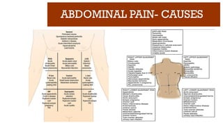
ABDOMINAL PAIN- CAUSES
Inflammation of a viscus
Perforation of a viscus
Obstruction of a viscus
Infarction of a viscus
Intra-abdominal hemorrhage or retroperitoneal hemorrhage
Extra-abdominal or medical causes for acute abdominal pain like lower lobe
pneumonia and inferior wall MI
6.
ABDOMINAL PAIN- HISTORY
SOCRATES= Nemonic
S - site
O - onset
C - characteristics
R - radiation
A – associated symptoms &
signs
T - timing
E - exacerbating/
alleviating
S - severity
“S” stands for “site”. Which region/quadrant? Is
it a general sense of overall discomfort? The site
of pain helps you fine tune your subsequent
physical exam and diagnostic decision making.
“O” stands for “onset”. When did the pain start?
Acute or insidious?
“C” stands for “characteristics”. The pain may
be sharp, dull, heavy, etc. or a combination of
descriptions.
7.
ABDOMINAL PAIN- HISTORY
SOCRATES= Nemonic
S - site
O - onset
C - characteristics
R - radiation
A – associated symptoms &
signs
T - timing
E - exacerbating/
alleviating
S - severity
“R”, which represents “radiation”. Ask if the
pain stays at the site they are describing or if it
travels somewhere else in the body. Ex:Ureteric
colic
A” stands for associated symptoms. What other
symptoms are present and associated with the
pain? Ask do they also have nausea and/or
vomiting?
"T" stands for timing. When does the pain
occur? Does it happen at specific times of the
day, or is it constant?
8.
ABDOMINAL PAIN- HISTORY
SOCRATES= Nemonic
S - site
O - onset
C - characteristics
R - radiation
A – associated symptoms &
signs
T - timing
E - exacerbating/
alleviating
S - severity
“E” represents “exacerbating” factors; grouped
within this is also alleviating factors. The
patient should be probed as to what makes their
pain better or worse. Certain physical positions,
medications, etc. These factors can all provide
historical clues about the root cause.
“S” stands for “severity”. In most hospitals this
is formulated on a 1 to 10 scale with 10 being
the most severe pain they’ve ever experienced.
9.
ABDOMINAL PAIN
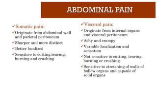
Somatic pain:
Originatefrom abdominal wall
and parietal peritoneum
Sharper and more distinct
Better localized
Sensitive to cutting,tearing,
burning and crushing
Visceral pain:
Originate from internal organs
and visceral peritoneum
Achy and crampy
Variable localization and
sensation
Not sensitive to cutting, tearing,
burning or crushing
Sensitive to stretching of walls of
hollow organs and capsule of
solid organs
10.
ABDOMINAL PAIN
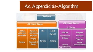
Shifting pain:Ex: Periumbilical pain shifting to RLQ in
Ac.appendicitis
Radiating pain: Ex: Pain radiating from loin to groin in ureteric
colic
Reffered pain: Ex: Pain felt at Lt shoulder in case of splenic
rupture
11.
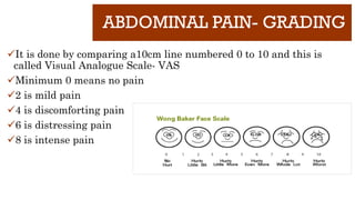
ABDOMINAL PAIN- GRADING
Itis done by comparing a10cm line numbered 0 to 10 and this is
called Visual Analogue Scale- VAS
Minimum 0 means no pain
2 is mild pain
4 is discomforting pain
6 is distressing pain
8 is intense pain
13.
CASE BASED LEARNING
ABDOMINALPAIN
RLQ PAIN
Dr.B.Selvaraj MS;Mch;FICS;
Professor of Surgery
Melaka Manipal Medical College
Melaka 75150 Malaysia
14.
ABDOMINAL PAIN-RLQ PAIN
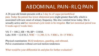
A28-year-old female presents with a 2-day hx of vague periumbilical
pain. Today the patient has lower abdominal pain (right greater than left), which is
associated with new onset of urinary frequency. She also vomited twice today. She is
sexually active and her menstrual cycle tends to be irregular. Her last menstrual period was
6 weeks ago (2 weeks late).
VS: T = 100.2, HR = 90, BP = 110/68.
Labs: H/H = 12.0/36.3, WBC = 11,300, U/A: 5 RBCs, 10–15 WBCs
Physical examination: RLQ tenderness, guarding, and rebound.
Pelvic examination without cervical motion tenderness
What would be your differential dx and plan for further evaluation?
ABDOMINAL PAIN-RLQ PAIN
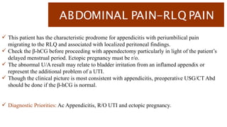
This patient has the characteristic prodrome for appendicitis with periumbilical pain
migrating to the RLQ and associated with localized peritoneal findings.
Check the β-hCG before proceeding with appendectomy particularly in light of the patient’s
delayed menstrual period. Ectopic pregnancy must be r/o.
The abnormal U/A result may relate to bladder irritation from an inflamed appendix or
represent the additional problem of a UTI.
Though the clinical picture is most consistent with appendicitis, preoperative USG/CT Abd
should be done if the β-hCG is normal.
Diagnostic Priorities: Ac Appendicitis, R/O UTI and ectopic pregnancy.
18.
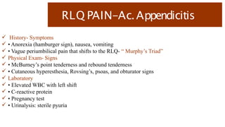
RLQ PAIN-Ac.Appendicitis
History-Symptoms
• Anorexia (hamburger sign), nausea, vomiting
• Vague periumbilical pain that shifts to the RLQ- “ Murphy’s Triad”
Physical Exam- Signs
• McBurney’s point tenderness and rebound tenderness
• Cutaneous hyperesthesia, Rovsing’s, psoas, and obturator signs
Laboratory
• Elevated WBC with left shift
• C-reactive protein
• Pregnancy test
• Urinalysis: sterile pyuria
19.
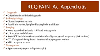
RLQ PAIN-Ac.Appendicitis
Diagnosis
• Oftentimes is a clinical diagnosis
Pathophysiology
• Closed-loop obstruction
• Fecolith in adults, lymphoid hyperplasia in children
Imaging
• None needed with classic H&P and leukocytosis
• US: women and children
• Avoid CT in children (increased risk of malignancy) and pregnancy (risk to fetus)
• CT: if diagnosis is equivocal in men and nonpregnant women
• MRI: pregnant women
Management
• Appendectomy (open or laparoscopic)
20.
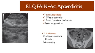
RLQ PAIN-Ac.Appendicitis
USGAbdomen:
Tubular structure
More than 6mm in diameter
Non compressible
CT Abdomen:
Thickened appendix
Fecolith
Fat stranding
CASE BASED LEARNING
ABDOMINALPAIN
RUQ PAIN-1
Dr.B.Selvaraj MS;Mch;FICS;
Professor of Surgery
Melaka Manipal Medical College
Melaka 75150 Malaysia
25.
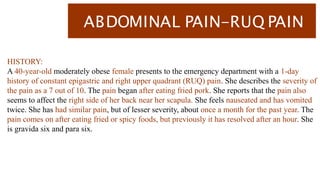
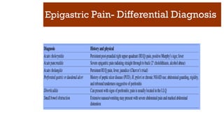
ABDOMINAL PAIN-RUQ PAIN
HISTORY:
A40-year-old moderately obese female presents to the emergency department with a 1-day
history of constant epigastric and right upper quadrant (RUQ) pain. She describes the severity of
the pain as a 7 out of 10. The pain began after eating fried pork. She reports that the pain also
seems to affect the right side of her back near her scapula. She feels nauseated and has vomited
twice. She has had similar pain, but of lesser severity, about once a month for the past year. The
pain comes on after eating fried or spicy foods, but previously it has resolved after an hour. She
is gravida six and para six.
26.
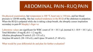
ABDOMINAL PAIN-RUQ PAIN
Onphysical examination, her temperature is 100 °F, heart rate is 110/min, and her blood
pressure is 120/80 mmHg. She has marked tenderness in the RUQ of the abdomen to palpation.
When the RUQ is palpated while she is taking a deep breath, she abruptly ceases inspiration
secondary to pain Murphy’s Sign+ve
Laboratory values are significant for WBC count of 14 × 10 3 /μL (normal 4.1–10.9 × 10 3 /μL),
Total Bilirubin 1.0 mg/dL (0.1–1.2 mg/dl),
Alkaline phosphatase70 units/L (33–131 u/L),
Amylase 60 units/L (30–110 u/L), and Lipase 30 units/L (7–60 u/L).
What would be your differential dx and plan for further evaluation?
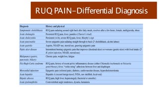
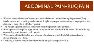
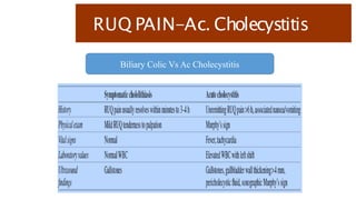
ABDOMINAL PAIN-RUQ PAIN
With her current history of severe persistent abdominal pain following ingestion of fatty
foods, nausea and vomiting, and associated right upper quadrant tenderness to palpation, the
etiology is most likely of biliary origin.
The patient’s prior history is consistent with symptomatic cholelithiasis
With a positive Murphy’s sign, fever, tachycardia, and elevated WBC count, the most likely
current diagnosis is acute cholecystitis.
With a normal total bilirubin and alkaline phosphatase, choledocholithiasis and acute
cholangitis are less likely.
Similarly, a normal amylase and lipase rule out gallstone pancreatitis.
29.
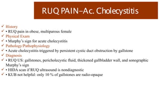
RUQ PAIN-Ac. Cholecystitis
History
• RUQ pain in obese, multiparous female
Physical Exam
• Murphy’s sign for acute cholecystitis
Pathology/Pathophysiology
• Acute cholecystitis triggered by persistent cystic duct obstruction by gallstone
Diagnosis
• RUQ US: gallstones, pericholecystic fluid, thickened gallbladder wall, and sonographic
Murphy’s sign
• HIDA scan if RUQ ultrasound is nondiagnostic
• KUB not helpful: only 10 % of gallstones are radio-opaque
RUQ PAIN-Ac. Cholecystitis
Management
• Asymptomatic gallstones: cholecystectomy not indicated
• Symptomatic cholelithiasis (biliary colic): elective lap cholecystectomy
• Acute cholecystitis: urgent (within 48 h) lap cholecystectomy
• Acute acalculous cholecystitis: cholecystostomy tube if critically ill
• Emphysematous cholecystitis: emergent cholecystectomy
• Gallstone ileus: remove large impacted gallstone from terminal ileum (leave gallbladder
alone)
Postoperative
• If a patient presents within the first week after cholecystectomy with abdominal pain,
distention, and anorexia, consider a biloma (cystic duct stump leak, CBD injury)
• Cystic duct stump leak readily treated with ERCP and stenting of the sphincter of Oddi
• CBD injury may require hepaticojejunostomy/choledochojejunostomy
33.
RUQ PAIN-Ac. Cholecystitis
Additional Important Facts
• Ursodeoxycholic acid could be employed as conservative management for patients with
cholelithiasis
• Calcified gallbladder (porcelain): increased risk of malignancy, perform cholecystectomy
• Choledochal cysts are congenital dilations of the biliary tree; prone to cholangitis, risk of
associated malignancy, need to excise (if intrahepatic ducts are involved (Caroli’s disease),
and may need liver transplantation
• Hemolytic anemia in childhood: high risk of black pigment gallstones
• Gallbladder cancer: associated with gallstones
• Gallbladder polyps: > 1 cm suspicious for cancer; >2 cm high likelihood of cancer
34.
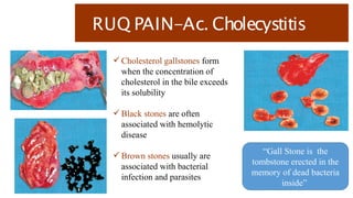
RUQ PAIN-Ac. Cholecystitis
Cholesterol gallstones form
when the concentration of
cholesterol in the bile exceeds
its solubility
Black stones are often
associated with hemolytic
disease
Brown stones usually are
associated with bacterial
infection and parasites
“Gall Stone is the
tombstone erected in the
memory of dead bacteria
inside”
35.
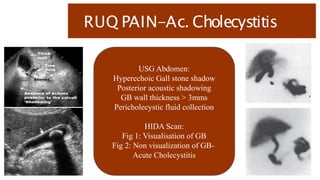
RUQ PAIN-Ac. Cholecystitis
USGAbdomen:
Hyperechoic Gall stone shadow
Posterior acoustic shadowing
GB wall thickness > 3mms
Pericholecystic fluid collection
HIDA Scan:
Fig 1: Visualisation of GB
Fig 2: Non visualization of GB-
Acute Cholecystitis
37.
CASE BASED LEARNING
ABDOMINALPAIN
RUQ PAIN-2
Dr.B.Selvaraj MS;Mch;FICS;
Professor of Surgery
Melaka Manipal Medical College
Melaka 75150 Malaysia
38.
ABDOMINAL PAIN-RUQ PAIN
HISTORY:
A40-year-old female presents with a 24 hour history of right upper quadrant (RUQ) and
epigastric pain, associated with nausea and vomiting. She has had similar pain in the past,
particularly after eating greasy foods. According to her family, over the last few hours, the
patient has become slightly confused. Past medical history is negative.
PHYSICAL EXAM:
Temperature of 102.5 °F, a heart rate of 110 beats/min, respiratory rate of 16/min, and a blood
pressure of 90/60mmHg. She is moderately tender in the RUQ to deep palpation. She has slight
scleral icterus. She has noted dark- colored Urine. The remainder of her abdominal exam is
negative.
39.
ABDOMINAL PAIN-RUQ PAIN
LaboratoryValues:
White blood count of 15 × 10 3 /μL (normal 4.1–10.9 × 10 3 /μL),
Total bilirubin of 4.0 mg/dl (0.1–1.2 mg/dl),
Alkaline phosphatase (AP) of 350 μ/L (33–131 μ/L),
Aspartate aminotransferase (AST) of 300 μ/L (5–35 μ/L)
Alanine aminotransferase (ALT) of 280 μ/L (7–56 μ/L),
Gamma-glutamyl transpeptidase (GGT) of 330 μ/L (8–88 μ/L),
Amylase of 100 μ/L (30–110 μ/L).
Urine is positive for bilirubin.
What would be your differential dx and plan for further evaluation?
ABDOMINAL PAIN-RUQ PAIN
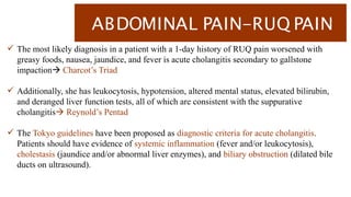
The most likely diagnosis in a patient with a 1-day history of RUQ pain worsened with
greasy foods, nausea, jaundice, and fever is acute cholangitis secondary to gallstone
impaction Charcot’s Triad
Additionally, she has leukocytosis, hypotension, altered mental status, elevated bilirubin,
and deranged liver function tests, all of which are consistent with the suppurative
cholangitis Reynold’s Pentad
The Tokyo guidelines have been proposed as diagnostic criteria for acute cholangitis.
Patients should have evidence of systemic inflammation (fever and/or leukocytosis),
cholestasis (jaundice and/or abnormal liver enzymes), and biliary obstruction (dilated bile
ducts on ultrasound).
42.
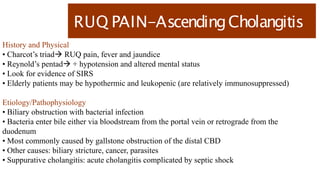
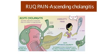
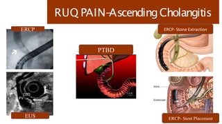
RUQ PAIN-AscendingCholangitis
History andPhysical
• Charcot’s triad RUQ pain, fever and jaundice
• Reynold’s pentad + hypotension and altered mental status
• Look for evidence of SIRS
• Elderly patients may be hypothermic and leukopenic (are relatively immunosuppressed)
Etiology/Pathophysiology
• Biliary obstruction with bacterial infection
• Bacteria enter bile either via bloodstream from the portal vein or retrograde from the
duodenum
• Most commonly caused by gallstone obstruction of the distal CBD
• Other causes: biliary stricture, cancer, parasites
• Suppurative cholangitis: acute cholangitis complicated by septic shock
43.
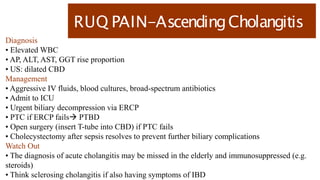
RUQ PAIN-AscendingCholangitis
Diagnosis
• ElevatedWBC
• AP, ALT, AST, GGT rise proportion
• US: dilated CBD
Management
• Aggressive IV fluids, blood cultures, broad-spectrum antibiotics
• Admit to ICU
• Urgent biliary decompression via ERCP
• PTC if ERCP fails PTBD
• Open surgery (insert T-tube into CBD) if PTC fails
• Cholecystectomy after sepsis resolves to prevent further biliary complications
Watch Out
• The diagnosis of acute cholangitis may be missed in the elderly and immunosuppressed (e.g.
steroids)
• Think sclerosing cholangitis if also having symptoms of IBD
CASE BASED LEARNING
ABDOMINALPAIN
EPIGASTRIC PAIN- 1
Dr.B.Selvaraj MS;Mch;FICS;
Professor of Surgery
Melaka Manipal Medical College
Melaka 75150 Malaysia
48.
ABDOMINAL PAIN-
EPIGASTRIC PAIN-1
HISTORY:
A 56-year-old male with a history of gastroesophageal reflux disease (GERD),
hypertension, and diabetes presents to the emergency room complaining of severe
upper central abdominal pain. The patient reports epigastric pain for months, but it
has just acutely become intolerable over the last 8 hrs. He states that the chronic
pain has been a “gnawing” pain that comes on after eating. He thought he was just
having some indigestion and would take some antacids for relief. Late last night,
the pain became excruciating and now he is having trouble moving.
49.
ABDOMINAL PAIN- RUQPAIN
On physical examination, blood pressure is 130/70 mmHg, heart rate is
110 bpm, and temperature is 101.5 °F. He appears to be in severe distress
secondary to pain. The patient refuses to straighten his legs because it hurts too
much. He almost jumps off of the table when you press on his abdomen. He has
diffuse guarding and rebound tenderness.
Laboratory values: WBC of 15 (normal 4.1–10.9 × 10 3 /μL), BUN of 35 (7–20
mg/dL), creatinine of 1.8 (0.5–1.4 mg/dL), serum amylase of 70 (30–110 μ/L), and
lipase of 60 (7–60 u/L).
An upright CXR demonstrates free air under the right diaphragm.
What would be your differential dx and plan for further evaluation?
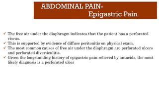
ABDOMINAL PAIN-
Epigastric Pain
✓The free air under the diaphragm indicates that the patient has a perforated
viscus.
✓ This is supported by evidence of diffuse peritonitis on physical exam.
✓ The most common causes of free air under the diaphragm are perforated ulcers
and perforated diverticulitis.
✓ Given the longstanding history of epigastric pain relieved by antacids, the most
likely diagnosis is a perforated ulcer
52.
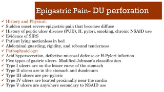
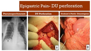
Epigastric Pain- DUperforation
✓ History and Physical:
✓ Sudden onset severe epigastric pain that becomes diffuse
✓ History of peptic ulcer disease (PUD), H. pylori, smoking, chronic NSAID use
✓ Evidence of SIRS
✓ Patient lying motionless in bed
✓ Abdominal guarding, rigidity, and rebound tenderness
✓ Pathophysiology:
✓ Acid hypersecretion, defective mucosal defense or H.Pylori infection
✓ Five types of gastric ulcers- Modified Johnson’s classification
✓ Type I ulcers are on the lesser curve of the stomach
✓ Type II ulcers are in the stomach and duodenum
✓ Type III ulcers are pre-pyloric
✓ Type IV ulcers are located proximally near the cardia
✓ Type V ulcers are anywhere secondary to NSAID use
53.
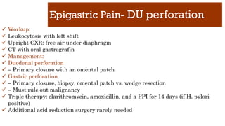
Epigastric Pain- DUperforation
✓ Workup:
✓ Leukocytosis with left shift
✓ Upright CXR: free air under diaphragm
✓ CT with oral gastrografin
✓ Management:
✓ Duodenal perforation
✓ – Primary closure with an omental patch
✓ Gastric perforation
✓ – Primary closure, biopsy, omental patch vs. wedge resection
✓ – Must rule out malignancy
✓ Triple therapy: clarithromycin, amoxicillin, and a PPI for 14 days (if H. pylori
positive)
✓ Additional acid reduction surgery rarely needed
54.
Epigastric Pain- DUperforation
Pneumoperitoneum DU Perforation Graham’s Patch- Omentopexy
56.
CASE BASED LEARNING
ABDOMINALPAIN
EPIGASTRIC PAIN- 2
Dr.B.Selvaraj MS;Mch;FICS;
Professor of Surgery
Melaka Manipal Medical College
Melaka 75150 Malaysia
57.
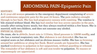
ABDOMINAL PAIN-Epigastric Pain
HISTORY:
A41-year-old woman presents to the emergency department complaining of severe
and continuous epigastric pain for the past 24 hours. The pain radiates straight
through to her back. She has had progressive nausea with vomiting. The vomitus is
bile stained without blood. She has had similar but less severe episodes in the past,
usually after eating heavy meals, but they always resolved within a few hours. She is
married, with two children, and does not consume any alcohol.
PHYSICAL EXAM:
On exam, she is afebrile, heart rate is 115/min, blood pressure is 128/86 mmHg, and
she has a normal respiratory rate. Her abdomen is not distended. She has no
surgical scars on her abdomen and no obvious masses visible. She has no bruising
around her umbilicus or along her flank. Bowel sounds are hypoactive. She has
marked tenderness to palpation in her epigastrium, without guarding or rebound.
The remainder of her abdomen is soft and non-tender to palpation. No masses or
organomegaly are appreciated.
58.
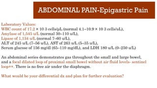
ABDOMINAL PAIN-Epigastric Pain
LaboratoryValues:
WBC count of 17.2 × 10 3 cells/μL (normal 4.1–10.9 × 10 3 cells/uL),
Amylase of 1,545 u/L (normal 30–110 u/L),
Lipase of 1,134 u/L (normal 7–60 u/L),
ALT of 245 u/L (7–56 u/L), AST of 263 u/L (5–35 u/L),
Serum glucose of 156 mg/dl (65–110 mg/dL), and LDH 180 u/L (0–250 u/L)
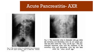
An abdominal series demonstrates gas throughout the small and large bowel,
and a focal dilated loop of proximal small bowel without air fluid levels- sentinel
loop++. There is no free air under the diaphragm.
What would be your differential dx and plan for further evaluation?
ABDOMINAL PAIN-Epigastric Pain
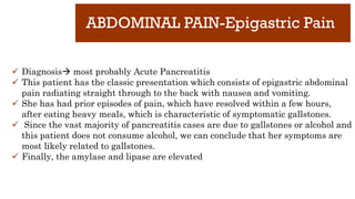
✓Diagnosis most probably Acute Pancreatitis
✓ This patient has the classic presentation which consists of epigastric abdominal
pain radiating straight through to the back with nausea and vomiting.
✓ She has had prior episodes of pain, which have resolved within a few hours,
after eating heavy meals, which is characteristic of symptomatic gallstones.
✓ Since the vast majority of pancreatitis cases are due to gallstones or alcohol and
this patient does not consume alcohol, we can conclude that her symptoms are
most likely related to gallstones.
✓ Finally, the amylase and lipase are elevated
61.
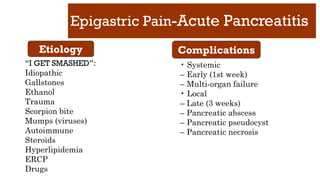
Epigastric Pain-Acute Pancreatitis
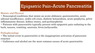
Historyand Physical
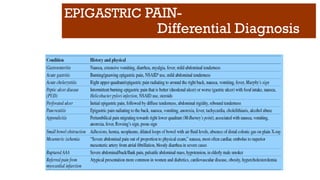
• Nonsurgical conditions that mimic an acute abdomen: gastroenteritis, acute
adrenal insufficiency, sickle cell crisis, diabetic ketoacidosis, acute porphyria, pelvic
inflammatory disease, kidney stones, and pyelonephritis
• Patients with pancreatitis typically present with epigastric pain radiating to the
back, nausea, vomiting, anorexia, fever,tachycardia.
Pathophysiology
• The initial event in pancreatitis is the inappropriate activation of pancreatic
enzymes
• Gallstones and alcohol are the most common causes of acute pancreatitis
62.
Epigastric Pain-Acute Pancreatitis
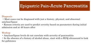
Diagnosis
•Most cases can be diagnosed with just a history, physical, and abnormal
amylase/lipase
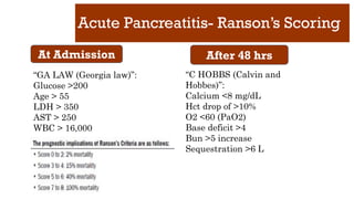
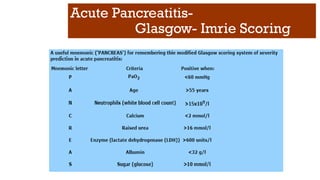
• Ranson criteria are used to predict severity based on parameters during initial
admission and at 48 hours after
Workup
• Amylase/lipase levels do not correlate with severity of pancreatitis
• In the absence of a history of alcohol abuse, start with a RUQ ultrasound to look
for gallstones
63.
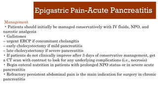
Epigastric Pain-Acute Pancreatitis
Management
•Patients should initially be managed conservatively with IV fluids, NPO, and
narcotic analgesia
• Gallstones
– urgent ERCP if concomitant cholangitis
– early cholecystectomy if mild pancreatitis
– late cholecystectomy if severe pancreatitis
• If patients do not clinically improve after 3 days of conservative management, get
a CT scan with contrast to look for any underlying complications (i.e., necrosis)
• Begin enteral nutrition in patients with prolonged NPO status or in severe acute
pancreatitis
• Refractory persistent abdominal pain is the main indication for surgery in chronic
pancreatitis
Acute Pancreatitis- Ranson’sScoring
At Admission After 48 hrs
“GA LAW (Georgia law)”:
Glucose >200
Age > 55
LDH > 350
AST > 250
WBC > 16,000
“C HOBBS (Calvin and
Hobbes)”:
Calcium <8 mg/dL
Hct drop of >10%
O2 <60 (PaO2)
Base deficit >4
Bun >5 increase
Sequestration >6 L
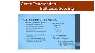
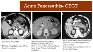
Acute Pancreatitis- CECT
CECT:Acute Pancreatitis
Diffusely enlarged pancreas with low
density from edema
Phlegmon / Inflammatory Mass
White arrowheads: Phlegmon
Black arrowhead: Pancreatic calcification
Large Arrow: Peripancreatic fascial
infiltration
Acute Pancreatitis / Pancreatic Necrosis
Arrow: No enhancement of pancreas
with IV contrast
Arrowheads: Normal enhancement in
the tail of Pancreas.
72.
CASE BASED LEARNING
ABDOMINALPAIN
LLQ Pain-1
Dr.B.Selvaraj MS;Mch;FICS;
Professor of Surgery
Melaka Manipal Medical College
Melaka 75150 Malaysia
73.
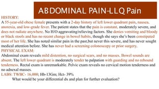
ABDOMINAL PAIN-
LLQ Pain
HISTORY:
A55-year-old obese female presents with a 2-day history of left lower quadrant pain, nausea,
anorexia, and low-grade fever. The patient states that the pain is constant, moderately severe, and
does not radiate anywhere. No H/O aggravating/relieving factors. She denies vomiting and bloody
or black stools and has no recent change in bowel habits, though she says she’s been constipated
most of her life. She has noted similar pain in the past,but never this severe, and has never sought
medical attention before. She has never had a screening colonoscopy or prior surgery.
PHYSICAL EXAM:
Abdominal exam reveals mild distention, no surgical scars, and no masses. Bowel sounds are
absent. The left lower quadrant is moderately tender to palpation with guarding and no rebound
tenderness. Rectal exam is unremarkable. Pelvic exam reveals no cervical motion tenderness and
no adnexal masses.
LABS: TWBC- 16,000; Hb-13Gm; Hct- 39%
What would be your differential dx and plan for further evaluation?
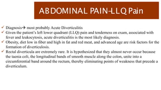
ABDOMINAL PAIN-
LLQ Pain
Diagnosis most probably Acute Diverticulitis
Given the patient’s left lower quadrant (LLQ) pain and tenderness on exam, associated with
fever and leukocytosis, acute diverticulitis is the most likely diagnosis.
Obesity, diet low in fiber and high in fat and red meat, and advanced age are risk factors for the
formation of diverticulosis.
Rectal diverticula are extremely rare. It is hypothesized that they almost never occur because
the taenia coli, the longitudinal bands of smooth muscle along the colon, unite into a
circumferential band around the rectum, thereby eliminating points of weakness that precede a
diverticulum.
76.
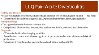
LLQ Pain-
AcuteDiverticulitis
History andPhysical
• Major risk factors are obesity, advanced age, and diet low in fiber, high in fat and red meat
• Diverticulitis is a clinical diagnosis (LLQ pain and tenderness, fever, leukocytosis)
Pathophysiology
• The sigmoid colon is the most common site
• Complicated diverticulitis: abscess, free perforation, fistula, stricture, and obstruction
Diagnosis
• CT scan is the first-line imaging modality
• Avoid barium enema and colonoscopy in acute presentation because of increased risk of
perforation
• Determine if complicated or uncomplicated and with or without SIRS
77.
LLQ Pain-
AcuteDiverticulitis
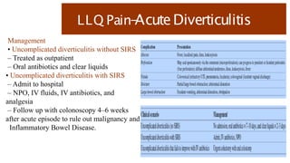
Management
• Uncomplicateddiverticulitis without SIRS
– Treated as outpatient
– Oral antibiotics and clear liquids
• Uncomplicated diverticulitis with SIRS
– Admit to hospital
– NPO, IV fluids, IV antibiotics, and
analgesia
– Follow up with colonoscopy 4–6 weeks
after acute episode to rule out malignancy and
Inflammatory Bowel Disease.
78.
LLQ Pain-
AcuteDiverticulitis
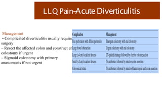
Management
• Complicateddiverticulitis usually requires
surgery
– Resect the affected colon and construct end
colostomy if urgent
– Sigmoid colectomy with primary
anastomosis if not urgent
79.
LLQ Pain-
AcuteDiverticulitis
-Etiopathogenesis
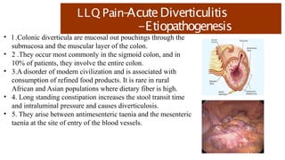
• l.Colonic diverticula are mucosal out pouchings through the
submucosa and the muscular layer of the colon.
• 2 .They occur most commonly in the sigmoid colon, and in
10% of patients, they involve the entire colon.
• 3.A disorder of modern civilization and is associated with
consumption of refined food products. It is rare in rural
African and Asian populations where dietary fiber is high.
• 4. Long standing constipation increases the stool transit time
and intraluminal pressure and causes diverticulosis.
• 5. They arise between antimesenteric taenia and the mesenteric
taenia at the site of entry of the blood vessels.
81.
CASE BASED LEARNING
ABDOMINALPAIN
LLQ PAIN-2
Dr.B.Selvaraj MS;Mch;FICS;
Professor of Surgery
Melaka Manipal Medical College
Melaka 75150 Malaysia
82.
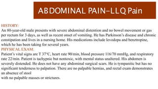
ABDOMINAL PAIN-LLQ Pain
HISTORY:
An80-year-old male presents with severe abdominal distention and no bowel movement or gas
per rectum for 3 days, as well as recent onset of vomiting. He has Parkinson’s disease and chronic
constipation and lives in a nursing home. His medications include levodopa and benztropine,
which he has been taking for several years.
PHYSICAL EXAM:
Patient’s vital signs are T 37°C, heart rate 90/min, blood pressure 116/70 mmHg, and respiratory
rate 22/min. Patient is tachypnic but nontoxic, with mental status unaltered. His abdomen is
severely distended. He does not have any abdominal surgical scars. He is tympanitic but has no
significant tenderness to palpation. There are no palpable hernias, and rectal exam demonstrates
an absence of stool
with no palpable masses or strictures.
83.
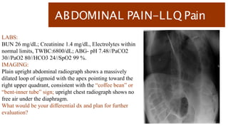
ABDOMINAL PAIN-LLQ Pain
LABS:
BUN26 mg/dL; Creatinine 1.4 mg/dL, Electrolytes within
normal limits, TWBC:6800/dL; ABG- pH 7.48//PaCO2
30//PaO2 80//HCO3 24//SpO2 99 %.
IMAGING:
Plain upright abdominal radiograph shows a massively
dilated loop of sigmoid with the apex pointing toward the
right upper quadrant, consistent with the “coffee bean” or
“bent-inner tube” sign; upright chest radiograph shows no
free air under the diaphragm.
What would be your differential dx and plan for further
evaluation?
ABDOMINAL PAIN-
LLQ Pain
Diagnosis most probably Sigmoid Volvulus
The massive, slowly progressive abdominal distention, combined with obstipation, and X-ray
findings are consistent with large bowel obstruction. The radiologic appearance is most
consistent with a sigmoid volvulus.
Large bowel obstruction-LBO- is more likely to be associated with more pronounced
distention, less or late onset vomiting, and decreased bowel sounds.
Small bowel obstruction- SBO- SBOs tend to be associated with more pronounced vomiting.
In an early SBO, bowel sounds are hyperactive, with “rushes and tinkles”- Borborygmi. In late
SBO- absent bowel sounds- silent abdomen.
86.
LLQ Pain-SigmoidVolvulus
History andPhysical
• LBO – gradual and severe abdominal distention, obstipation, and vomiting
• Uncomplicated volvulus – normal vitals, normal mental status, and non-tender abdomen
• Complicated volvulus – severe abdominal pain, fever, tachycardia, toxic appearance, peritoneal
signs, and leukocytosis
• Look for abdominal scars and hernias and perform a rectal exam to assess other differential
diagnoses
Etiology/Risk Factors:
• Most common causes of LBO: Cancer, Diverticulitis & Volvulus
• Sigmoid Volvulus – acquired stretching of the sigmoid
– Neuropsychiatric disease, institutionalization, chronic constipation, long-term anticholinergic
use, highfiber diet, and pregnancy
• Cecal Volvulus – congenital failure of fixation of the cecum
87.
LLQ Pain-SigmoidVolvulus
Diagnosis
• Comparedto LBO, SBO has faster onset and more likely to cause vomiting and high-pitched
bowel sounds
• Patients with Ogilvie’s syndrome are more likely to be already hospitalized and bedridden,
often in the postoperative setting
• Abdominal X-ray
– Sigmoid volvulus –“coffee bean”, “omega”, or “bent inner tube”, “kidney-bean” sign
– Cecal volvulus –“comma” or “kidney bean” sign- Human embryo sign
• CT scan if equivocal X-ray findings
• Contrast enema may be diagnostic (“bird’s beak” sign) and therapeutic in reducing the volvulus
– Water-soluble contrast (Gastrografin) rather than barium, to avoid peritonitis and scarring in
case of perforation
– Bowel wall thickening, mesenteric edema, pneumatosis, and portal venous gas suggest
ischemic bowel
88.
LLQ Pain-SigmoidVolvulus
Management
• Therapydiffers based on the location and severity of complication
– Uncomplicated sigmoid volvulus – endoscopic detorsion followed by semi-elective resection
– Complicated sigmoid volvulus – no detorsion attempted; emergent laparotomy with resection
– Cecal volvulus – no detorsion attempted; take to OR for right colectomy
• Complications of surgery – wound infection, anastomotic leak, and recurrence. Without
detorsion or resection – ischemia,
perforation, and sepsis