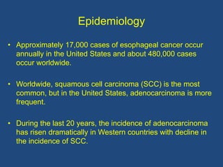
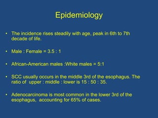
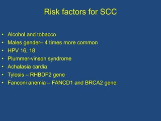
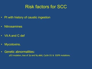
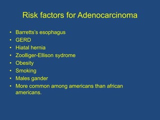
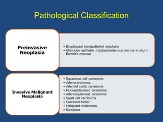
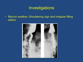
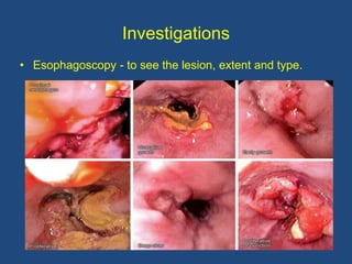
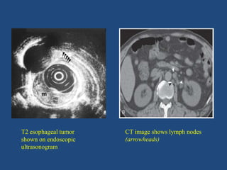
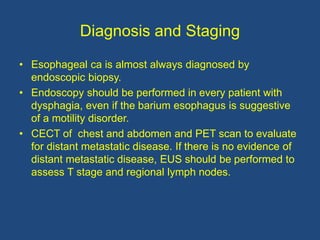
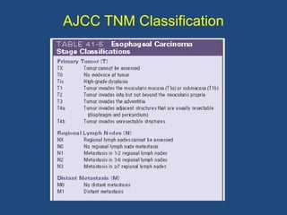
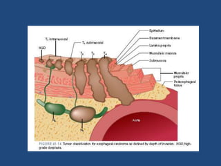
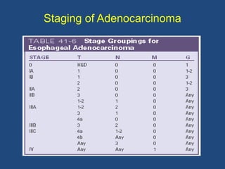
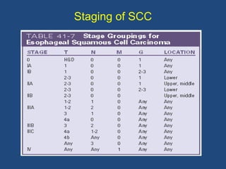
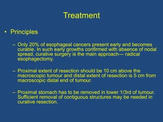
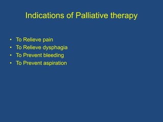
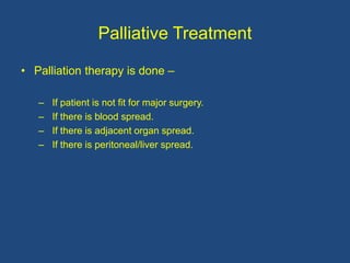
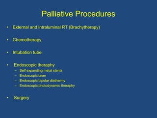
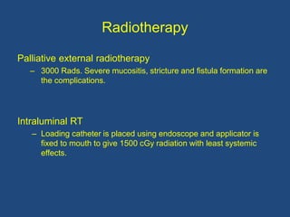
3. Introduction
• Carcinoma esophagus is common in China, South Africa and
Asian countries. In India, it is common in Karnataka and Orissa.
• It is less common in America and European countries.
• It is 6th most common cancer in the world.
• It constitutes less than 1% of all cancers. Accounts 7% of all GI
malignancies.
• Dysphagia is most common symptom at presentation. By this
time of presentation, it becomes advanced and inoperable and
only palliative is treatment of choice.
5. Anatomical Specialties
• Lacks serosa (other structure without serosa is rectum).
• Contains 2 different types of muscles (striated and
smooth at proximal 1/3 and distal 2/3 respectively)
• Contains 2 different types of epithelium.
• Segmental blood supply.
• Only part of GIT which shows very thinly scattered
Meissner’s plexus.
• Longitudinal arrangement of veins and lymphatics.
6. Epidemiology
• Approximately 17,000 cases of esophageal cancer occur
annually in the United States and about 480,000 cases
occur worldwide.
• Worldwide, squamous cell carcinoma (SCC) is the most
common, but in the United States, adenocarcinoma is more
frequent.
• During the last 20 years, the incidence of adenocarcinoma
has risen dramatically in Western countries with decline in
the incidence of SCC.
7. Epidemiology
• The incidence rises steadily with age, peak in 6th to 7th
decade of life.
• Male : Female = 3.5 : 1
• African-American males :White males = 5:1
• SCC usually occurs in the middle 3rd of the esophagus. The
ratio of upper : middle : lower is 15 : 50 : 35.
• Adenocarcinoma is most common in the lower 3rd of the
esophagus, accounting for 65% of cases.
8. Risk factors for SCC
• Alcohol and tobacco
• Males gender– 4 times more common
• HPV 16, 18
• Plummer-vinson syndrome
• Achalasia cardia
• Tylosis – RHBDF2 gene
• Fanconi anemia – FANCD1 and BRCA2 gene
9. Risk factors for SCC
• Pt with history of caustic ingestion
• Nitrosamines
• Vit A and C def
• Mycotoxins.
• Genetic abnormalities:
p53 mutation, loss of 3p and 9q alleli, Cyclin D1 & EGFR mutations.
10. Risk factors for Adenocarcinoma
• Barretts’s esophagus
• GERD
• Hiatal hernia
• Zoolliger-Ellison sydrome
• Obesity
• Smoking
• Males gander
• More common among americans than african
americans.
12. Spread of Ca Esophagus
• Direct
• Lymphatic
• Haematogenous
13. Direct Spread
• Lack of serosal layer in esophagus favours local extension.
• In upper third it spreads through muscular layer and get adherent
to left main bronchus, trachea, and left recurrent laryngeal nerve
(causes hoarseness), aorta or its branches (causes fatal
haemorrhage).
• It may perforate and cause mediastinitis.
• It may get adherent to pleura.
• Broncho-esophageal, tracheo-esophageal, esophageal-aortic
fistulas can occur in advanced cases.
14. Lymphatic Spread
• It spreads by lymphatic permeation and lymphatic
embolization.
• It can cause satellite nodules in the esophagus, away from
the main tumour.
• Above in the neck, it spreads to supraclavicular
lymphnodes.
• In thorax, it spreads to para-esophageal, tracheobronchial
lymph nodes to sub diaphragmatic lymph nodes.
• In abdomen, it spreads to coeliac lymph nodes.
• Blood spread occurs to liver, lungs, brain and bones.
15. Clinical Features
• Recent dysphagia is the commonest feature. Two-third
of the lumen should be occluded to cause dysphagia.
• Regurgitation.
• Anorexia and loss of weight (severe), cachexia.
• Substernal or abdomen pain.
• Ascites due 2nd in liver.
• Bronchopneumonia
• Melaena.
16. Clinical Features
• Features of broncho-oesophageal fistula in carcinoma of
upper third esophagus .
• Left supraclavicular lymph nodes may be palpable.
• Hoarseness of voice due to involvement of recurrent
laryngeal nerve.
• Hiccough, due to phrenic nerve involvement.
• Back pain—due to nodal spread
(paraoesophageal/coeliac nodes).
19. Investigations
• Biopsy - for histological type and confirmation.
• Chest X-ray - to look for aspiration pneumonia.
• Bronchoscopy - to see invasion in upper third growth.
• Laryngoscopy - To identify vocal cord palsy.
20. Investigations
• Esophageal ultrasonography -
To look for the depth of the tumor, involvement of
nodes, cardia and left lobe of the liver. Nodes smaller
than 5 mm can be very well visualized by EUS which
may be missed in CT scan.
• CT scan –
To look for local extension, nodal status,
perioesophageal, diaphragmatic, pericardial vascular
infiltration, obliteration of mediastinal fat and status of
tracheobronchial tree in case of upper third growth.
22. Investigations
• U/S abdomen—to look for liver and lymph nodes status
in abdomen.
• Endoscopic esophageal staining with labelled iodine -
Here normal mucosa is stained brown and carcinoma
remains pale (as mucosa in carcinoma will not take up
iodine).
23. Investigations
• Laparoscopy –
– It is useful to see peritoneal spread, liver spread and
nodal spread. It is the only reliable method to detect
peritoneal seedlings. Biopsy from different places can
also be taken. It will prevent unnecessary laparotomy.
• PET with CT scan is used for staging and to see
response for therapy.
• Video assisted thoracoscopic approach—to stage
oesophageal carcinoma.
24. Investigations
• Endoscopic mucosal resection (EMR) –
– It is basically a diagnostic biopsy tool, but can be therapeutic in
early and premalignant lesion.
– T1a tumors are resected by EMR, as the risk of lymphnode
metastasis is very low.
– Endoscopic submucosal dissection removes the lesion up to
muscularis propria.
25. Investigations
• Chemoendoscopy, magnification endocsopies-
• They are newer methods.
• Local topical application of different strains will improve the
tumour localization, features and diagnosis.
• Biopsy is done in this areas.
27. Diagnosis and Staging
• Esophageal ca is almost always diagnosed by
endoscopic biopsy.
• Endoscopy should be performed in every patient with
dysphagia, even if the barium esophagus is suggestive
of a motility disorder.
• CECT of chest and abdomen and PET scan to evaluate
for distant metastatic disease. If there is no evidence of
distant metastatic disease, EUS should be performed to
assess T stage and regional lymph nodes.
33. Treatment
• Principles
– Only 20% of esophageal cancers present early and becomes
curable. In such early growths confirmed with absence of nodal
spread, curative surgery is the main approach— radical
esophagectomy.
– Proximal extent of resection should be 10 cm above the
macroscopic tumour and distal extent of resection is 5 cm from
macroscopic distal end of tumour.
– Proximal stomach has to be removed in lower 1/3rd of tumour.
Sufficient removal of contiguous structures may be needed in
curative resection.
34. Treatment
• Principles
– If nodes are present, then multimodal approach should be
used like—curative resection; radiotherapy and
chemotherapy. Outcome of surgery depends on location of
tumour; number, location and size of nodes; tumour
grading.
– Neoadjuvant therapy by chemotherapy and/or
radiotherapy prior to surgery may improve the survival.
35. Treatment
• Principles
– Aggressive chemoradiation also may be used as curative
therapy in some patients especially upper 1/3rd growths and
in patients who are unfit for surgery.
– Palliation therapy is done if patient is not fit for major surgery,
if there is blood spread, if there is spread to adjacent organ
and if there is peritoneal/liver spread. It is to relieve pain and
dysphagia and also to prevent aspiration and bleeding.
36. Indications for Curative Treatment
• Early growth when patient is fit.
• When there is no involvement of adjacent perioesophageal
structures, bronchus, liver or distant organs.
37. Approaches for Different Level Tumours
• Post cricoid tumour (Squamous cell carcinoma):
• Treated mainly by radiotherapy. Radical radiotherapy—
5000-6000 rads.
• Often pharyngolaryngectomy is done along with gastric or
colonic transposition. But complications are more in this
procedure. Free jejunal transfer is the other option.
38. Approaches for Different Level Tumours
Upper third growth (Squamous cell carcinoma):
– Treated mainly by radiotherapy.
– Commonly it invades left recurrent laryngeal nerve and
bronchus.
– In early and operable, McKeown three phased
esophagectomy and anastomosis is done in the neck.
Initially laparotomy is done to mobilise the stomach. Then
thoracotomy through right 5th space is done and
esophagus is mobilised. Through right side neck,
esophagus with growth is removed. Anastomosis between
pharynx and stomach is done in the neck.
39. Approaches for Different Level Tumours
Middle third growth (SCC):
– Ivor Lewis operation (Lewis-Tanner two-phased esophagectomy): By
laparotomy stomach is mobilised and Pyloroplasty is done. Through
right 5th space thoracotomy is done and growth with tumour is
mobilised. Partial esophagectomy and esophagogastric anastomosis
is done in the thorax.
– If the growth is inoperable, palliative radiotherapy is given.
40. Approaches for Different Level Tumors
Lower third growth (SCC and Adeno Ca):
– Here through left thoracoabdominal approach, partial
esophagogastrectomy is done with esophagogastric
anastomosis.Often jejunal Roux-en-Y loop anastomosis is done.
– Orringer approach, i.e. transhiatal blind total esophagectomy with
anastomosis in the left side of the neck. Through laparotomy, stomach
and lower part of the esophagus are mobilised. Through left sided
neck approach, upper part of the esophagus is mobilised using finger.
Blind dissection is completed by meeting both fingers above and
below in the thorax. Later esophagus is pulled up out through the neck
wound and removed.
41. Other Approaches
• Thoracoscopic-laparoscopic esophagectomy and
lymphadenectomy is becoming popular, safer and
effective.
• Radical esophagectomy with 3-field clearance of
abdominal/thoracic and cervical nodes is also practiced
in many centres.
42. Esophageal Substitutes
• Stomach: It is preferred one . But postprandial symptoms
are more.
• Colon: It is better as there is less postprandial problems.
Complications are leak, fistula formation.
• Jejunum: It is last option.
43. Indications of Palliative therapy
• 80% of pts have advanced tumor at the time of
presentation and so they are amenable for only palliative
treatment.
• Nodes greater than 5 involvement
• Invasive, poorly differentiated grade.
• Length of involvement >8cm.
• Abnormal esophageal axis in barium study.
• Horner’s syndrome
• Loss of wt >20%
• Metastatic disease
44. Indications of Palliative therapy
• To Relieve pain
• To Relieve dysphagia
• To Prevent bleeding
• To Prevent aspiration
45. Palliative Treatment
• Palliation therapy is done –
– If patient is not fit for major surgery.
– If there is blood spread.
– If there is adjacent organ spread.
– If there is peritoneal/liver spread.
47. Radiotherapy
Palliative external radiotherapy
– 3000 Rads. Severe mucositis, stricture and fistula formation are
the complications.
Intraluminal RT
– Loading catheter is placed using endoscope and applicator is
fixed to mouth to give 1500 cGy radiation with least systemic
effects.
48. Chemotherapy
• Cisplatin
• Methotrexate
• 5 FU
• Palcitaxel
• Etoposide
• Bleomycin
• Platinum based chemotherapy is beneficial especially in
advanced adenocarcinoma of esophagus.
49. Intubation
• Here guidewire is passed across the growth under X-ray
screening or C-arm guidance; flexible introducer and
prosthetic tube is pushed across the tumor along the
guidewire.
• It carries 90% success rate.
• Problems are tube intolerance, poor drainage, airway
compression, reflux, aspiration, displacement, food
blockage, tumor overgrowth beyond the prosthesis
causing its failure.
• Perforation chance is 10%.
50. Endoscopic therapy
• Self-expanding metal stents (SEMS) are passed through
endoscope under C-arm guidance. It is the ideal method
of palliation. Advantage – perforation is minimal.
• Problems of stents are—aspiration, displacement,
erosion, bleeding, tumor growth across or beyond mesh,
food bolus obstruction, retrosternal pain, need for
reinsertion (40%). Mortality is 1-2%.
51. Endoscopic laser
• It is used to core a channel through the tumor to improve
dysphagia. It causes thermal destruction of tumor. Nd
YAG laser and Diode laser are used.
• Success rate of palliation is 85%.
• Problems are—fever, chest pain, mortality, perforation
and fistula formation.
52. Endoscopic photodynamic therapy (PDT)
– It is used to destruct tumor and to relieve dysphagia.
It is often used as a therapy in early cancer.
Photosensitive haematoporphyrin agent is injected
intravenously 48 hours before endoscopy. It is
activated over tumour using laser. Sunburn, fever,
perforation, pleural effusions are complications. It is
effective only to superficial cancers.
53. Pallative Surgeries
• Transhiatal Orringer’s blind oesophagectomy is a
palliative surgical procedure.
• Kirschner palliative gastric bypass done in advanced
carcinoma esophagus wherein mobilised stomach is
brought to neck via retrosternal or subcutaneous route
and anastomosed to divided cervical oesophagus.Lower
cut end of oesophagus is anastomosed to a jejunal loop.
Here oesophagus is left alone.
54. Complications of esophagectomy
• 5-10% mortality
• Haemorrhage
• Respiratory infection
• Chylothorax, injury to thoracic duct
• Anastomotic leak—thoracic leak is most dangerous (5-10%)
• Hoarseness due to recurrent laryngeal nerve palsy
• Stricture formation (40%)
• GERD
• Conduit necrosis due to ischaemia to stomach or colon
• Colonic dysmotility causing partial obstruction in colon
transfer
55. Prognosis
• Not good because of early spread, longitudinal lymphatics,
aggressiveness, difficult approach, late presentation.
• Nodal involvement carries bad prognosis.
• 5-year survival rate is only 10%.
56. Summary
• Esophagus has no serosal covering, so direct invasion of
adjacent structures occurs early.
• Commonly spread by lymphatics (70%).
• Most common symptom at presentation is dysphagia.
• Often diagnosed late, so most therapeutic approaches are
palliative.
• During the last 20 years, the incidence of adenocarcinoma
has risen dramatically in Western countries.