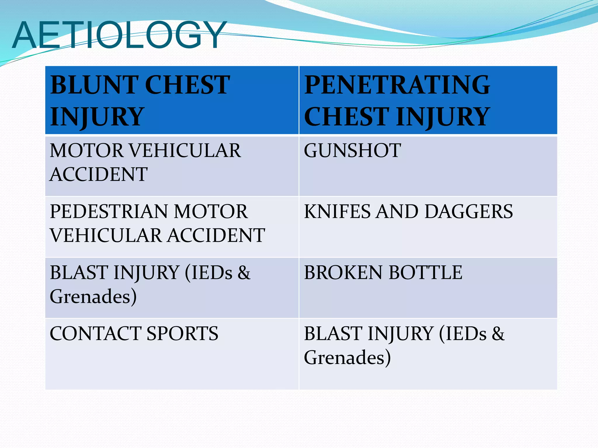
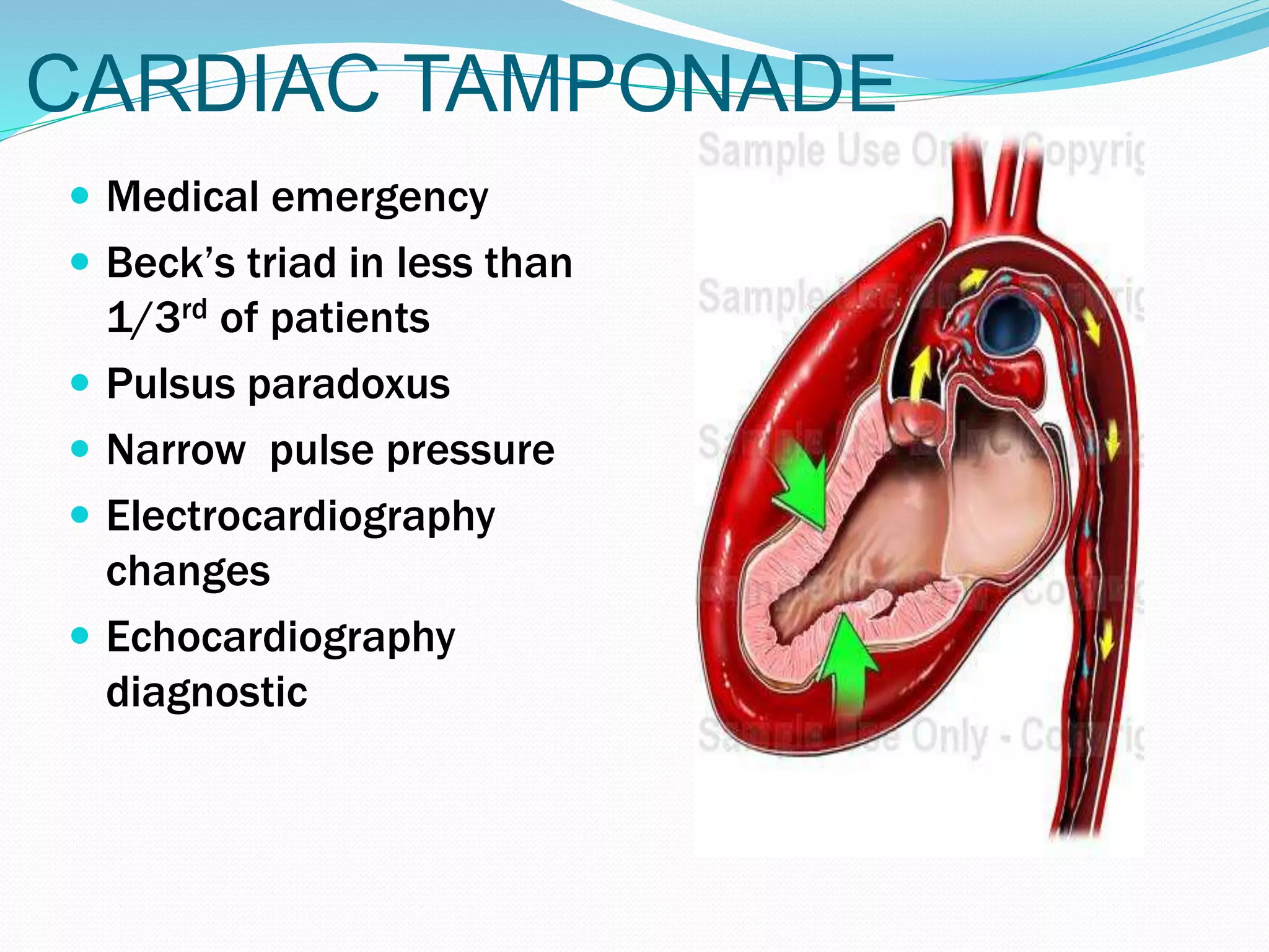
MANAGEMENT OF
CHEST INJURIES
BY
DR. LAWAL GBENGA D.
REGISTRAR, SURGERY DEPT.
NATIONAL HOSPITAL ABUJA
28/09/15
OUTLINE
 INTRODUCTION
 EPIDEMIOLOGY
 RELEVANT ANATOMY
 AETIOLOGY
 PATHOPHYSIOLOGY
 MANAGEMENT
 COMPLICATIONS
 CONCLUSION
 REFERENCES
INTRODUCTION
 Chest trauma is a significant source of morbidity and
mortality in developed and developing nations
 Thoracic injuries account for 20-25% of deaths due to
trauma
 They are either blunt or penetrating
 Less than 15% of chest trauma patients will require any
procedure more invasive than the insertion of a chest tube
Epidemiology
 Trauma is the 4rd commonest cause of death in US
 45% to 50% of unrestrained drivers involved in an
accident have thoracic injury
 25% of Motor vehicular accident (MVA) driver death
due to thoracic injury
 Penetrating trauma, 30% have thoracic injury
 Blunt trauma, 43% have thoracic injury
 Associated extra thoracic Injuries occur in 68%-85%
of chest trauma
Epidemiology
Thomas o.m. & ogunleye o.e.
• Penetrating chest injuries; gunshots 60.1%, traffic accidents 27.3%
Adegboye v.o. , Ladipo j.k. , Brimmo i.o. , adebo i.o.
• Blunt chest injuries 69%
• Majority of the blunt chest injuries were minor chest wall injuries
• 7.6% had major but stable chest wall injuries,
• 10.8% had flail chest injuries.
• Thoracic injuries without fractures of bony chest wall occurred in
13.6%.
• 59.1% had associated extra-thoracic injuries,
RELEVANT ANATOMY
APPROACHES TO CHEST
CAVITY
• Median sternotomy
• Posterolateral thoracotomy
• Left anterolateral thoracotomy
• Clam-shell
THORACOTOMY
INCISIONS
AETIOLOGY
BLUNT CHEST
INJURY
PENETRATING
CHEST INJURY
MOTOR VEHICULAR
ACCIDENT
GUNSHOT
PEDESTRIAN MOTOR
VEHICULAR ACCIDENT
KNIFES AND DAGGERS
BLAST INJURY (IEDs &
Grenades)
BROKEN BOTTLE
CONTACT SPORTS BLAST INJURY (IEDs &
Grenades)
PATHOPHYSIOLOGY
MECHANISM OF INJURY
 PENETRATING INJURY: lacerations, contusions,
disruption and destruction of tissue.
 BLUNT INJURY: rupture, in addition to above
making them more deadly
 For penetrating injuries, the trajectory defines the
anatomical injury
 Low, medium or high velocity
PATHOPHYSIOLOGY
CHEST WALL INJURY
Grouped into three categories:
1. chest wall fractures, dislocations, and barotrauma
(including diaphragmatic injuries)
2. injuries of the pleurae, lungs, and aerodigestive tracts
3. injuries of the heart, great arteries, veins, and
lymphatics.
PATHOPHYSIOLOGY
CHEST WALL INJURY
1. CHEST WALL FRACTURES, DISLOCATIONS, AND
BAROTRAUMA (INCLUDING DIAPHRAGMATIC INJURIES):
i. Rib fractures and flail chest
ii. Clavicular, sternal and scapular fractures and
sternoclavicular dislocations
iii. Diaphragmatic rupture
PATHOPHYSIOLOGY
2. INJURIES OF THE PLEURAE, LUNGS, AND
AERODIGESTIVE TRACTS:
i. Simple / Tension / Open pneumothorax
ii. Haemothorax
iii. Pulmonary contusion and parenchymal injuries
iv. Tracheobronchial injuries
v. Oesophageal injuries
PATHOPHYSIOLOGY
3. INJURIES OF THE HEART, GREAT ARTERIES, VEINS, AND
LYMPHATICS:
i. Cardiac tamponade
ii. Cardial contusion
iii. Myocardial laceration /chamber rupture
iv. Intracardial injury: septal / valvular structural damage
v. Coronary artery contusion / laceration
vi. Aorta, arch branches, SVC, IVC, brachiocephalic veins,
pulmonary arteries and veins
vii. Commotio cordis
MAJOR THORACIC INJURY
LETHAL SIX HIDDEN SIX
AIRWAY OBSTRUCTION CONTAINED TRAUMATIC
AORTIC RUPTURE
FLAIL CHEST DIAPHRAGMATIC TEAR
TENSION PNEUMOTHORAX ESOPHAGEAL PERFORATION
OPEN PNEUMOTHORAX MAJOR TRACHEOBRONCHIAL
DISRUPTION
CARDIAC TAMPONADE BLUNT CARDIAC INJURY
MASSIVE HEMOTHORAX PULMONARY CONTUSION
PATHOPHYSIOLOGY
 Derangements in the ventilation, perfusion, or both in combination.
 The pain associated with these injuries can make breathing difficult,
and this may compromise ventilation.
 Shunting and dead space ventilation produced by these injuries can
also impair oxygenation.
 Space-occupying lesion, such as pneumothorax, interferes with
oxygenation and ventilation by compressing otherwise healthy lung
parenchyma and can result in decreased blood return to the heart,
circulatory compromise, and shock.
 Immediate and devastating exsanguination or loss of cardiac pump
function can cause hypovolemic or cardiogenic shock and death.
MANAGEMENT
 ATLS protocol : Primary survey and
Resuscitation;
Secondary survey; Definitive Care
i. History and examination
ii. Investigations
iii. Treatment
INVESTIGATIONS
 Laboratory
i. Full blood count
ii. Serum chemistry
iii. Lactate levels
iv. Arterial blood gases
v. Group and crossmatch
INVESTIGATIONS
 Imaging
i. Trauma series; cervical spine X-ray, chest x-ray,
pelvic x-ray
ii. Chest CT scan
iii. MRI
iv. Thoracic Ultrasound
v. FAST
vi. Contrast Studies; Angiography and
Esophagogram
vii. Echocardiography
INVESTIGATIONS
i. 12 lead ECG
ii. Bronchoscopy
iii. Esophagoscopy
iv. Thoracoscopy
Cardiac tamponade
Diaphragm rupture
Tension pneumothorax
CLINICAL FEATURES
KEY FEATURES OF CHEST WALL TRAUMA
 Respiratory distress
 Bruises on the chest wall
 Marks from seat belts or car tires
 Subcutaneous emphysema
 Sucking chest wound
 Paradoxical breathing
CLINICAL FEATURES
Key Features of Major Vascular Injuries
 10% fortuitously reach the hospital
 Sentinel bleeding
 Radiological findings;
►mediastinal widening
►blurring of aortic outline
►tracheal shift to the right
►depression of left main bronchus
TREATMENT
 Mostly conservative
 Adequate analgesia
 Antibiotics
 Oxygen supplementation
 Tube thoracostomy - often required
 Chest physiotherapy
 Treat associated ailments
Flail chest
 Flail chest is traditionally described as the paradoxical
movement of a segment of chest wall caused by fractures of
3 or more consecutive ribs anteriorly and posteriorly within
each rib.
 Paradoxical breathing
 Variations include
-Posterior flail segments,
-Anterior flail segments,
-Flail including the sternum with ribs
on both sides of the thoracic cage
fractured.
Mechanics of Flail Chest
Flail chest
 The degree of respiratory insufficiency is typically related to the
underlying lung injury, rather than the chest wall abnormality.
 Investigations must include chest x-ray and arterial blood
gases
 Treatment is mainly by adequate pain control and pulmonary
toilet
 Mechanical ventilation is reserved for patients with respiratory
insufficiency
 In general, operative fixation is most commonly performed in
patients requiring a thoracotomy for other reasons or in cases
of gross chest wall deformity
Tension pneumothorax
 EMERGENCY
 Respiratory distress
 Tracheal
(Mediastinal) shift
 Hyperresonant
percussion
 Decreased air entry
 Immediate needle
decompression
 Subsequent tube
thoracostomy
CARDIAC TAMPONADE
 Medical emergency
 Beck’s triad in less than
1/3rd of patients
 Pulsus paradoxus
 Narrow pulse pressure
 Electrocardiography
changes
 Echocardiography
diagnostic
CARDIAC TAMPONADE
MANAGEMENT:
 Oxygen
 Volume expansion
 Bed rest with elevation of lower limbs
 Inotropic drugs
 Avoid positive pressure mechanical ventilation
 Pericardiocentesis or pericardiotomy (Creation of
pericardial window)
 Open thoracotomy and/or pericardiotomy
PERICARDIOCENTESIS
BLUNT CARDIAC INJURY
Spectrum of Disease ranging from:
• concussion - manifested by arrhythmias, to
cardiac rupture
• Cardiac Contusion
–ECG evidence
–Ultrasound evidence
–Technetium Scan
--Troponin
THORACIC AORTIC
INJURY
Spectrum of Injury
•Intimal Tear
•Tear of Intima and Media
•Free Rupture
THORACIC AORTIC
INJURY
Ruptured Thoracic Aorta
•90% of patients dead at the scene
•50% of the patients who arrive at the hospital are dead
within 24 hours without proper diagnosis and Rx.
THORACIC AORTIC
INJURY
Radiologic Signs Suggesting Ruptured Thoracic Aorta
•Widened Mediastinum
•Blurring of the Aorta Knob
•Depression of left main stem bronchus
•Ng tube shifted to the right
•1st and 2nd rib fractures
•Fractured sternum/scapula
THORACIC AORTIC
INJURY
Diagnosis and Rx of Ruptured Thoracic Aorta
•High Index of Suspicion
–Mechanism of Injury
–Associated Radiologic Findings
•Arterial Line
•Beta Blockade
•Additional blood pressure control
THORACIC AORTIC
INJURY
Methods of Diagnosis
 Arteriogram
 Helical CT Scan
THORACIC AORTIC
INJURY
Methods of Treatment
•Observation with blood pressure and wall tension
control
•Repair
–With or without graft
–With or without cardiopulmonary bypass
•Stent placement
TREATMENT (in general)
 Chest wall injury; Techniques involving closure with
autogenous tissue of myocutaneous flaps; where these
fail prosthetic material (e.g., polypropylene mesh,) may
be used.
 DIAPHRAGMATIC INJURIES – Primary repair.
*Approach – abdominal (in acute conditions) or
thoracotomy incision
INDICATIONS FOR
THORACOTOMY
CLINICAL
 Massive hemothorax from tube of 1.5 litres or
more
 Tube hourly drainage of 200ml per hour for 3 or
more hours
 Cardiac tamponade
 Acute hemodynamic deterioration/cardiac arrest
at trauma center
 Vascular injury at thoracic outlet
 Traumatic thoracotomy
 Massive air leak
INDICATIONS FOR
THORACOTOMY
RADIOLOGICAL
 Endoscopic/x-ray evidence of significant
tracheal/bronchial injury
 Endoscopic/x-ray evidence of esophageal injury
 X-ray of great vessel injury
 Significant missile embolism to the heart or
pulmonary artery
 Diaphragmatic rupture with herniation of
abdominal contents.
EMERGENCY
THORACOTOMY
 Thoracotomy performed at the emergency room
 Objectives:
 Release pericardial tamponade
 Control cardiac hemorrhage
 Control intrathoracic bleeding
 Evacuate massive air embolism
 Perform open cardiac massage
 Temporarily occlude descending aorta
POST OPERATIVE CARE
 ICU Admission AND MONITORING
 Analgesics and antibiotics
 Mechanical ventilation
 Inotropic support
 Extra Corporal Membrane Oxygenation
 Chest physiotherapy
COMPLICATIONS
 Wound
Wound infection
Wound dehiscence - particularly problematic in
sternal wounds
 Cardiac
Myocardial infarction
Arrhythmias
Pericarditis
Ventricular aneurysm formation
Septal defects
Valvular insufficiency
COMPLICATIONS
Pulmonary and bronchial
Atelectasis
Pneumonia
Pulmonary abscess
Empyema
Pneumatocele, lung cyst
Clotted hemothorax
Fibrothorax
Bronchial repair disruption
Bronchopleural fistula
 Vascular
 Pseudoaneurysm
 Deep venous
thrombosis
 Pulmonary
embolism
 Missile embolism
 Graft infection
 Graft thrombosis
COMPLICATIONS
 Oesophageal
Leakage of repair
Mediastinitis
Esophageal fistula
Oesophageal stricture
 Bony skeleton
Skeletal deformity
Chronic pain
Impaired pulmonary mechanics
PROGNOSIS
 The outcome and prognosis for the great majority of patients with
blunt chest trauma are excellent.
 Most (>80%) require either no invasive therapy or, at most, a tube
thoracostomy
 The most important determinant of outcome is the presence or
absence of significant associated injuries of the central nervous
system, abdomen, and pelvis.
CONCLUSION
 Chest injuries is common following incidence of
trauma
 Most of these, arising from blunt injuries
 Management follows the ATLS protocol, with an index
of suspicion needed for hidden but deadly injuries that
may involve intrathoracic vital organs
 Non operative management (CTTD and oxygen
therapy) is sufficient for most patients, with < 15%
requiring surgical interventions
REFERENCES
 Schwartz principles of surgery, 10th ed.
 Principles of surgery, Bailey & Loves, 25th ed.
 surgery in the tropics Badoe et al, 4th ed.
 Sabiston textbook of surgery, 19th ed.
 Postgraduate surgery Al fallouji, 2nd ed.
 www.ncbi.nlm.nih.gov.pubmed.
 www.emedicine.medscape.com
 ATLS student manual 9th ed.
Chest injuries

Chest injuries

  • 1.
    MANAGEMENT OF CHEST INJURIES BY DR.LAWAL GBENGA D. REGISTRAR, SURGERY DEPT. NATIONAL HOSPITAL ABUJA 28/09/15
  • 2.
    OUTLINE  INTRODUCTION  EPIDEMIOLOGY RELEVANT ANATOMY  AETIOLOGY  PATHOPHYSIOLOGY  MANAGEMENT  COMPLICATIONS  CONCLUSION  REFERENCES
  • 3.
    INTRODUCTION  Chest traumais a significant source of morbidity and mortality in developed and developing nations  Thoracic injuries account for 20-25% of deaths due to trauma  They are either blunt or penetrating  Less than 15% of chest trauma patients will require any procedure more invasive than the insertion of a chest tube
  • 4.
    Epidemiology  Trauma isthe 4rd commonest cause of death in US  45% to 50% of unrestrained drivers involved in an accident have thoracic injury  25% of Motor vehicular accident (MVA) driver death due to thoracic injury  Penetrating trauma, 30% have thoracic injury  Blunt trauma, 43% have thoracic injury  Associated extra thoracic Injuries occur in 68%-85% of chest trauma
  • 5.
    Epidemiology Thomas o.m. &ogunleye o.e. • Penetrating chest injuries; gunshots 60.1%, traffic accidents 27.3% Adegboye v.o. , Ladipo j.k. , Brimmo i.o. , adebo i.o. • Blunt chest injuries 69% • Majority of the blunt chest injuries were minor chest wall injuries • 7.6% had major but stable chest wall injuries, • 10.8% had flail chest injuries. • Thoracic injuries without fractures of bony chest wall occurred in 13.6%. • 59.1% had associated extra-thoracic injuries,
  • 6.
  • 7.
    APPROACHES TO CHEST CAVITY •Median sternotomy • Posterolateral thoracotomy • Left anterolateral thoracotomy • Clam-shell
  • 8.
  • 9.
    AETIOLOGY BLUNT CHEST INJURY PENETRATING CHEST INJURY MOTORVEHICULAR ACCIDENT GUNSHOT PEDESTRIAN MOTOR VEHICULAR ACCIDENT KNIFES AND DAGGERS BLAST INJURY (IEDs & Grenades) BROKEN BOTTLE CONTACT SPORTS BLAST INJURY (IEDs & Grenades)
  • 10.
    PATHOPHYSIOLOGY MECHANISM OF INJURY PENETRATING INJURY: lacerations, contusions, disruption and destruction of tissue.  BLUNT INJURY: rupture, in addition to above making them more deadly  For penetrating injuries, the trajectory defines the anatomical injury  Low, medium or high velocity
  • 11.
    PATHOPHYSIOLOGY CHEST WALL INJURY Groupedinto three categories: 1. chest wall fractures, dislocations, and barotrauma (including diaphragmatic injuries) 2. injuries of the pleurae, lungs, and aerodigestive tracts 3. injuries of the heart, great arteries, veins, and lymphatics.
  • 12.
    PATHOPHYSIOLOGY CHEST WALL INJURY 1.CHEST WALL FRACTURES, DISLOCATIONS, AND BAROTRAUMA (INCLUDING DIAPHRAGMATIC INJURIES): i. Rib fractures and flail chest ii. Clavicular, sternal and scapular fractures and sternoclavicular dislocations iii. Diaphragmatic rupture
  • 14.
    PATHOPHYSIOLOGY 2. INJURIES OFTHE PLEURAE, LUNGS, AND AERODIGESTIVE TRACTS: i. Simple / Tension / Open pneumothorax ii. Haemothorax iii. Pulmonary contusion and parenchymal injuries iv. Tracheobronchial injuries v. Oesophageal injuries
  • 15.
    PATHOPHYSIOLOGY 3. INJURIES OFTHE HEART, GREAT ARTERIES, VEINS, AND LYMPHATICS: i. Cardiac tamponade ii. Cardial contusion iii. Myocardial laceration /chamber rupture iv. Intracardial injury: septal / valvular structural damage v. Coronary artery contusion / laceration vi. Aorta, arch branches, SVC, IVC, brachiocephalic veins, pulmonary arteries and veins vii. Commotio cordis
  • 16.
    MAJOR THORACIC INJURY LETHALSIX HIDDEN SIX AIRWAY OBSTRUCTION CONTAINED TRAUMATIC AORTIC RUPTURE FLAIL CHEST DIAPHRAGMATIC TEAR TENSION PNEUMOTHORAX ESOPHAGEAL PERFORATION OPEN PNEUMOTHORAX MAJOR TRACHEOBRONCHIAL DISRUPTION CARDIAC TAMPONADE BLUNT CARDIAC INJURY MASSIVE HEMOTHORAX PULMONARY CONTUSION
  • 17.
    PATHOPHYSIOLOGY  Derangements inthe ventilation, perfusion, or both in combination.  The pain associated with these injuries can make breathing difficult, and this may compromise ventilation.  Shunting and dead space ventilation produced by these injuries can also impair oxygenation.  Space-occupying lesion, such as pneumothorax, interferes with oxygenation and ventilation by compressing otherwise healthy lung parenchyma and can result in decreased blood return to the heart, circulatory compromise, and shock.  Immediate and devastating exsanguination or loss of cardiac pump function can cause hypovolemic or cardiogenic shock and death.
  • 18.
    MANAGEMENT  ATLS protocol: Primary survey and Resuscitation; Secondary survey; Definitive Care i. History and examination ii. Investigations iii. Treatment
  • 19.
    INVESTIGATIONS  Laboratory i. Fullblood count ii. Serum chemistry iii. Lactate levels iv. Arterial blood gases v. Group and crossmatch
  • 20.
    INVESTIGATIONS  Imaging i. Traumaseries; cervical spine X-ray, chest x-ray, pelvic x-ray ii. Chest CT scan iii. MRI iv. Thoracic Ultrasound v. FAST vi. Contrast Studies; Angiography and Esophagogram vii. Echocardiography
  • 21.
    INVESTIGATIONS i. 12 leadECG ii. Bronchoscopy iii. Esophagoscopy iv. Thoracoscopy
  • 22.
  • 23.
  • 24.
  • 25.
    CLINICAL FEATURES KEY FEATURESOF CHEST WALL TRAUMA  Respiratory distress  Bruises on the chest wall  Marks from seat belts or car tires  Subcutaneous emphysema  Sucking chest wound  Paradoxical breathing
  • 26.
    CLINICAL FEATURES Key Featuresof Major Vascular Injuries  10% fortuitously reach the hospital  Sentinel bleeding  Radiological findings; ►mediastinal widening ►blurring of aortic outline ►tracheal shift to the right ►depression of left main bronchus
  • 27.
    TREATMENT  Mostly conservative Adequate analgesia  Antibiotics  Oxygen supplementation  Tube thoracostomy - often required  Chest physiotherapy  Treat associated ailments
  • 28.
    Flail chest  Flailchest is traditionally described as the paradoxical movement of a segment of chest wall caused by fractures of 3 or more consecutive ribs anteriorly and posteriorly within each rib.  Paradoxical breathing  Variations include -Posterior flail segments, -Anterior flail segments, -Flail including the sternum with ribs on both sides of the thoracic cage fractured.
  • 30.
  • 31.
    Flail chest  Thedegree of respiratory insufficiency is typically related to the underlying lung injury, rather than the chest wall abnormality.  Investigations must include chest x-ray and arterial blood gases  Treatment is mainly by adequate pain control and pulmonary toilet  Mechanical ventilation is reserved for patients with respiratory insufficiency  In general, operative fixation is most commonly performed in patients requiring a thoracotomy for other reasons or in cases of gross chest wall deformity
  • 32.
    Tension pneumothorax  EMERGENCY Respiratory distress  Tracheal (Mediastinal) shift  Hyperresonant percussion  Decreased air entry  Immediate needle decompression  Subsequent tube thoracostomy
  • 33.
    CARDIAC TAMPONADE  Medicalemergency  Beck’s triad in less than 1/3rd of patients  Pulsus paradoxus  Narrow pulse pressure  Electrocardiography changes  Echocardiography diagnostic
  • 34.
    CARDIAC TAMPONADE MANAGEMENT:  Oxygen Volume expansion  Bed rest with elevation of lower limbs  Inotropic drugs  Avoid positive pressure mechanical ventilation  Pericardiocentesis or pericardiotomy (Creation of pericardial window)  Open thoracotomy and/or pericardiotomy
  • 35.
  • 36.
    BLUNT CARDIAC INJURY Spectrumof Disease ranging from: • concussion - manifested by arrhythmias, to cardiac rupture • Cardiac Contusion –ECG evidence –Ultrasound evidence –Technetium Scan --Troponin
  • 38.
    THORACIC AORTIC INJURY Spectrum ofInjury •Intimal Tear •Tear of Intima and Media •Free Rupture
  • 39.
    THORACIC AORTIC INJURY Ruptured ThoracicAorta •90% of patients dead at the scene •50% of the patients who arrive at the hospital are dead within 24 hours without proper diagnosis and Rx.
  • 40.
    THORACIC AORTIC INJURY Radiologic SignsSuggesting Ruptured Thoracic Aorta •Widened Mediastinum •Blurring of the Aorta Knob •Depression of left main stem bronchus •Ng tube shifted to the right •1st and 2nd rib fractures •Fractured sternum/scapula
  • 41.
    THORACIC AORTIC INJURY Diagnosis andRx of Ruptured Thoracic Aorta •High Index of Suspicion –Mechanism of Injury –Associated Radiologic Findings •Arterial Line •Beta Blockade •Additional blood pressure control
  • 42.
    THORACIC AORTIC INJURY Methods ofDiagnosis  Arteriogram  Helical CT Scan
  • 43.
    THORACIC AORTIC INJURY Methods ofTreatment •Observation with blood pressure and wall tension control •Repair –With or without graft –With or without cardiopulmonary bypass •Stent placement
  • 44.
    TREATMENT (in general) Chest wall injury; Techniques involving closure with autogenous tissue of myocutaneous flaps; where these fail prosthetic material (e.g., polypropylene mesh,) may be used.  DIAPHRAGMATIC INJURIES – Primary repair. *Approach – abdominal (in acute conditions) or thoracotomy incision
  • 45.
    INDICATIONS FOR THORACOTOMY CLINICAL  Massivehemothorax from tube of 1.5 litres or more  Tube hourly drainage of 200ml per hour for 3 or more hours  Cardiac tamponade  Acute hemodynamic deterioration/cardiac arrest at trauma center  Vascular injury at thoracic outlet  Traumatic thoracotomy  Massive air leak
  • 46.
    INDICATIONS FOR THORACOTOMY RADIOLOGICAL  Endoscopic/x-rayevidence of significant tracheal/bronchial injury  Endoscopic/x-ray evidence of esophageal injury  X-ray of great vessel injury  Significant missile embolism to the heart or pulmonary artery  Diaphragmatic rupture with herniation of abdominal contents.
  • 47.
    EMERGENCY THORACOTOMY  Thoracotomy performedat the emergency room  Objectives:  Release pericardial tamponade  Control cardiac hemorrhage  Control intrathoracic bleeding  Evacuate massive air embolism  Perform open cardiac massage  Temporarily occlude descending aorta
  • 48.
    POST OPERATIVE CARE ICU Admission AND MONITORING  Analgesics and antibiotics  Mechanical ventilation  Inotropic support  Extra Corporal Membrane Oxygenation  Chest physiotherapy
  • 49.
    COMPLICATIONS  Wound Wound infection Wounddehiscence - particularly problematic in sternal wounds  Cardiac Myocardial infarction Arrhythmias Pericarditis Ventricular aneurysm formation Septal defects Valvular insufficiency
  • 50.
    COMPLICATIONS Pulmonary and bronchial Atelectasis Pneumonia Pulmonaryabscess Empyema Pneumatocele, lung cyst Clotted hemothorax Fibrothorax Bronchial repair disruption Bronchopleural fistula  Vascular  Pseudoaneurysm  Deep venous thrombosis  Pulmonary embolism  Missile embolism  Graft infection  Graft thrombosis
  • 51.
    COMPLICATIONS  Oesophageal Leakage ofrepair Mediastinitis Esophageal fistula Oesophageal stricture  Bony skeleton Skeletal deformity Chronic pain Impaired pulmonary mechanics
  • 52.
    PROGNOSIS  The outcomeand prognosis for the great majority of patients with blunt chest trauma are excellent.  Most (>80%) require either no invasive therapy or, at most, a tube thoracostomy  The most important determinant of outcome is the presence or absence of significant associated injuries of the central nervous system, abdomen, and pelvis.
  • 53.
    CONCLUSION  Chest injuriesis common following incidence of trauma  Most of these, arising from blunt injuries  Management follows the ATLS protocol, with an index of suspicion needed for hidden but deadly injuries that may involve intrathoracic vital organs  Non operative management (CTTD and oxygen therapy) is sufficient for most patients, with < 15% requiring surgical interventions
  • 54.
    REFERENCES  Schwartz principlesof surgery, 10th ed.  Principles of surgery, Bailey & Loves, 25th ed.  surgery in the tropics Badoe et al, 4th ed.  Sabiston textbook of surgery, 19th ed.  Postgraduate surgery Al fallouji, 2nd ed.  www.ncbi.nlm.nih.gov.pubmed.  www.emedicine.medscape.com  ATLS student manual 9th ed.