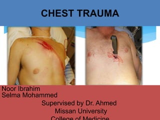
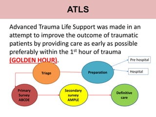
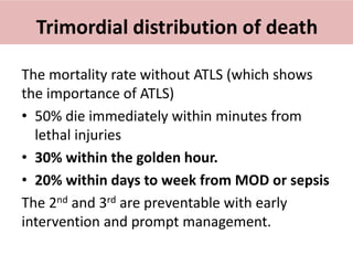
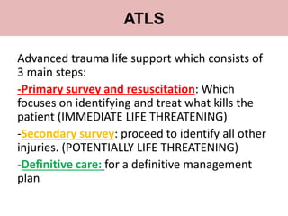
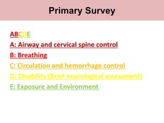
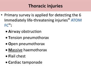
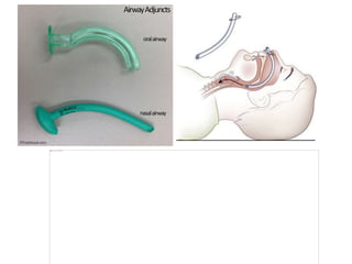
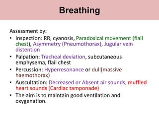
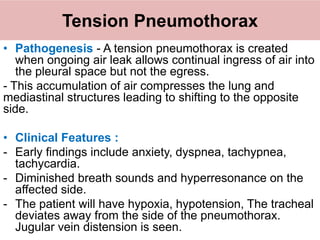
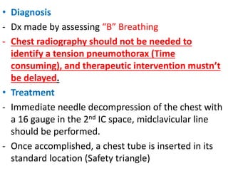
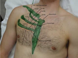
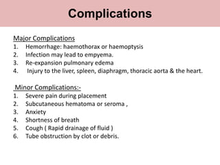
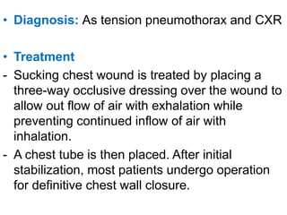
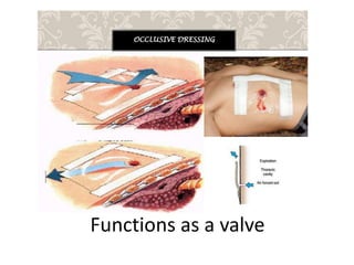
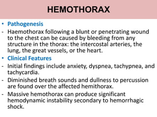
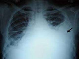
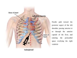
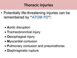
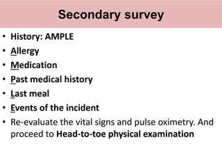
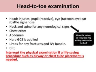
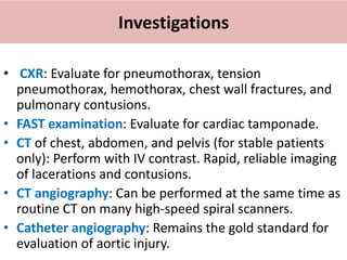
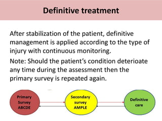
CHEST TRAUMA
Noor Ibrahim
Selma Mohammed
Supervised by Dr. Ahmed
Missan University
Thoracic anatomy
12 Pair of ribs
• Ribs 1-7: Join at sternum with cartilage end-points
• Ribs 8-10: Join sternum with combined cartilage at
7th rib
• Ribs 11-12: No anterior attachment
Sternum:
• Manubrium: has the jugular notch and joins to
clavicle and 1st rib
• Body: has sternal angle (Angle of Louis) is formed
by junction of the manubrium with the sternal body
• Xiphoid process: distal portion of sternum
Mediastinum
• Anterior mediastinum
- Thymus, fat, lymphatics
• Posterior mediastinum
- Descending aorta, esophagus, azygos veins,
autonomics, thoracic duct.
• Middle mediastinum
- Heart, pericardium, aorta, trachea.
Introduction
• All multiple trauma patients have chest trauma
till proven otherwise
• If there is head injury and abdominal trauma
there is also chest trauma. (Therefore if CT
scanning the other two scan the chest as well)
• Most injuries to the chest, both blunt and
penetrating, do not require surgery.
• Only about 10% of chest injuries actually
require operative management.
• All cardiothoracic injuries should be considered
lethal until proven otherwise
Mechanism of chest injury
1. Body acceleration and deceleration (organ
behind skeletal acceleration or deceleration)
Eg: RTA
2. Body compression (force > the strength of
skeleton) Eg: Crush injury and falls
3. Penetrating wounds (open pneumothorax
and organ injury) Eg: Assaults- Stab and Missile
injuries
Types of Chest Injury
1. Blunt Chest Injury (Closed Chest Injury)
-RTA, Fall, Crush injury
- Associated with multiple injuries such as
head, limb, abdomen
2. Penetrating chest injury (Open chest injury)
- Mostly by assault
- Associated with chest wall damage, open
pneumothorax and organ injury.
Clinical Approach
• The most common injuries to the chest wall
fractures of the ribs, sternum, and clavicle, are
rarely life threatening
• They may be a sign for a more significant underlying
visceral or neurovascular injury.
• The primary survey (ABCs) of the ATLS algorithm
will direct you to evaluate for the six conditions that
results from immediate life threatening injuries.
Advanced Trauma Life Support was made in an
attempt to improve the outcome of traumatic
patients by providing care as early as possible
preferably within the 1st hour of trauma
(GOLDEN HOUR).
Primary
Survey
ABCDE
Secondary
survey
AMPLE
Definitive
care
Preparation
Triage
ATLS
Pre hospital
Hospital
Trimordial distribution of death
The mortality rate without ATLS (which shows
the importance of ATLS)
• 50% die immediately within minutes from
lethal injuries
• 30% within the golden hour.
• 20% within days to week from MOD or sepsis
The 2nd and 3rd are preventable with early
intervention and prompt management.
Advanced trauma life support which consists of
3 main steps:
-Primary survey and resuscitation: Which
focuses on identifying and treat what kills the
patient (IMMEDIATE LIFE THREATENING)
-Secondary survey: proceed to identify all other
injuries. (POTENTIALLY LIFE THREATENING)
-Definitive care: for a definitive management
plan
ATLS
Primary survey
ABCDE
A: Airway and cervical spine control
B: Breathing
C: Circulation and hemorrhage control
D: Disability (Brief neurological assessment)
E: Exposure and Environment
Primary Survey
Thoracic injuries
• Primary survey is applied for detecting the 6
Immediately life-threatening injuries” ATOM
FC”:
 Airway obstruction
 Tension pneumothorax
 Open pneumothorax
 Massive haemothorax
 Flail chest
 Cardiac tamponade
Primary survey
A
A: Airway and cervical spine
control
Primary Survey
If patient is able to speak airway is patent.
If patient is unconscious or has an abnormal
voice need airway management.
Cervical spine is secured by a neck collar.
Airway and cervical spine control
1)Open airway: Head tilt and chin lift or jaw
thrust.
2)Clear airway: Remove vomit, blood or FB by
suction.
3)Protect “maintain” airway: by oropharyngeal
or nasopharyngeal airway (fallen tongue), NGT
or ETT (definitive)
NOTE: the simplest, quickest method with no
theater required CRICOTHYROIDOTOMY.
Airway obstruction
C/I in case of suspected
cervical spine injury!!!
ETT indicated in:
• Airway injury by neck or maxillofacial trauma (NGT Contraindicated)
• Airway obstruction: Coma, stridor, apnea
• Impending obstruction: burn, expanding neck hematoma
Primary survey
B
B: Breathing
Primary Survey
Assessment by:
• Inspection: RR, cyanosis, Paradoxical movement (flail
chest), Asymmetry (Pneumothorax), Jugular vein
distention
• Palpation: Tracheal deviation, subcutaneous
emphysema, flail chest
• Percussion: Hyperresonance or dull(massive
haemothorax)
• Auscultation: Decreased or Absent air sounds, muffled
heart sounds (Cardiac tamponade)
• The aim is to maintain good ventilation and
oxygenation.
Breathing
Tension Pneumothorax
• Pathogenesis - A tension pneumothorax is created
when ongoing air leak allows continual ingress of air into
the pleural space but not the egress.
- This accumulation of air compresses the lung and
mediastinal structures leading to shifting to the opposite
side.
• Clinical Features :
- Early findings include anxiety, dyspnea, tachypnea,
tachycardia.
- Diminished breath sounds and hyperresonance on the
affected side.
- The patient will have hypoxia, hypotension, The tracheal
deviates away from the side of the pneumothorax.
Jugular vein distension is seen.
• Diagnosis
- Dx made by assessing “B” Breathing
- Chest radiography should not be needed to
identify a tension pneumothorax (Time
consuming), and therapeutic intervention mustn’t
be delayed.
• Treatment
- Immediate needle decompression of the chest with
a 16 gauge in the 2nd IC space, midclavicular line
should be performed.
- Once accomplished, a chest tube is inserted in its
standard location (Safety triangle)
• Definition: Is a flexible hollow
plastic tube that is inserted
through the chest wall into the
pleural space at the safety
triangle and connected to a
bedside under water seal
drainage container.
Chest tube
Drainage Instillation
Fluid Air
Effusion
hydrothorax
Pus(Empye
ma)
Blood
(hemothora
x)
Chyle
Pneumothorax
Chemical
pleurodesis
Bleomycin
Talc
Tetracycline
Indications
• Refractory coagulopathy.
• Lack of cooperation by the patient
• Diaphragmatic Hernia
• Lobar Emphysema
• Surgical Emphysema without underlying
pneumothorax.
Contraindications
• Indications of removal:
• The tube remains in place until the lung is re-
expanded and the fluid is drained. (less than
25 ml for 2 consecutive days)
Chest tube
Major Complications
1. Hemorrhage: haemothorax or haemoptysis
2. Infection may lead to empyema.
3. Re-expansion pulmonary edema
4. Injury to the liver, spleen, diaphragm, thoracic aorta & the heart.
Minor Complications:-
1. Severe pain during placement
2. Subcutaneous hematoma or seroma ,
3. Anxiety
4. Shortness of breath
5. Cough ( Rapid drainage of fluid )
6. Tube obstruction by clot or debris.
Complications
Open Pneumothorax
• Pathogenesis
- More common in penetrating wounds, open pneumothorax may
occur with blunt thoracic trauma also - Pathophysiology is
similar to that of a tension pneumothorax however, the chest
wall is compromised, and the pleural cavity is in communication
with the atmosphere.
• Clinical Features
- Patients typically present with respiratory distress due to
collapse of the lung on the affected side. Physical examination
should reveal an obvious chest wall defect.
- Auscultation reveals complete or near complete loss of breath
sounds.
• Diagnosis: As tension pneumothorax and CXR
• Treatment
- Sucking chest wound is treated by placing a
three-way occlusive dressing over the wound to
allow out flow of air with exhalation while
preventing continued inflow of air with
inhalation.
- A chest tube is then placed. After initial
stabilization, most patients undergo operation
for definitive chest wall closure.
Functions as a valve
FLAIL CHEST
• Pathogenesis
- Flail chest is an injury that involves 3 or more
consecutive rib fractures in two or more locations,
producing a free-floating, unstable bony segment that is
detached from the remainder of the chest wall.
- Associated injuries are common such as pulmonary
contusion leading to atelectasis.
• Clinical Features
- Respiratory distress is the common initial presentation.
Dyspnea, tachycardia, tachypnea, pain, and tenderness
usually are present.
- The flail segment moves paradoxically, compared to the
rest of the hemithorax.
• Diagnosis
- Dx is made by physical examination and CXR.
- CT may help in identification of early pulmonary
contusion.
• Treatment
- Pain control, pulmonary hygiene, and
supplemental oxygen are the primary therapies
for pulmonary contusions.
- Flail injuries and associated contusions may
require endotracheal intubation and PEEP
mechanical ventilation to prevent alveolar
collapse) with strong analgesia.
Primary survey
C
C: Circulation and hemorrhage
control
Primary Survey
• Assessment for:
Shock which may be
1. Hemorrhagic,
2. Cardiogenic (tamponade) or
3. Neurogenic (spinal cord injury)
May be assessed by examining the vital signs,
skin and the rest of the body for any signs of
external bleeding.
Circulation
• Management:
1) Control bleeding by pressure if possible.
2) 2 wide bore peripheral IV cannula (green or gray
or orange colored)
3) Draw blood and send for blood group, cross
match, HCT, CBC.
4) IV fluid given is Ringer’s lactate (except in head
trauma) or 0.9% N/S then blood.
More than 1.5 L of crystalloid is associated with
increased mortality.
Circulation
HEMOTHORAX
• Pathogenesis
- Haemothorax following a blunt or penetrating wound
to the chest can be caused by bleeding from any
structure in the thorax: the intercostal arteries, the
lung, the great vessels, or the heart.
• Clinical Features
- Initial findings include anxiety, dyspnea, tachypnea, and
tachycardia.
- Diminished breath sounds and dullness to percussion
are found over the affected hemithorax.
- Massive hemothorax can produce significant
hemodynamic instability secondary to hemorrhagic
shock.
• Diagnosis : By physical examination and CXR
• Treatment
- Place a chest tube.
- Findings of 1,500 mL of blood initially, or more than
200 mL/hour for 2 to 4 hours, generally mandate a
thoracotomy to control bleeding.
- Witnessing any loss of vital signs is an indication for
ED thoracotomy
Cardiac Tamponade
• Pathogenesis:
- Pericardium is a two layered membrane surrounding the
heart that normally contains 20 to 50 mL of fluid.
- Rapid accumulation of as little as 150 mL of fluid after trauma
can produce cardiac tamponade and hypotension. cause
reduction in systemic venous return, diastolic filling, and
cardiac output
- If untreated, cardiac tamponade can produce cardiovascular
collapse and death. Beck’s triad (arterial hypotension,
venous hypertension, and muffled heart tones)
- The classic presentation of tamponade. Narrowing of pulse
pressures and pulsus paradoxus, a change of greater than 10
mmHg in the systolic pressure between inspiration and
expiration.
• Diagnosis:
- By assessing “B” and “C” muffled hearts sounds
on auscultation.
- If patient is on a 12 lead ECG then low voltage
will be seen.
• Treatment-:
- By needle pericardiocentesis if it’s due to blunt
trauma
- If it’s due to penetrating trauma operative repair of
the source of bleeding should be done immediately
- Fluid resuscitation is needed to maintain cardiac
output during transport to the operation room.
Primary survey
D
D: Disability (Brief neurological
assessment)
Primary Survey
• Neurological
• Look for any cause for altered LOC (head trauma,
hypoxia, shock)
• AVPU evaluation for rapid assessment
• A: Alert and interactive.
• V: Vocal stimuli elicit a response.
• P: Painful stimuli elicit a response. (GCS ˂8)
• U: Unresponsive.
Followed by GCS in 2nd survey.
Disability
Primary survey
E
E: Exposure and Environment
Primary Survey
• Completely expose for any additional wounds.
• Prevent hypothermia by removing any wet
clothes, cover the patient with blankets and
warm ER room.
Exposure and Environment
• Blood tests: CBC, GUE, clotting screen, glucose, cross
match
• ECG, pulse oximeter, ABG
• Foley catheter to monitor UOP except if there is:
• Bleeding from external meatus, scrotal hematoma,
high riding prostate on PR exam and patient wants to
void but is unable to.
• NG tube for gastric decompression and to prevent
aspiration.
• Imaging for head, cervical, chest, pelvis. (xray, Ct scan)
Adjuncts to Primary Survey
• Doesn’t begin until 1st survey is completed
and the immediately life threatening injuries
are dealt with.
• It consists of a rapid but thorough head-to-toe
examination to identify potentially life
threatening injuries.
Secondary Survey
Thoracic injuries
• Potentially life-threatening injuries can be
remembered by “ATOM PD”:
 Aortic disruption
 Tracheobronchial injury
 Oesophageal injury
 Myocardial contusion
 Pulmonary contusion and pneumothorax
 Diaphragmatic rupture
Secondary survey
• History: AMPLE
• Allergy
• Medication
• Past medical history
• Last meal
• Events of the incident
• Re-evaluate the vital signs and pulse oximetry. And
proceed to Head-to-toe physical examination
• Head: injuries, pupil (reactive), eye (raccoon eye) ear
(battle sign) nose
• Neck and spine for any neurological signs.
• Chest exam
• Abdomen
• Here GCS is applied
• Limbs for any fractures and NV bundle.
• DRE
Interrupt the physical examination if a life-saving
procedure such as airway or chest tube placement is
needed.
Head-to-toe examination
Move the patient
as one piece (log
rolling) to examine
the back
Investigations
• CXR: Evaluate for pneumothorax, tension
pneumothorax, hemothorax, chest wall fractures, and
pulmonary contusions.
• FAST examination: Evaluate for cardiac tamponade.
• CT of chest, abdomen, and pelvis (for stable patients
only): Perform with IV contrast. Rapid, reliable imaging
of lacerations and contusions.
• CT angiography: Can be performed at the same time as
routine CT on many high-speed spiral scanners.
• Catheter angiography: Remains the gold standard for
evaluation of aortic injury.
After stabilization of the patient, definitive
management is applied according to the type of
injury with continuous monitoring.
Note: Should the patient’s condition deterioate
any time during the assessment then the
primary survey is repeated again.
Definitive treatment
Chest trauma .pptx

Chest trauma .pptx

  • 1.
    CHEST TRAUMA Noor Ibrahim SelmaMohammed Supervised by Dr. Ahmed Missan University
  • 2.
    Thoracic anatomy 12 Pairof ribs • Ribs 1-7: Join at sternum with cartilage end-points • Ribs 8-10: Join sternum with combined cartilage at 7th rib • Ribs 11-12: No anterior attachment Sternum: • Manubrium: has the jugular notch and joins to clavicle and 1st rib • Body: has sternal angle (Angle of Louis) is formed by junction of the manubrium with the sternal body • Xiphoid process: distal portion of sternum
  • 4.
    Mediastinum • Anterior mediastinum -Thymus, fat, lymphatics • Posterior mediastinum - Descending aorta, esophagus, azygos veins, autonomics, thoracic duct. • Middle mediastinum - Heart, pericardium, aorta, trachea.
  • 5.
    Introduction • All multipletrauma patients have chest trauma till proven otherwise • If there is head injury and abdominal trauma there is also chest trauma. (Therefore if CT scanning the other two scan the chest as well) • Most injuries to the chest, both blunt and penetrating, do not require surgery. • Only about 10% of chest injuries actually require operative management. • All cardiothoracic injuries should be considered lethal until proven otherwise
  • 6.
    Mechanism of chestinjury 1. Body acceleration and deceleration (organ behind skeletal acceleration or deceleration) Eg: RTA 2. Body compression (force > the strength of skeleton) Eg: Crush injury and falls 3. Penetrating wounds (open pneumothorax and organ injury) Eg: Assaults- Stab and Missile injuries
  • 7.
    Types of ChestInjury 1. Blunt Chest Injury (Closed Chest Injury) -RTA, Fall, Crush injury - Associated with multiple injuries such as head, limb, abdomen 2. Penetrating chest injury (Open chest injury) - Mostly by assault - Associated with chest wall damage, open pneumothorax and organ injury.
  • 8.
    Clinical Approach • Themost common injuries to the chest wall fractures of the ribs, sternum, and clavicle, are rarely life threatening • They may be a sign for a more significant underlying visceral or neurovascular injury. • The primary survey (ABCs) of the ATLS algorithm will direct you to evaluate for the six conditions that results from immediate life threatening injuries.
  • 9.
    Advanced Trauma LifeSupport was made in an attempt to improve the outcome of traumatic patients by providing care as early as possible preferably within the 1st hour of trauma (GOLDEN HOUR). Primary Survey ABCDE Secondary survey AMPLE Definitive care Preparation Triage ATLS Pre hospital Hospital
  • 10.
    Trimordial distribution ofdeath The mortality rate without ATLS (which shows the importance of ATLS) • 50% die immediately within minutes from lethal injuries • 30% within the golden hour. • 20% within days to week from MOD or sepsis The 2nd and 3rd are preventable with early intervention and prompt management.
  • 11.
    Advanced trauma lifesupport which consists of 3 main steps: -Primary survey and resuscitation: Which focuses on identifying and treat what kills the patient (IMMEDIATE LIFE THREATENING) -Secondary survey: proceed to identify all other injuries. (POTENTIALLY LIFE THREATENING) -Definitive care: for a definitive management plan ATLS
  • 12.
    Primary survey ABCDE A: Airwayand cervical spine control B: Breathing C: Circulation and hemorrhage control D: Disability (Brief neurological assessment) E: Exposure and Environment Primary Survey
  • 13.
    Thoracic injuries • Primarysurvey is applied for detecting the 6 Immediately life-threatening injuries” ATOM FC”:  Airway obstruction  Tension pneumothorax  Open pneumothorax  Massive haemothorax  Flail chest  Cardiac tamponade
  • 14.
    Primary survey A A: Airwayand cervical spine control Primary Survey
  • 15.
    If patient isable to speak airway is patent. If patient is unconscious or has an abnormal voice need airway management. Cervical spine is secured by a neck collar. Airway and cervical spine control
  • 16.
    1)Open airway: Headtilt and chin lift or jaw thrust. 2)Clear airway: Remove vomit, blood or FB by suction. 3)Protect “maintain” airway: by oropharyngeal or nasopharyngeal airway (fallen tongue), NGT or ETT (definitive) NOTE: the simplest, quickest method with no theater required CRICOTHYROIDOTOMY. Airway obstruction
  • 18.
    C/I in caseof suspected cervical spine injury!!!
  • 19.
    ETT indicated in: •Airway injury by neck or maxillofacial trauma (NGT Contraindicated) • Airway obstruction: Coma, stridor, apnea • Impending obstruction: burn, expanding neck hematoma
  • 20.
  • 21.
    Assessment by: • Inspection:RR, cyanosis, Paradoxical movement (flail chest), Asymmetry (Pneumothorax), Jugular vein distention • Palpation: Tracheal deviation, subcutaneous emphysema, flail chest • Percussion: Hyperresonance or dull(massive haemothorax) • Auscultation: Decreased or Absent air sounds, muffled heart sounds (Cardiac tamponade) • The aim is to maintain good ventilation and oxygenation. Breathing
  • 22.
    Tension Pneumothorax • Pathogenesis- A tension pneumothorax is created when ongoing air leak allows continual ingress of air into the pleural space but not the egress. - This accumulation of air compresses the lung and mediastinal structures leading to shifting to the opposite side. • Clinical Features : - Early findings include anxiety, dyspnea, tachypnea, tachycardia. - Diminished breath sounds and hyperresonance on the affected side. - The patient will have hypoxia, hypotension, The tracheal deviates away from the side of the pneumothorax. Jugular vein distension is seen.
  • 23.
    • Diagnosis - Dxmade by assessing “B” Breathing - Chest radiography should not be needed to identify a tension pneumothorax (Time consuming), and therapeutic intervention mustn’t be delayed. • Treatment - Immediate needle decompression of the chest with a 16 gauge in the 2nd IC space, midclavicular line should be performed. - Once accomplished, a chest tube is inserted in its standard location (Safety triangle)
  • 25.
    • Definition: Isa flexible hollow plastic tube that is inserted through the chest wall into the pleural space at the safety triangle and connected to a bedside under water seal drainage container. Chest tube
  • 26.
  • 27.
    • Refractory coagulopathy. •Lack of cooperation by the patient • Diaphragmatic Hernia • Lobar Emphysema • Surgical Emphysema without underlying pneumothorax. Contraindications
  • 28.
    • Indications ofremoval: • The tube remains in place until the lung is re- expanded and the fluid is drained. (less than 25 ml for 2 consecutive days) Chest tube
  • 29.
    Major Complications 1. Hemorrhage:haemothorax or haemoptysis 2. Infection may lead to empyema. 3. Re-expansion pulmonary edema 4. Injury to the liver, spleen, diaphragm, thoracic aorta & the heart. Minor Complications:- 1. Severe pain during placement 2. Subcutaneous hematoma or seroma , 3. Anxiety 4. Shortness of breath 5. Cough ( Rapid drainage of fluid ) 6. Tube obstruction by clot or debris. Complications
  • 31.
    Open Pneumothorax • Pathogenesis -More common in penetrating wounds, open pneumothorax may occur with blunt thoracic trauma also - Pathophysiology is similar to that of a tension pneumothorax however, the chest wall is compromised, and the pleural cavity is in communication with the atmosphere. • Clinical Features - Patients typically present with respiratory distress due to collapse of the lung on the affected side. Physical examination should reveal an obvious chest wall defect. - Auscultation reveals complete or near complete loss of breath sounds.
  • 33.
    • Diagnosis: Astension pneumothorax and CXR • Treatment - Sucking chest wound is treated by placing a three-way occlusive dressing over the wound to allow out flow of air with exhalation while preventing continued inflow of air with inhalation. - A chest tube is then placed. After initial stabilization, most patients undergo operation for definitive chest wall closure.
  • 34.
  • 35.
    FLAIL CHEST • Pathogenesis -Flail chest is an injury that involves 3 or more consecutive rib fractures in two or more locations, producing a free-floating, unstable bony segment that is detached from the remainder of the chest wall. - Associated injuries are common such as pulmonary contusion leading to atelectasis. • Clinical Features - Respiratory distress is the common initial presentation. Dyspnea, tachycardia, tachypnea, pain, and tenderness usually are present. - The flail segment moves paradoxically, compared to the rest of the hemithorax.
  • 36.
    • Diagnosis - Dxis made by physical examination and CXR. - CT may help in identification of early pulmonary contusion. • Treatment - Pain control, pulmonary hygiene, and supplemental oxygen are the primary therapies for pulmonary contusions. - Flail injuries and associated contusions may require endotracheal intubation and PEEP mechanical ventilation to prevent alveolar collapse) with strong analgesia.
  • 38.
    Primary survey C C: Circulationand hemorrhage control Primary Survey
  • 39.
    • Assessment for: Shockwhich may be 1. Hemorrhagic, 2. Cardiogenic (tamponade) or 3. Neurogenic (spinal cord injury) May be assessed by examining the vital signs, skin and the rest of the body for any signs of external bleeding. Circulation
  • 40.
    • Management: 1) Controlbleeding by pressure if possible. 2) 2 wide bore peripheral IV cannula (green or gray or orange colored) 3) Draw blood and send for blood group, cross match, HCT, CBC. 4) IV fluid given is Ringer’s lactate (except in head trauma) or 0.9% N/S then blood. More than 1.5 L of crystalloid is associated with increased mortality. Circulation
  • 41.
    HEMOTHORAX • Pathogenesis - Haemothoraxfollowing a blunt or penetrating wound to the chest can be caused by bleeding from any structure in the thorax: the intercostal arteries, the lung, the great vessels, or the heart. • Clinical Features - Initial findings include anxiety, dyspnea, tachypnea, and tachycardia. - Diminished breath sounds and dullness to percussion are found over the affected hemithorax. - Massive hemothorax can produce significant hemodynamic instability secondary to hemorrhagic shock.
  • 42.
    • Diagnosis :By physical examination and CXR • Treatment - Place a chest tube. - Findings of 1,500 mL of blood initially, or more than 200 mL/hour for 2 to 4 hours, generally mandate a thoracotomy to control bleeding. - Witnessing any loss of vital signs is an indication for ED thoracotomy
  • 44.
    Cardiac Tamponade • Pathogenesis: -Pericardium is a two layered membrane surrounding the heart that normally contains 20 to 50 mL of fluid. - Rapid accumulation of as little as 150 mL of fluid after trauma can produce cardiac tamponade and hypotension. cause reduction in systemic venous return, diastolic filling, and cardiac output - If untreated, cardiac tamponade can produce cardiovascular collapse and death. Beck’s triad (arterial hypotension, venous hypertension, and muffled heart tones) - The classic presentation of tamponade. Narrowing of pulse pressures and pulsus paradoxus, a change of greater than 10 mmHg in the systolic pressure between inspiration and expiration.
  • 45.
    • Diagnosis: - Byassessing “B” and “C” muffled hearts sounds on auscultation. - If patient is on a 12 lead ECG then low voltage will be seen. • Treatment-: - By needle pericardiocentesis if it’s due to blunt trauma - If it’s due to penetrating trauma operative repair of the source of bleeding should be done immediately - Fluid resuscitation is needed to maintain cardiac output during transport to the operation room.
  • 47.
    Primary survey D D: Disability(Brief neurological assessment) Primary Survey
  • 48.
    • Neurological • Lookfor any cause for altered LOC (head trauma, hypoxia, shock) • AVPU evaluation for rapid assessment • A: Alert and interactive. • V: Vocal stimuli elicit a response. • P: Painful stimuli elicit a response. (GCS ˂8) • U: Unresponsive. Followed by GCS in 2nd survey. Disability
  • 49.
    Primary survey E E: Exposureand Environment Primary Survey
  • 50.
    • Completely exposefor any additional wounds. • Prevent hypothermia by removing any wet clothes, cover the patient with blankets and warm ER room. Exposure and Environment
  • 51.
    • Blood tests:CBC, GUE, clotting screen, glucose, cross match • ECG, pulse oximeter, ABG • Foley catheter to monitor UOP except if there is: • Bleeding from external meatus, scrotal hematoma, high riding prostate on PR exam and patient wants to void but is unable to. • NG tube for gastric decompression and to prevent aspiration. • Imaging for head, cervical, chest, pelvis. (xray, Ct scan) Adjuncts to Primary Survey
  • 52.
    • Doesn’t beginuntil 1st survey is completed and the immediately life threatening injuries are dealt with. • It consists of a rapid but thorough head-to-toe examination to identify potentially life threatening injuries. Secondary Survey
  • 53.
    Thoracic injuries • Potentiallylife-threatening injuries can be remembered by “ATOM PD”:  Aortic disruption  Tracheobronchial injury  Oesophageal injury  Myocardial contusion  Pulmonary contusion and pneumothorax  Diaphragmatic rupture
  • 54.
    Secondary survey • History:AMPLE • Allergy • Medication • Past medical history • Last meal • Events of the incident • Re-evaluate the vital signs and pulse oximetry. And proceed to Head-to-toe physical examination
  • 55.
    • Head: injuries,pupil (reactive), eye (raccoon eye) ear (battle sign) nose • Neck and spine for any neurological signs. • Chest exam • Abdomen • Here GCS is applied • Limbs for any fractures and NV bundle. • DRE Interrupt the physical examination if a life-saving procedure such as airway or chest tube placement is needed. Head-to-toe examination Move the patient as one piece (log rolling) to examine the back
  • 56.
    Investigations • CXR: Evaluatefor pneumothorax, tension pneumothorax, hemothorax, chest wall fractures, and pulmonary contusions. • FAST examination: Evaluate for cardiac tamponade. • CT of chest, abdomen, and pelvis (for stable patients only): Perform with IV contrast. Rapid, reliable imaging of lacerations and contusions. • CT angiography: Can be performed at the same time as routine CT on many high-speed spiral scanners. • Catheter angiography: Remains the gold standard for evaluation of aortic injury.
  • 57.
    After stabilization ofthe patient, definitive management is applied according to the type of injury with continuous monitoring. Note: Should the patient’s condition deterioate any time during the assessment then the primary survey is repeated again. Definitive treatment