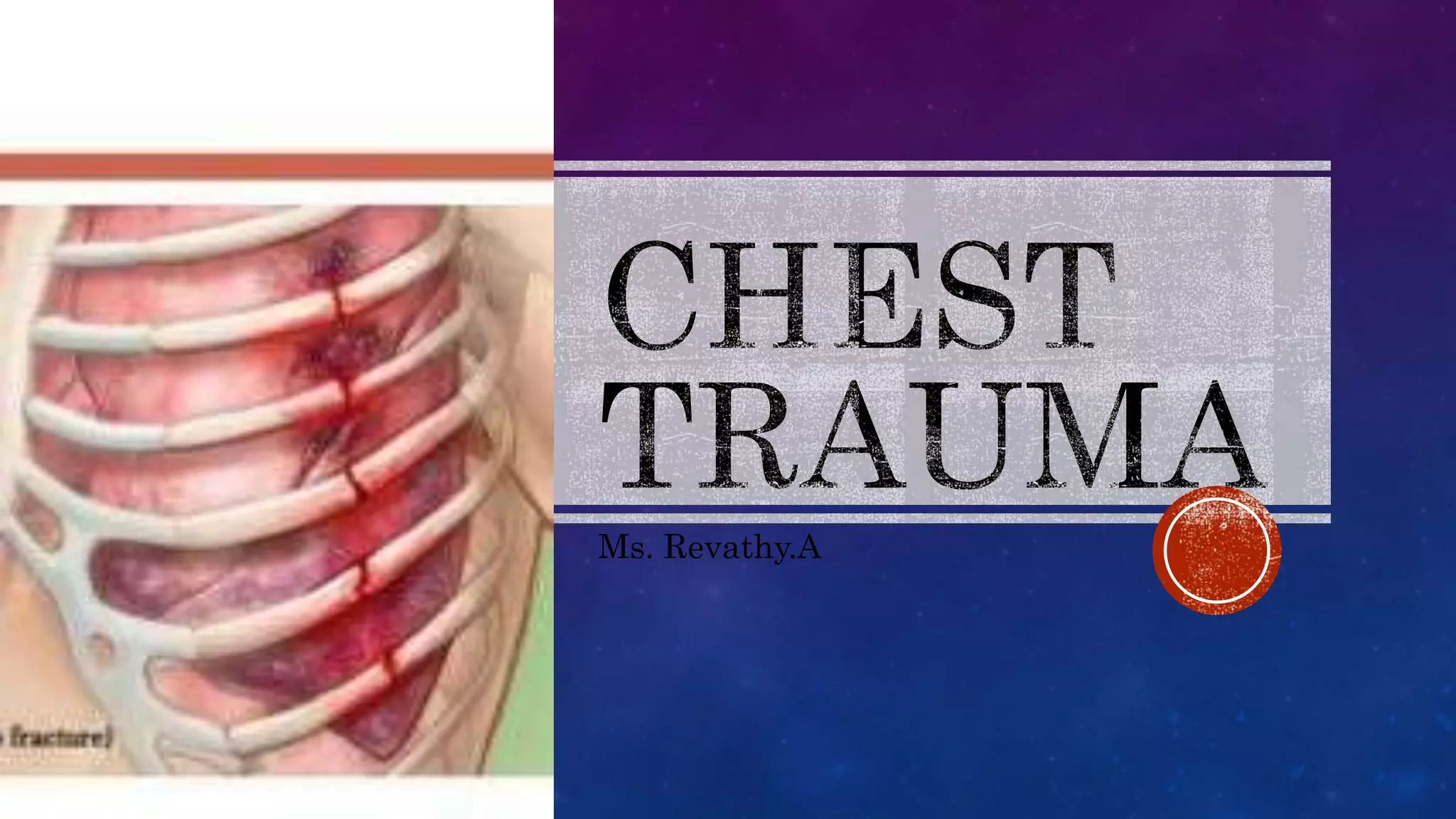
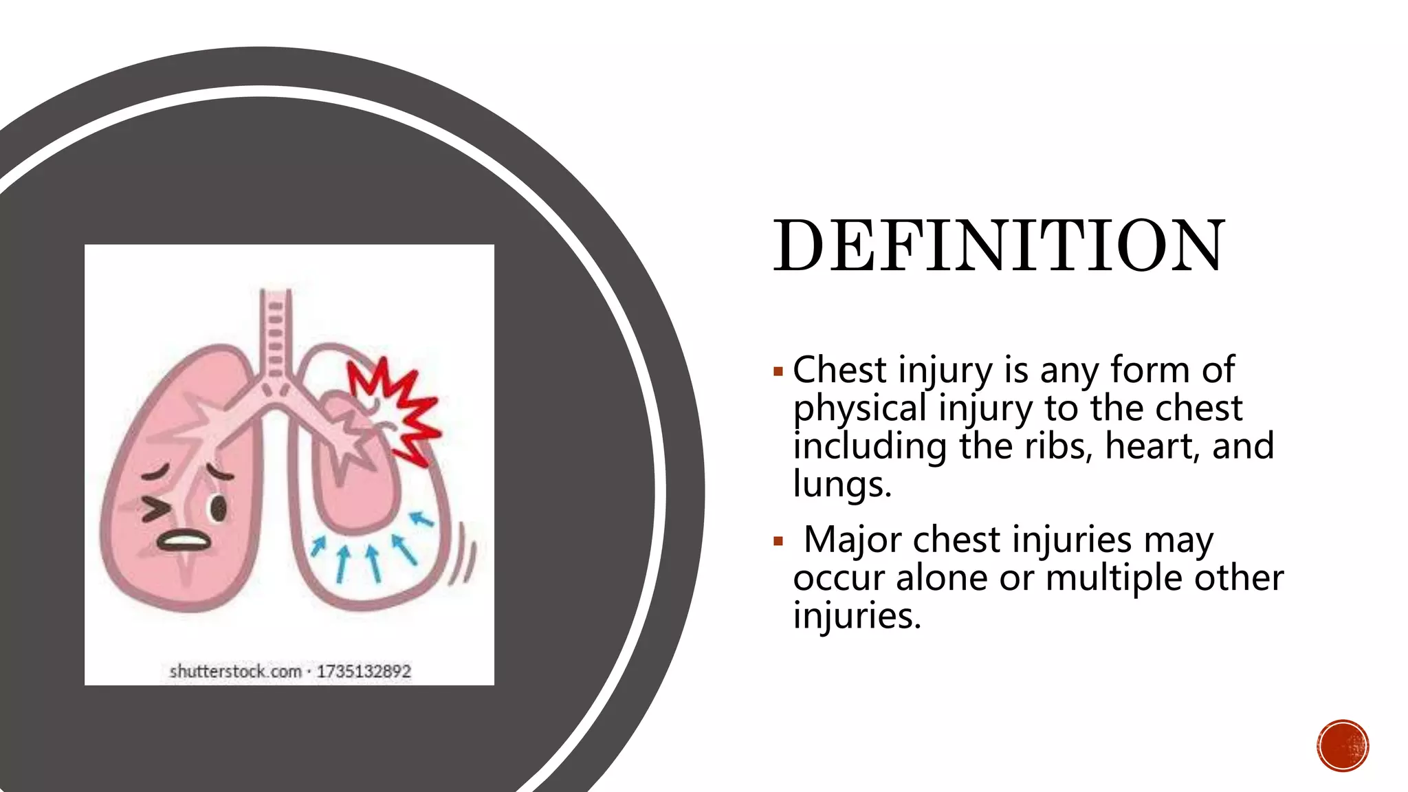
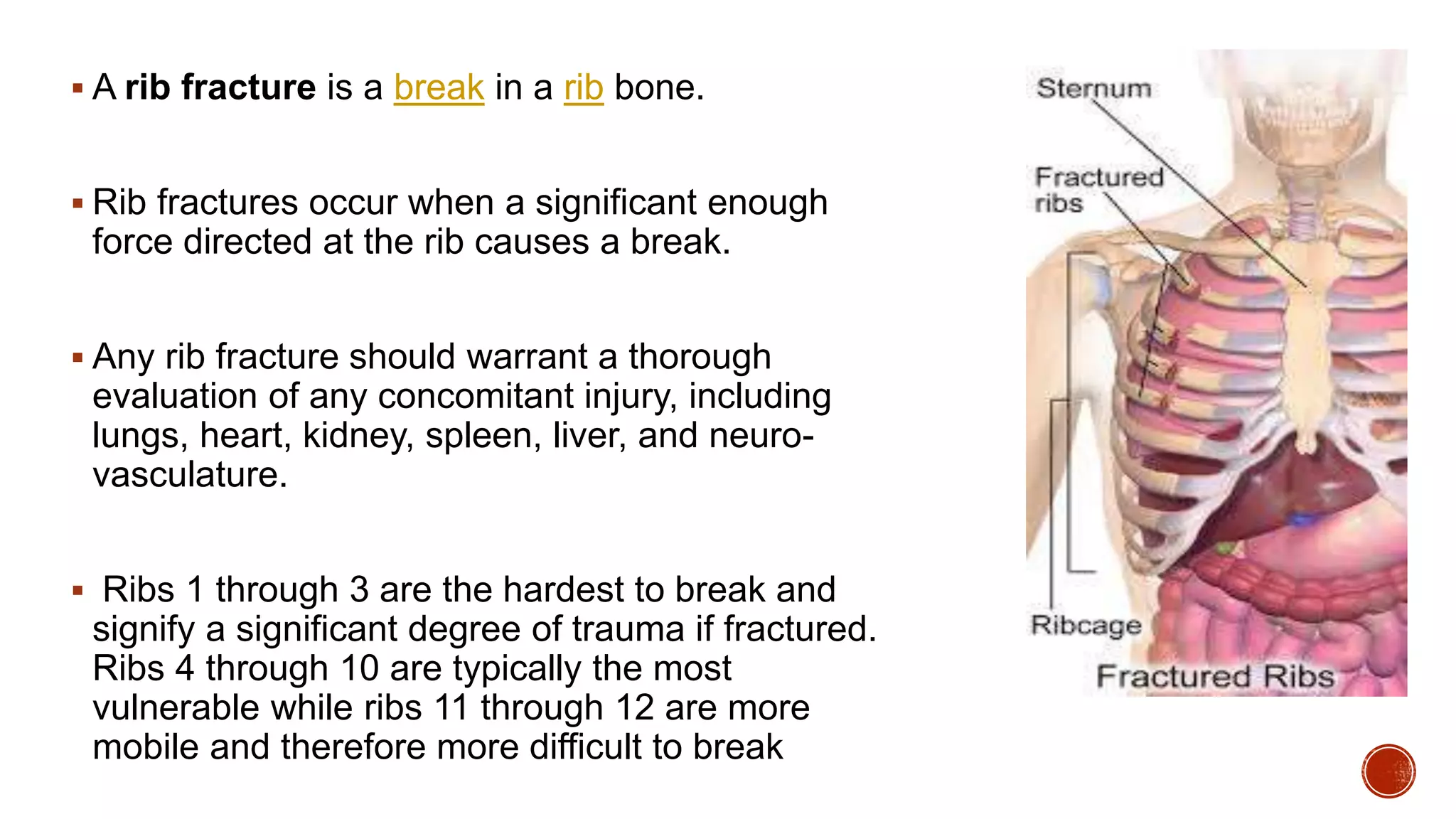
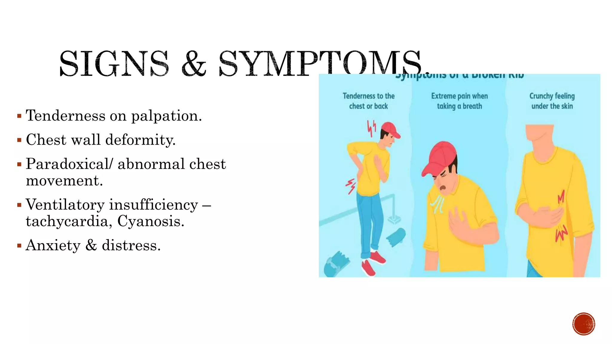
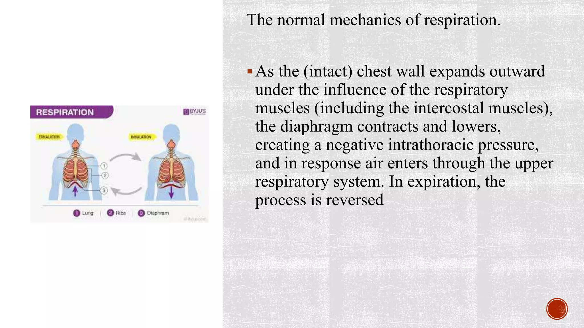
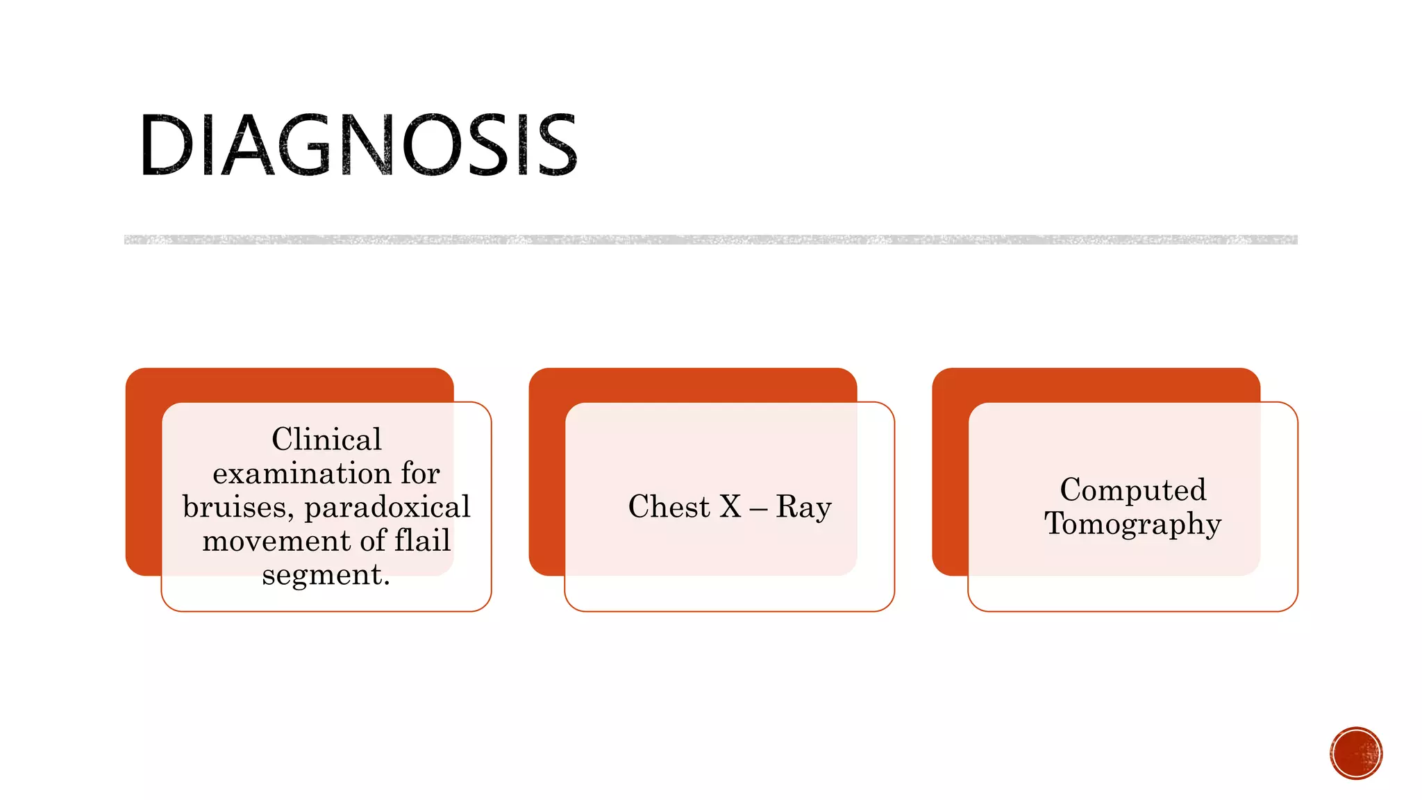
This document discusses chest injuries and their management. It begins by introducing chest trauma as life-threatening due to injuries to the heart, lungs, and blood vessels in the chest. It then defines chest injury and provides epidemiological data showing thoracic trauma is a leading cause of trauma deaths. Specific injuries discussed in detail include rib fractures, flail chest, pneumothorax, and hemothorax. For each injury, the document covers causes, signs/symptoms, diagnostic tests, and treatment approaches including analgesics, ventilator support, chest tube insertion, and surgery. Nursing assessments and interventions are also reviewed.
Hemopneumothorax, or haemopneumothorax is the condition of having air in the chest cavity (pneumothorax) and blood in the chest cavity (hemothorax). A hemothorax, pneumothorax, or the combination of both can occur due to an injury to the lung or chest.
Pleural effusion, sometimes referred to as “water on the lungs,” is the build-up of excess fluid between the layers of the pleura outside the lungs. The pleura are thin membranes that line the lungs and the inside of the chest cavity and act to lubricate and facilitate breathing.
CHEST INJURY- BLUNT/ Trauma Surgery
Dear viewers,
Greetings from “Surgical Educator”
Today I have uploaded a video on CHEST INJURY- BLUNT- an important topic in trauma. Even the blunt chest trauma can turn into penetrating one because of jagged edges of the broken ribs. I haven’t talked elaborately but have included the essential minimum an undergraduate medical student should know. I have talked about pathophysiology, clinical approach, symptoms, signs, investigations, different individual types of Chest injuries and management of all the varieties of Chest injuries. My aim is, after watching this video all of you should be able to arrive at a correct working diagnosis of the type of chest injury and should also be able to institute immediate lifesaving treatment to the patients if there is a need. You can watch the video in the following links:
surgicaleducator.blogspot.com
youtube.com/c/surgicaleducator
Thank you for watching the video.
Cor pulmonale is alteration in the structure and function of the right ventricle (RV) of the heart. The overall five-year survival rate for cor pulmonale complicating COPD is approximately 50%.
Normally, the pleural space contains a small amount of fluid (5 to 15 mL), which acts as a lubricant that allows the pleural surfaces to move without friction.
But if fluid builds up from either increased production or inadequate removal pleural effusion results.
Pleural effusion B/L or unilateral (parapneumonic process)
Refers to any significant collection of fluid within pleural space.
Any imbalance in formation, absorption lead accumulation of pleural fluid. Common condition:
CHF
Bacterial pneumonia
Malignancy(chest tumor)
Pulmonary embolism
Pleura effusion is a condition refers to a collection of fluid in the pleural space. It is almost secondary to other conditions.
Respiratory Disorders
Disease Condition Pneumothorax, Causes, Sign and Symptoms, Pathophysiology, Types, Assessment and Dignostic Test, Management
By HIREN GEHLOTH For Nursing Students Medical Surgical Nursing
LAUGH A LOT IT CLEARS THE LUNGS
TEACHING IS ONE PROFESSION THAT CREATE ALL OTHER PROFESSION
injury to thorax and abdomen. tension pneumothorax , cardiac tamponade, rupture of kidney, rupture of liver, intestinal perforation, foreign body in rectum
NVBDCP.pptx Nation vector borne disease control programSapna Thakur
NVBDCP was launched in 2003-2004 . Vector-Borne Disease: Disease that results from an infection transmitted to humans and other animals by blood-feeding arthropods, such as mosquitoes, ticks, and fleas. Examples of vector-borne diseases include Dengue fever, West Nile Virus, Lyme disease, and malaria.
Local Advanced Lung Cancer: Artificial Intelligence, Synergetics, Complex Sys...Oleg Kshivets
Overall life span (LS) was 1671.7±1721.6 days and cumulative 5YS reached 62.4%, 10 years – 50.4%, 20 years – 44.6%. 94 LCP lived more than 5 years without cancer (LS=2958.6±1723.6 days), 22 – more than 10 years (LS=5571±1841.8 days). 67 LCP died because of LC (LS=471.9±344 days). AT significantly improved 5YS (68% vs. 53.7%) (P=0.028 by log-rank test). Cox modeling displayed that 5YS of LCP significantly depended on: N0-N12, T3-4, blood cell circuit, cell ratio factors (ratio between cancer cells-CC and blood cells subpopulations), LC cell dynamics, recalcification time, heparin tolerance, prothrombin index, protein, AT, procedure type (P=0.000-0.031). Neural networks, genetic algorithm selection and bootstrap simulation revealed relationships between 5YS and N0-12 (rank=1), thrombocytes/CC (rank=2), segmented neutrophils/CC (3), eosinophils/CC (4), erythrocytes/CC (5), healthy cells/CC (6), lymphocytes/CC (7), stick neutrophils/CC (8), leucocytes/CC (9), monocytes/CC (10). Correct prediction of 5YS was 100% by neural networks computing (error=0.000; area under ROC curve=1.0).
Title: Sense of Taste
Presenter: Dr. Faiza, Assistant Professor of Physiology
Qualifications:
MBBS (Best Graduate, AIMC Lahore)
FCPS Physiology
ICMT, CHPE, DHPE (STMU)
MPH (GC University, Faisalabad)
MBA (Virtual University of Pakistan)
Learning Objectives:
Describe the structure and function of taste buds.
Describe the relationship between the taste threshold and taste index of common substances.
Explain the chemical basis and signal transduction of taste perception for each type of primary taste sensation.
Recognize different abnormalities of taste perception and their causes.
Key Topics:
Significance of Taste Sensation:
Differentiation between pleasant and harmful food
Influence on behavior
Selection of food based on metabolic needs
Receptors of Taste:
Taste buds on the tongue
Influence of sense of smell, texture of food, and pain stimulation (e.g., by pepper)
Primary and Secondary Taste Sensations:
Primary taste sensations: Sweet, Sour, Salty, Bitter, Umami
Chemical basis and signal transduction mechanisms for each taste
Taste Threshold and Index:
Taste threshold values for Sweet (sucrose), Salty (NaCl), Sour (HCl), and Bitter (Quinine)
Taste index relationship: Inversely proportional to taste threshold
Taste Blindness:
Inability to taste certain substances, particularly thiourea compounds
Example: Phenylthiocarbamide
Structure and Function of Taste Buds:
Composition: Epithelial cells, Sustentacular/Supporting cells, Taste cells, Basal cells
Features: Taste pores, Taste hairs/microvilli, and Taste nerve fibers
Location of Taste Buds:
Found in papillae of the tongue (Fungiform, Circumvallate, Foliate)
Also present on the palate, tonsillar pillars, epiglottis, and proximal esophagus
Mechanism of Taste Stimulation:
Interaction of taste substances with receptors on microvilli
Signal transduction pathways for Umami, Sweet, Bitter, Sour, and Salty tastes
Taste Sensitivity and Adaptation:
Decrease in sensitivity with age
Rapid adaptation of taste sensation
Role of Saliva in Taste:
Dissolution of tastants to reach receptors
Washing away the stimulus
Taste Preferences and Aversions:
Mechanisms behind taste preference and aversion
Influence of receptors and neural pathways
Impact of Sensory Nerve Damage:
Degeneration of taste buds if the sensory nerve fiber is cut
Abnormalities of Taste Detection:
Conditions: Ageusia, Hypogeusia, Dysgeusia (parageusia)
Causes: Nerve damage, neurological disorders, infections, poor oral hygiene, adverse drug effects, deficiencies, aging, tobacco use, altered neurotransmitter levels
Neurotransmitters and Taste Threshold:
Effects of serotonin (5-HT) and norepinephrine (NE) on taste sensitivity
Supertasters:
25% of the population with heightened sensitivity to taste, especially bitterness
Increased number of fungiform papillae
Knee anatomy and clinical tests 2024.pdfvimalpl1234
This includes all relevant anatomy and clinical tests compiled from standard textbooks, Campbell,netter etc..It is comprehensive and best suited for orthopaedicians and orthopaedic residents.
Muktapishti is a traditional Ayurvedic preparation made from Shoditha Mukta (Purified Pearl), is believed to help regulate thyroid function and reduce symptoms of hyperthyroidism due to its cooling and balancing properties. Clinical evidence on its efficacy remains limited, necessitating further research to validate its therapeutic benefits.
263778731218 Abortion Clinic /Pills In Harare ,sisternakatoto
263778731218 Abortion Clinic /Pills In Harare ,ABORTION WOMEN’S CLINIC +27730423979 IN women clinic we believe that every woman should be able to make choices in her pregnancy. Our job is to provide compassionate care, safety,affordable and confidential services. That’s why we have won the trust from all generations of women all over the world. we use non surgical method(Abortion pills) to terminate…Dr.LISA +27730423979women Clinic is committed to providing the highest quality of obstetrical and gynecological care to women of all ages. Our dedicated staff aim to treat each patient and her health concerns with compassion and respect.Our dedicated group ABORTION WOMEN’S CLINIC +27730423979 IN women clinic we believe that every woman should be able to make choices in her pregnancy. Our job is to provide compassionate care, safety,affordable and confidential services. That’s why we have won the trust from all generations of women all over the world. we use non surgical method(Abortion pills) to terminate…Dr.LISA +27730423979women Clinic is committed to providing the highest quality of obstetrical and gynecological care to women of all ages. Our dedicated staff aim to treat each patient and her health concerns with compassion and respect.Our dedicated group of receptionists, nurses, and physicians have worked together as a teamof receptionists, nurses, and physicians have worked together as a team wwww.lisywomensclinic.co.za/
Title: Sense of Smell
Presenter: Dr. Faiza, Assistant Professor of Physiology
Qualifications:
MBBS (Best Graduate, AIMC Lahore)
FCPS Physiology
ICMT, CHPE, DHPE (STMU)
MPH (GC University, Faisalabad)
MBA (Virtual University of Pakistan)
Learning Objectives:
Describe the primary categories of smells and the concept of odor blindness.
Explain the structure and location of the olfactory membrane and mucosa, including the types and roles of cells involved in olfaction.
Describe the pathway and mechanisms of olfactory signal transmission from the olfactory receptors to the brain.
Illustrate the biochemical cascade triggered by odorant binding to olfactory receptors, including the role of G-proteins and second messengers in generating an action potential.
Identify different types of olfactory disorders such as anosmia, hyposmia, hyperosmia, and dysosmia, including their potential causes.
Key Topics:
Olfactory Genes:
3% of the human genome accounts for olfactory genes.
400 genes for odorant receptors.
Olfactory Membrane:
Located in the superior part of the nasal cavity.
Medially: Folds downward along the superior septum.
Laterally: Folds over the superior turbinate and upper surface of the middle turbinate.
Total surface area: 5-10 square centimeters.
Olfactory Mucosa:
Olfactory Cells: Bipolar nerve cells derived from the CNS (100 million), with 4-25 olfactory cilia per cell.
Sustentacular Cells: Produce mucus and maintain ionic and molecular environment.
Basal Cells: Replace worn-out olfactory cells with an average lifespan of 1-2 months.
Bowman’s Gland: Secretes mucus.
Stimulation of Olfactory Cells:
Odorant dissolves in mucus and attaches to receptors on olfactory cilia.
Involves a cascade effect through G-proteins and second messengers, leading to depolarization and action potential generation in the olfactory nerve.
Quality of a Good Odorant:
Small (3-20 Carbon atoms), volatile, water-soluble, and lipid-soluble.
Facilitated by odorant-binding proteins in mucus.
Membrane Potential and Action Potential:
Resting membrane potential: -55mV.
Action potential frequency in the olfactory nerve increases with odorant strength.
Adaptation Towards the Sense of Smell:
Rapid adaptation within the first second, with further slow adaptation.
Psychological adaptation greater than receptor adaptation, involving feedback inhibition from the central nervous system.
Primary Sensations of Smell:
Camphoraceous, Musky, Floral, Pepperminty, Ethereal, Pungent, Putrid.
Odor Detection Threshold:
Examples: Hydrogen sulfide (0.0005 ppm), Methyl-mercaptan (0.002 ppm).
Some toxic substances are odorless at lethal concentrations.
Characteristics of Smell:
Odor blindness for single substances due to lack of appropriate receptor protein.
Behavioral and emotional influences of smell.
Transmission of Olfactory Signals:
From olfactory cells to glomeruli in the olfactory bulb, involving lateral inhibition.
Primitive, less old, and new olfactory systems with different path
Tom Selleck Health: A Comprehensive Look at the Iconic Actor’s Wellness Journeygreendigital
Tom Selleck, an enduring figure in Hollywood. has captivated audiences for decades with his rugged charm, iconic moustache. and memorable roles in television and film. From his breakout role as Thomas Magnum in Magnum P.I. to his current portrayal of Frank Reagan in Blue Bloods. Selleck's career has spanned over 50 years. But beyond his professional achievements. fans have often been curious about Tom Selleck Health. especially as he has aged in the public eye.
Follow us on: Pinterest
Introduction
Many have been interested in Tom Selleck health. not only because of his enduring presence on screen but also because of the challenges. and lifestyle choices he has faced and made over the years. This article delves into the various aspects of Tom Selleck health. exploring his fitness regimen, diet, mental health. and the challenges he has encountered as he ages. We'll look at how he maintains his well-being. the health issues he has faced, and his approach to ageing .
Early Life and Career
Childhood and Athletic Beginnings
Tom Selleck was born on January 29, 1945, in Detroit, Michigan, and grew up in Sherman Oaks, California. From an early age, he was involved in sports, particularly basketball. which played a significant role in his physical development. His athletic pursuits continued into college. where he attended the University of Southern California (USC) on a basketball scholarship. This early involvement in sports laid a strong foundation for his physical health and disciplined lifestyle.
Transition to Acting
Selleck's transition from an athlete to an actor came with its physical demands. His first significant role in "Magnum P.I." required him to perform various stunts and maintain a fit appearance. This role, which he played from 1980 to 1988. necessitated a rigorous fitness routine to meet the show's demands. setting the stage for his long-term commitment to health and wellness.
Fitness Regimen
Workout Routine
Tom Selleck health and fitness regimen has evolved. adapting to his changing roles and age. During his "Magnum, P.I." days. Selleck's workouts were intense and focused on building and maintaining muscle mass. His routine included weightlifting, cardiovascular exercises. and specific training for the stunts he performed on the show.
Selleck adjusted his fitness routine as he aged to suit his body's needs. Today, his workouts focus on maintaining flexibility, strength, and cardiovascular health. He incorporates low-impact exercises such as swimming, walking, and light weightlifting. This balanced approach helps him stay fit without putting undue strain on his joints and muscles.
Importance of Flexibility and Mobility
In recent years, Selleck has emphasized the importance of flexibility and mobility in his fitness regimen. Understanding the natural decline in muscle mass and joint flexibility with age. he includes stretching and yoga in his routine. These practices help prevent injuries, improve posture, and maintain mobilit
micro teaching on communication m.sc nursing.pdfAnurag Sharma
Microteaching is a unique model of practice teaching. It is a viable instrument for the. desired change in the teaching behavior or the behavior potential which, in specified types of real. classroom situations, tends to facilitate the achievement of specified types of objectives.
Basavarajeeyam is a Sreshta Sangraha grantha (Compiled book ), written by Neelkanta kotturu Basavaraja Virachita. It contains 25 Prakaranas, First 24 Chapters related to Rogas& 25th to Rasadravyas.
Integrating Ayurveda into Parkinson’s Management: A Holistic ApproachAyurveda ForAll
Explore the benefits of combining Ayurveda with conventional Parkinson's treatments. Learn how a holistic approach can manage symptoms, enhance well-being, and balance body energies. Discover the steps to safely integrate Ayurvedic practices into your Parkinson’s care plan, including expert guidance on diet, herbal remedies, and lifestyle modifications.
2. INTRODU
CTION
Chest is large exposed portion of the body that is
vulnerable to impact injuries.
Because chest houses heart, lungs, and great vessels
chest trauma is frequently life threatening .
Injuries to thoracic cage and its content can restrict the
hearts ability to pump blood or lungs ability to exchange
air and oxygenated blood.
Major danger with chest injuries is internal bleeding and
organ puncture
3.
4. DEFINITION
Chest injury is any form of
physical injury to the chest
including the ribs, heart, and
lungs.
Major chest injuries may
occur alone or multiple other
injuries.
5. EPIDEMIOL
OGY
The number of accidental deaths
in India is even higher than in
the Western World.
Thoracic trauma contributes
heavily to these figures besides
head injury, abdominal injury
and orthopedic injuries.
Approximately one quarter of
civilian trauma deaths are caused
by thoracic trauma
6.
7.
8. A rib fracture is a break in a rib bone.
Rib fractures occur when a significant enough
force directed at the rib causes a break.
Any rib fracture should warrant a thorough
evaluation of any concomitant injury, including
lungs, heart, kidney, spleen, liver, and neuro-
vasculature.
Ribs 1 through 3 are the hardest to break and
signify a significant degree of trauma if fractured.
Ribs 4 through 10 are typically the most
vulnerable while ribs 11 through 12 are more
mobile and therefore more difficult to break
11. For simple, isolated rib fractures, conservative therapy is usually
adequate which includes appropriate analgesia, rest, and ice.
The use of an incentive spirometer should be encouraged to prevent
pulmonary atelectasis and splinting.
Intercostal nerve blocks can also be applied to aid in pain control.
When conservative management fails or for more severe rib
fractures, surgical stabilization can be an option.
12. The nurse instructs the patient to use an incentive spirometer every hour.
Provide rest and avoid sternuous activities for several days.
Assess the respiratory patterns every hour for several days.
13.
14. Flail chest is a life-threatening medical condition
that occurs when a segment of the rib cage breaks
due to trauma and becomes detached from the rest
of the chest wall.
Flail chest is a thoracic injury resulting in
paradoxical motion of the chestwall segment.
Flail chest is a traumatic condition of the thorax. It
may occur when 3 or more ribs are broken in at
least 2 places.
A flail chest arises when these injuries cause a
segment of the chest wall to move independently of
the rest of the chest wall.
A flail chest can create a significant disturbance to
respiratory physiology. This disturbance in
respiratory function is important in patients who are
older or who have chronic lung disease.
Flail chest is an important injury with significant
complications.
15.
16. The normal mechanics of respiration.
As the (intact) chest wall expands outward
under the influence of the respiratory
muscles (including the intercostal muscles),
the diaphragm contracts and lowers,
creating a negative intrathoracic pressure,
and in response air enters through the upper
respiratory system. In expiration, the
process is reversed
17. A flail segment of the chest wall will
negatively affect respiration in three ways:
ineffective ventilation,
pulmonary contusion, and
hypoventilation with atelectasis.
27. A pneumothorax is an abnormal collection
of air in the pleural space between
the lung and the chest wall.
A pneumothorax is defined as a collection
of air outside the lung but within the
pleural cavity.
It occurs when air accumulates between
the parietal and visceral pleurae inside
the chest.
The air accumulation can apply pressure
on the lung and make it collapse.
The degree of collapse determines the
clinical presentation of pneumothorax.
34. The definition of large vs. small
pneumothorax is by the
distance between the lung margin
and chest wall:
Small pneumothorax: the
presence of a visible rim of less
than 2 cm between the lung margin
and the chest wall
Large pneumothorax: the
presence of a visible rim of greater
than 2 cm between the lung margin
and the chest wall
35.
36.
37.
38. Chest pain – severe, sharp/stabbing,
pleuritic and radiates to ipsilateral
shoulder/arm.
Shortness of breath (64 to 85%).
Anxiety and cough
Absent breath sounds on the
affected side.
39.
40. The management is guided by the etiology, clinical presentation,
and risk stratification.
The principles of treatment of pneumothorax are :
1. air elimination,
2. reduction of air leakage,
3. healing of pleural fistula, promoting re-expansion of the lung,
4. prevention of future recurrences.
41. Chest tube.
A chest tube, also known as a thoracostomy
tube, is a flexible tube that can be inserted
through the chest wall between the ribs into
the pleural space.
Thoracostomy tubes are commonly made
from PVC or silicone.
They range in from 6 French to 40 French.
The majority are fenestrated along the sides
of the insertion end, and the tubes have a
radiopaque stripe.
After placement, the distal end of the tube is
connected to a pleura-evac system.
42. A thoracostomy tube is usually
placed between the mid to
anterior axillary line in the
fourth or fifth intercostal space
tracking above the rib so as not
to injure the intercostal bundle
(artery, vein, nerve).
43. Maintain a closed chest drainage system.
Be sure to tape all connections, and secure the tube carefully
at the insertion site with adhesive bandages.
Regulate suction according to the chest tube system
directions; generally, suction does not exceed 20 to 25 cm
H2O negative pressure
44. Autotransfusion.
Autotransfusion involves taking the patient’s own blood that
has been drained from the chest, filtering it, and then
transfusing it back into the vascular system.
Antibiotics.
Antibiotics are usually prescribed to combat infection from
contamination.
Oxygen therapy.
The patient with possible tension pneumothorax should
immediately be given a high concentration of supplemental
oxygen to treat the hypoxemia.
46. Haemothorax is an
accumulation of blood in the
intrapleural space.
It is frequently found in
association with Open
pneumothorax and is then
called a haemopneumothorax.
53. Perform initial resuscitation and management of a trauma patient.
Every patient should have two large bore IVs access
Connect to 12 lead ECG
Decompression needle thoracostomy, and/or emergent tube thoracostomy
57. To perform tube thoracostomy, a
large-bore tube (32F to 40F) should
be placed in the 4th or 5th intercostal
space at the anterior axillary line,
and connected to water seal and
suction (20-30 mL H20).
Re-expansion of the lung
parenchyma and resolution of the
hemothorax should be monitored
with serial CXRs.
58.
59. Pain management
Epidural analgesia
Systemic analgesics
Intrapleural
Other techniques
Management of fluid electrolytes
Intercostal catheter
Physiotherapy & Early ambulation
DVT prophylaxis.
60. NURSING ASSESSMENT
The nurse should assess the following:
Tracheal alignment.
Expansion of the chest.
Breath sounds.
Percussion of the chest.