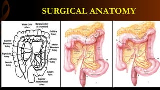
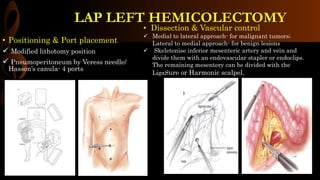
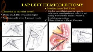
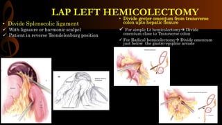
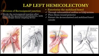
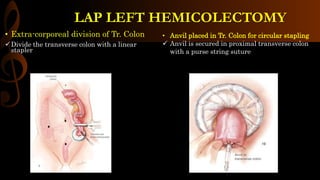
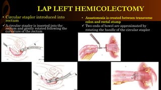
2. LAP LEFT HEMICOLECTOMY
• INDICATIONS:
Malignant tumors in descending colon and splenic
flexure
Diverticulitis, Polyps, Inflammatory bowel disease and
colonic hemorrhage
• ANESTHESIA:
GA/ETT
• POSITION:
Supine/modified lithotomy
• Diagnosis
Confirmed by colonoscopic biopsy
Staging by CECT
• Informed consent- risks of surgery:
Anastomotic leak- 2%
Hemorrhage- 1%
Bowel obstruction- 2%
Wound infection- 2 to 10%
Splenic injury- < 5%
Injury to ureter and gonadal vessels- 1%
• Pre-op preparation
Adequate mechanical bowel preparation the day before surgery
with orthograde enema
Prophylactic IV broad spectrum antibiotics
VTE prophylaxis with LMWH and pneumatic stockings
Tattooing of the tumor endoscopically or by localization on a barium
enema.
4. • Positioning & Port placement
Modified lithotomy position
Pneumoperitoneum by Veress needle/
Hasson’s canula- 4 ports
• Dissection & Vascular control
Medial to lateral approach- for malignant tumors;
Lateral to medial approach- for benign lesions
Skeletonise inferior mesenteric artery and vein and
divide them with an endovascular stapler or endoclips.
The remaining mesentery can be divided with the
LigaSure or Harmonic scalpel.
LAP LEFT HEMICOLECTOMY
5. • Dissection & Vascular control
Divide IMA & IMV by vascular stapler
Avoid injuring Lt ureter & gonadal vessels
• Mobilisation of Left Colon
Mobilise sigmoid & descending colon by
incising the lateral peritoneal reflection and
pulling it towards the midline. Patient in
Trendelenburg position.
This mobilisation is Mattox Maneuver
LAP LEFT HEMICOLECTOMY
6. • Divide greter omentum from transverse
colon upto hepatic flexure
For simple Lt hemicolectomy Divide
omentum close to Transverse colon
For Radical hemicolectomy Divide omentum
just below the gastro-epiploic arcade
• Divide Splenocolic ligament
With ligasure or harmonic scalpel
Patient in reverse Trendelenburg position
LAP LEFT HEMICOLECTOMY
7. • Division of Rectosigmoid junction
Divide the rectosigmoid junction after
clearing the mesenteric attachments, with
laparoscopic linear stapling device
• Exteriorise the mobilised bowel
Through infraumbilical midline or LLQ incision
Place Alexis wound protector
Extract the devascularised and mobilised bowel
outside
LAP LEFT HEMICOLECTOMY
8. • Extra-corporeal division of Tr. Colon
Divide the transverse colon with a linear
stapler
LAP LEFT HEMICOLECTOMY
• Anvil placed in Tr. Colon for circular stapling
Anvil is secured in proximal transverse colon
with a purse string suture
9. • Circular stapler introduced into
rectum
A circular stapler is inserted into the
rectum and gently rotated following the
curvature of the rectum
LAP LEFT HEMICOLECTOMY
• Anastomosis is created between transverse
colon and rectal stump
Two ends of bowel are approximated by
rotating the handle of the circular stapler
stapling device.
10. Post-op Care
Adherence to a postoperative colorectal clinical pathway ensures standardization of care and
facilitates timely discharge from the hospital.
Adequate pain control is achieved using patient-controlled analgesia, Inj Tramadol which can be
replaced with oral analgesics on 2 nd post-op day
Stress ulcer prophylaxis should be made for patients with symptoms or history of
gastroesophageal reflux disease (GERD) or peptic ulcer disease (PUD).
Prophylaxis for deep venous thrombosis, consisting of sequential compression devices while in
bed, and heparin 5000 U subcutaneously every 8 hours or enoxaparin 40 mg subcutaneously every
morning, starting within 24 hours after surgery.
Adequate IV fluid should be administered with monitoring of urine output. The Foley catheter
may be removed on postoperative day 1.
Clear liquid diet is started on postoperative day 1 and gradually advanced to normal diet
Early ambulation should be started on postoperative day 1.
LAP LEFT HEMICOLECTOMY
11. Pearls & Pitfalls
Prophylactic antibiotic for colorectal surgery, ertapenem (Invanz) 1 g intravenously, before surgery, requires
only a single dose for 24-hour coverage. Furthermore, it lasts for the duration of the procedure and does not
require additional dosing
Watch out for injury to the splenic capsule by traction on the colon during the entire dissection
Smaller lesions in the colon should be marked with tattoo ink for confirmation of location, which will assist
in removal of the primary lesion with adequate 5-cm margin and areas of lymphatic drainage.
Ensure the correct orientation of the bowel ends to be anastomosed- position proximal colon so that the
mesentery lies to the right of the anastomosis- “meso to meso”
Placement of Sepra-film under the midline incision minimizes adhesions on re-entry for subsequent
operations. This should be considered, especially for indications such as Crohn’s disease and colon cancer.
LAP LEFT HEMICOLECTOMY