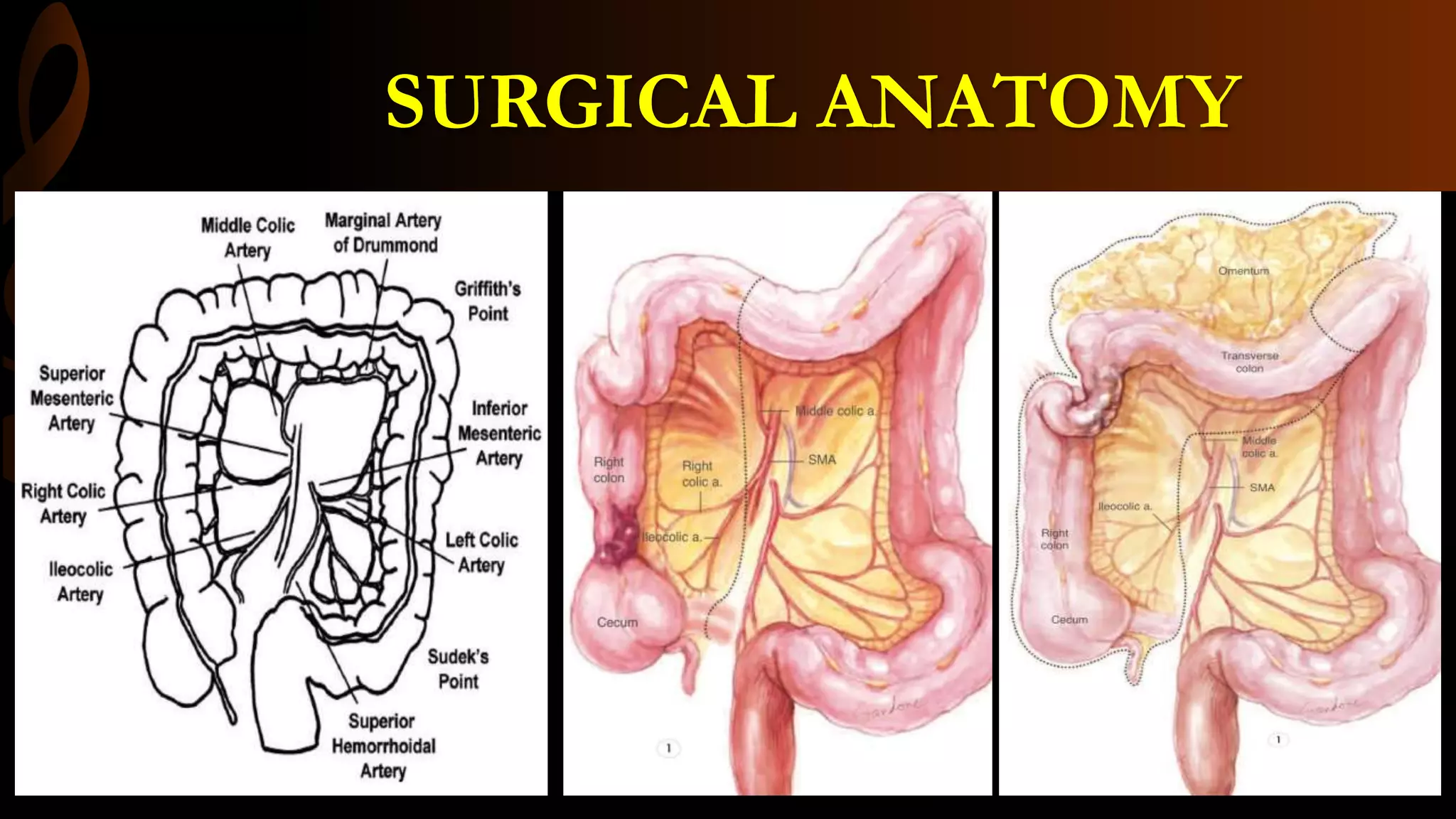
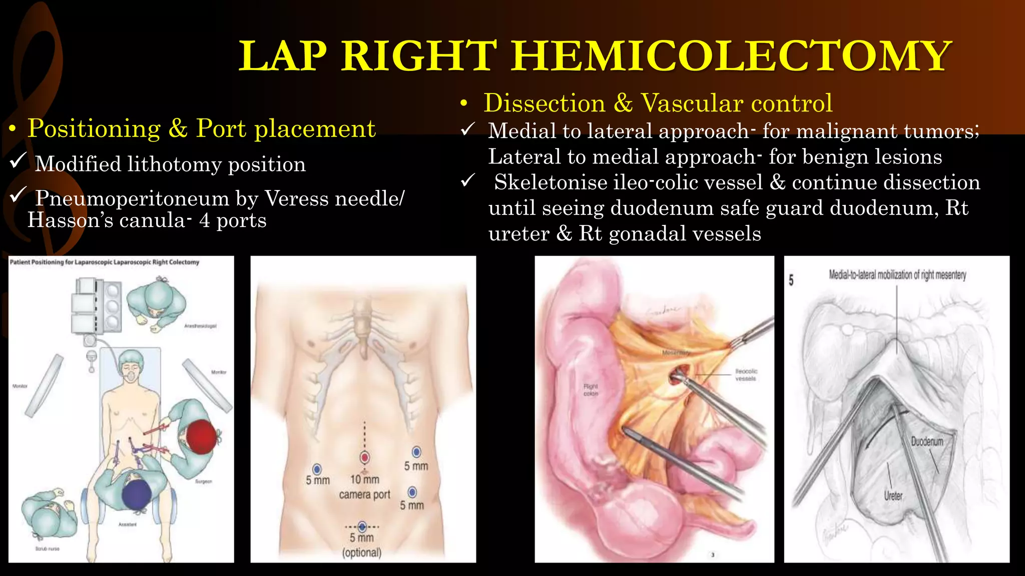
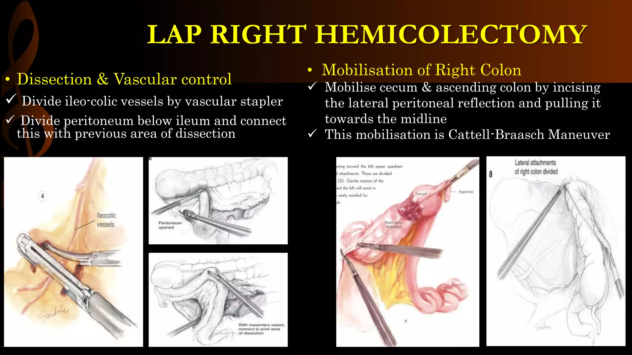
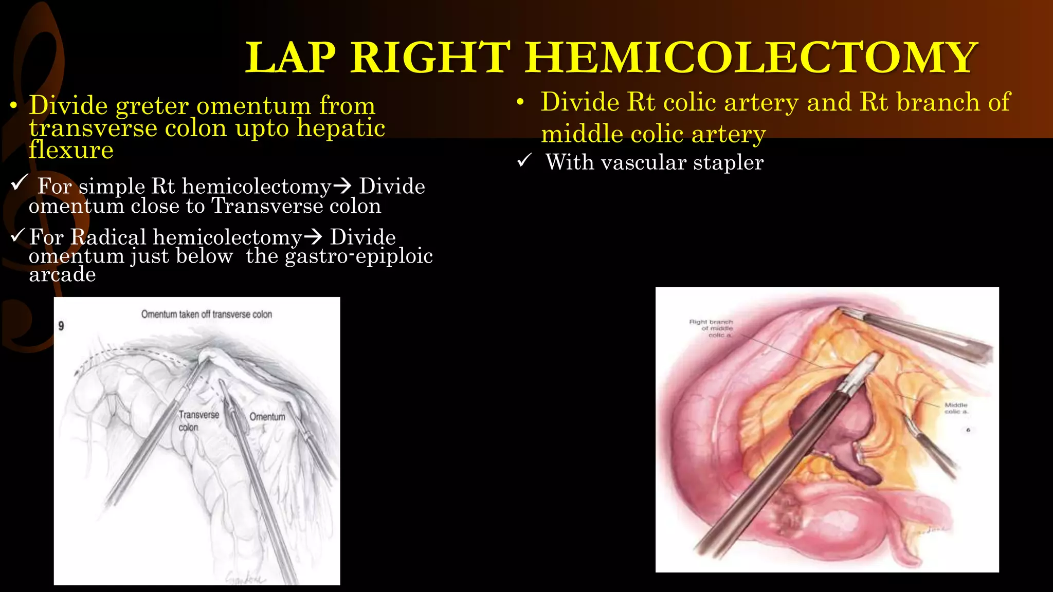
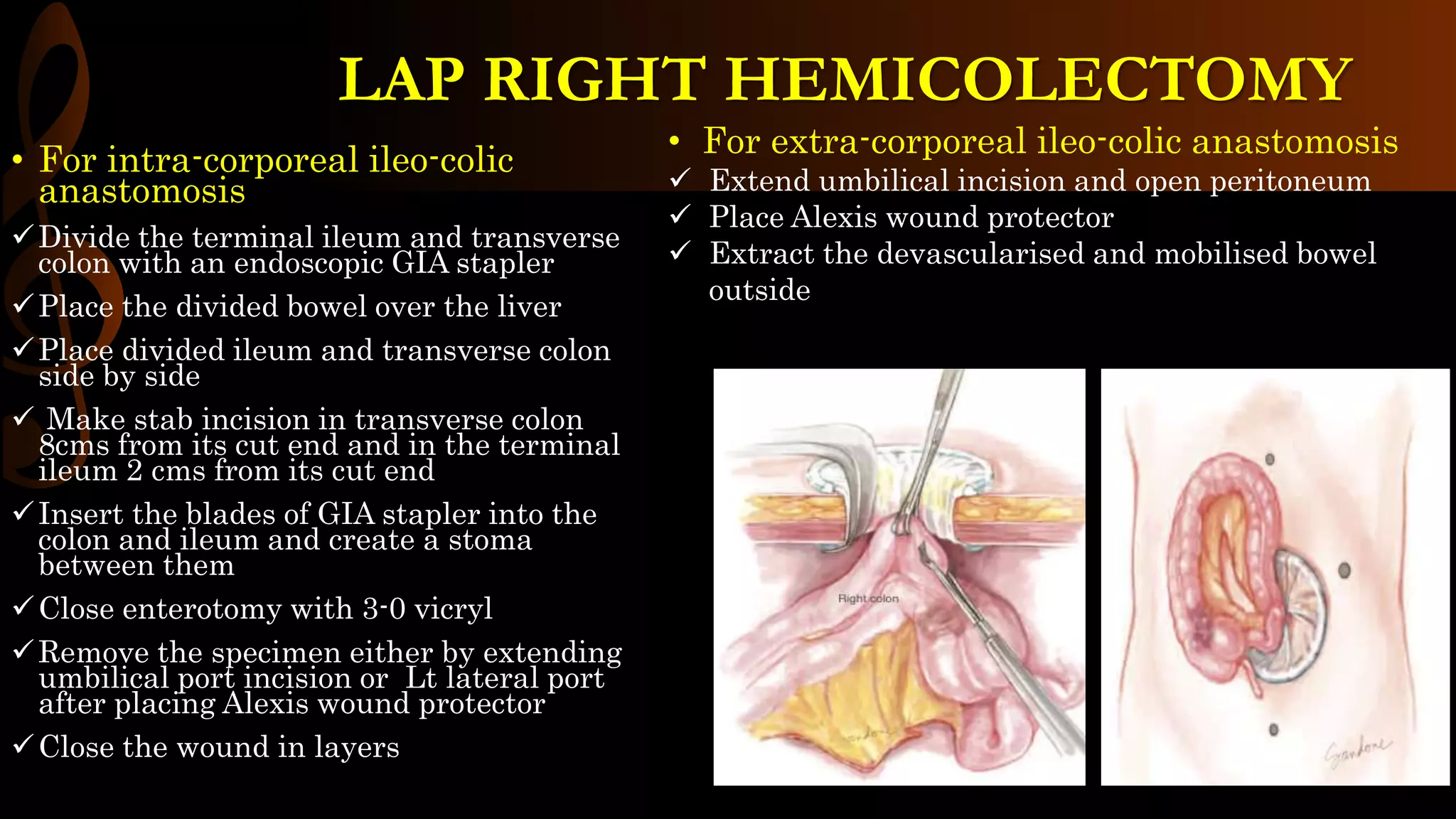
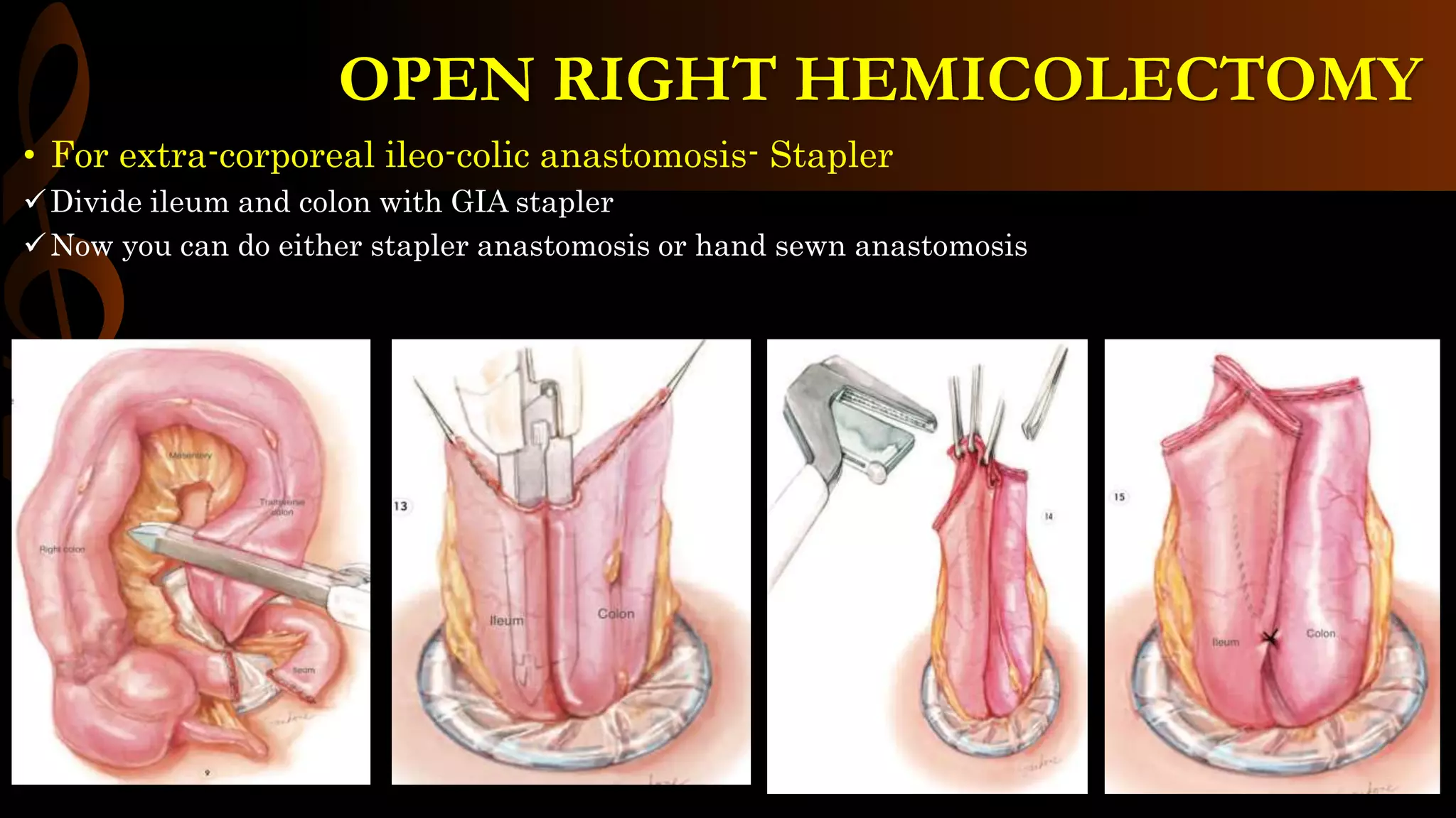
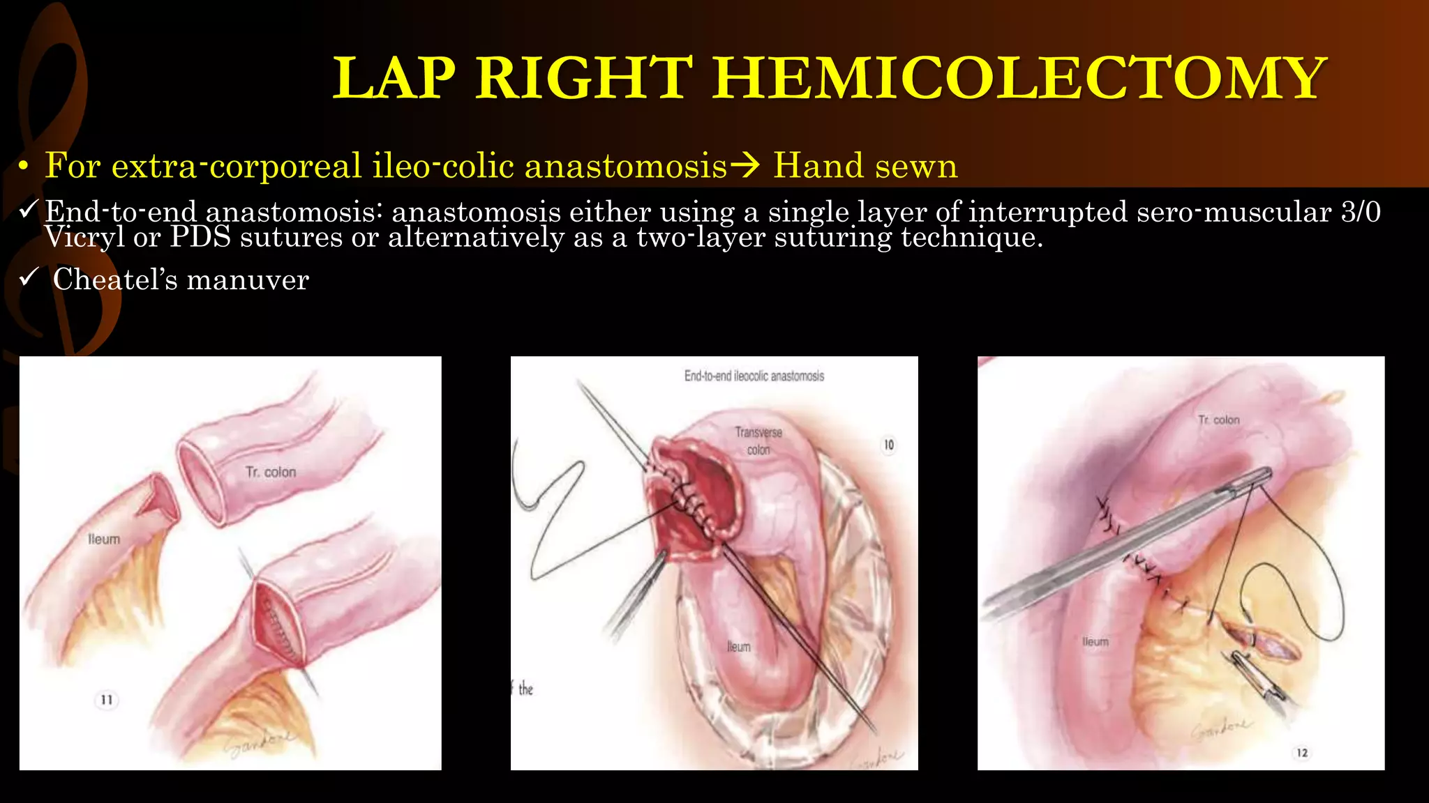
LAP RIGHT
HEMICOLECTOMY
DR.B.Selvaraj MS; Mch; FICS;
“ Surgical Educator”
Malaysia
OPERATIVE SURGERY
LAP RIGHT HEMICOLECTOMY
• INDICATIONS:
 Malignant tumors in Ileocecal region, ascending
colon and hepatic flexure
 Adenomatous polyps in Rt Colon
 Ileocecal TB, IBD, Cecal diverticulosis, bleeding
Vascular ectasia & Cecal volvulus
• ANESTHESIA:
 GA/ETT
• POSITION:
 Supine/modified lithotomy
• Diagnosis
 Confirmed by colonoscopic biopsy
 Staging by CECT
• Informed consent- risks of surgery:
 Anastomotic leak- 2%
 Hemorrhage- 1%
 Bowel obstruction- 2%
 Wound infection- 2 to 10%
 Injury to ureter and duodenum- 1%
• Pre-op preparation
 Adequate mechanical bowel preparation the day before
surgery with orthograde enema
 Prophylactic IV broad spectrum antibiotics
 VTE prophylaxis with LMWH and pneumatic stockings
 Tattooing of the tumor endoscopically or by localization on
a barium enema.
SURGICAL ANATOMY
• Positioning & Port placement
 Modified lithotomy position
 Pneumoperitoneum by Veress needle/
Hasson’s canula- 4 ports
• Dissection & Vascular control
 Medial to lateral approach- for malignant tumors;
Lateral to medial approach- for benign lesions
 Skeletonise ileo-colic vessel & continue dissection
until seeing duodenum safe guard duodenum, Rt
ureter & Rt gonadal vessels
LAP RIGHT HEMICOLECTOMY
• Dissection & Vascular control
 Divide ileo-colic vessels by vascular stapler
 Divide peritoneum below ileum and connect
this with previous area of dissection
• Mobilisation of Right Colon
 Mobilise cecum & ascending colon by incising
the lateral peritoneal reflection and pulling it
towards the midline
 This mobilisation is Cattell-Braasch Maneuver
LAP RIGHT HEMICOLECTOMY
• Divide greter omentum from
transverse colon upto hepatic
flexure
 For simple Rt hemicolectomy Divide
omentum close to Transverse colon
For Radical hemicolectomy Divide
omentum just below the gastro-epiploic
arcade
• Divide Rt colic artery and Rt branch of
middle colic artery
 With vascular stapler
LAP RIGHT HEMICOLECTOMY
• For intra-corporeal ileo-colic
anastomosis
Divide the terminal ileum and transverse
colon with an endoscopic GIA stapler
Place the divided bowel over the liver
Place divided ileum and transverse colon
side by side
 Make stab incision in transverse colon
8cms from its cut end and in the terminal
ileum 2 cms from its cut end
Insert the blades of GIA stapler into the
colon and ileum and create a stoma
between them
Close enterotomy with 3-0 vicryl
Remove the specimen either by extending
umbilical port incision or Lt lateral port
after placing Alexis wound protector
Close the wound in layers
• For extra-corporeal ileo-colic anastomosis
 Extend umbilical incision and open peritoneum
 Place Alexis wound protector
 Extract the devascularised and mobilised bowel
outside
LAP RIGHT HEMICOLECTOMY
• For extra-corporeal ileo-colic anastomosis- Stapler
Divide ileum and colon with GIA stapler
Now you can do either stapler anastomosis or hand sewn anastomosis
OPEN RIGHT HEMICOLECTOMY
• For extra-corporeal ileo-colic anastomosis Hand sewn
End-to-end anastomosis: anastomosis either using a single layer of interrupted sero-muscular 3/0
Vicryl or PDS sutures or alternatively as a two-layer suturing technique.
 Cheatel’s manuver
LAP RIGHT HEMICOLECTOMY
Post-op Care
Adherence to a postoperative colorectal clinical pathway ensures standardization of care and
facilitates timely discharge from the hospital.
Adequate pain control is achieved using patient-controlled analgesia, Inj Tramadol which can be
replaced with oral analgesics on 2 nd post-op day
Stress ulcer prophylaxis should be made for patients with symptoms or history of
gastroesophageal reflux disease (GERD) or peptic ulcer disease (PUD).
Prophylaxis for deep venous thrombosis, consisting of sequential compression devices while in
bed, and heparin 5000 U subcutaneously every 8 hours or enoxaparin 40 mg subcutaneously every
morning, starting within 24 hours after surgery.
Adequate IV fluid should be administered with monitoring of urine output. The Foley catheter
may be removed on postoperative day 1.
Clear liquid diet is started on postoperative day 1 and gradually advanced to normal diet
Early ambulation should be started on postoperative day 1.
LAP RIGHT HEMICOLECTOMY
Pearls & Pitfalls
 Prophylactic antibiotic for colorectal surgery, ertapenem (Invanz) 1 g intravenously, before surgery, requires
only a single dose for 24-hour coverage. Furthermore, it lasts for the duration of the procedure and does not
require additional dosing
 The hepatic flexure suspensory ligaments should be divided with caution, because there are often large
veins here. Careful dissection and the use of energy ligatures should strongly be considered to avoid
uncontrollable bleeding and subsequent conversion to open laparotomy.
 Smaller lesions in the colon should be marked with tattoo ink for confirmation of location, which will assist
in removal of the primary lesion with adequate 5-cm margin and areas of lymphatic drainage.
 Placement of Seprafilm under the midline incision minimizes adhesions on re-entry for subsequent
operations. This should be considered, especially for indications such as Crohn’s disease and colon cancer.
LAP RIGHT HEMICOLECTOMY
MINDMAP
THANK YOU

LAP RIGHT HEMICOLECTOMY-STEP BY STEP Operative Surgery.pptx

  • 1.
    LAP RIGHT HEMICOLECTOMY DR.B.Selvaraj MS;Mch; FICS; “ Surgical Educator” Malaysia OPERATIVE SURGERY
  • 2.
    LAP RIGHT HEMICOLECTOMY •INDICATIONS:  Malignant tumors in Ileocecal region, ascending colon and hepatic flexure  Adenomatous polyps in Rt Colon  Ileocecal TB, IBD, Cecal diverticulosis, bleeding Vascular ectasia & Cecal volvulus • ANESTHESIA:  GA/ETT • POSITION:  Supine/modified lithotomy • Diagnosis  Confirmed by colonoscopic biopsy  Staging by CECT • Informed consent- risks of surgery:  Anastomotic leak- 2%  Hemorrhage- 1%  Bowel obstruction- 2%  Wound infection- 2 to 10%  Injury to ureter and duodenum- 1% • Pre-op preparation  Adequate mechanical bowel preparation the day before surgery with orthograde enema  Prophylactic IV broad spectrum antibiotics  VTE prophylaxis with LMWH and pneumatic stockings  Tattooing of the tumor endoscopically or by localization on a barium enema.
  • 3.
  • 4.
    • Positioning &Port placement  Modified lithotomy position  Pneumoperitoneum by Veress needle/ Hasson’s canula- 4 ports • Dissection & Vascular control  Medial to lateral approach- for malignant tumors; Lateral to medial approach- for benign lesions  Skeletonise ileo-colic vessel & continue dissection until seeing duodenum safe guard duodenum, Rt ureter & Rt gonadal vessels LAP RIGHT HEMICOLECTOMY
  • 5.
    • Dissection &Vascular control  Divide ileo-colic vessels by vascular stapler  Divide peritoneum below ileum and connect this with previous area of dissection • Mobilisation of Right Colon  Mobilise cecum & ascending colon by incising the lateral peritoneal reflection and pulling it towards the midline  This mobilisation is Cattell-Braasch Maneuver LAP RIGHT HEMICOLECTOMY
  • 6.
    • Divide greteromentum from transverse colon upto hepatic flexure  For simple Rt hemicolectomy Divide omentum close to Transverse colon For Radical hemicolectomy Divide omentum just below the gastro-epiploic arcade • Divide Rt colic artery and Rt branch of middle colic artery  With vascular stapler LAP RIGHT HEMICOLECTOMY
  • 7.
    • For intra-corporealileo-colic anastomosis Divide the terminal ileum and transverse colon with an endoscopic GIA stapler Place the divided bowel over the liver Place divided ileum and transverse colon side by side  Make stab incision in transverse colon 8cms from its cut end and in the terminal ileum 2 cms from its cut end Insert the blades of GIA stapler into the colon and ileum and create a stoma between them Close enterotomy with 3-0 vicryl Remove the specimen either by extending umbilical port incision or Lt lateral port after placing Alexis wound protector Close the wound in layers • For extra-corporeal ileo-colic anastomosis  Extend umbilical incision and open peritoneum  Place Alexis wound protector  Extract the devascularised and mobilised bowel outside LAP RIGHT HEMICOLECTOMY
  • 8.
    • For extra-corporealileo-colic anastomosis- Stapler Divide ileum and colon with GIA stapler Now you can do either stapler anastomosis or hand sewn anastomosis OPEN RIGHT HEMICOLECTOMY
  • 9.
    • For extra-corporealileo-colic anastomosis Hand sewn End-to-end anastomosis: anastomosis either using a single layer of interrupted sero-muscular 3/0 Vicryl or PDS sutures or alternatively as a two-layer suturing technique.  Cheatel’s manuver LAP RIGHT HEMICOLECTOMY
  • 10.
    Post-op Care Adherence toa postoperative colorectal clinical pathway ensures standardization of care and facilitates timely discharge from the hospital. Adequate pain control is achieved using patient-controlled analgesia, Inj Tramadol which can be replaced with oral analgesics on 2 nd post-op day Stress ulcer prophylaxis should be made for patients with symptoms or history of gastroesophageal reflux disease (GERD) or peptic ulcer disease (PUD). Prophylaxis for deep venous thrombosis, consisting of sequential compression devices while in bed, and heparin 5000 U subcutaneously every 8 hours or enoxaparin 40 mg subcutaneously every morning, starting within 24 hours after surgery. Adequate IV fluid should be administered with monitoring of urine output. The Foley catheter may be removed on postoperative day 1. Clear liquid diet is started on postoperative day 1 and gradually advanced to normal diet Early ambulation should be started on postoperative day 1. LAP RIGHT HEMICOLECTOMY
  • 11.
    Pearls & Pitfalls Prophylactic antibiotic for colorectal surgery, ertapenem (Invanz) 1 g intravenously, before surgery, requires only a single dose for 24-hour coverage. Furthermore, it lasts for the duration of the procedure and does not require additional dosing  The hepatic flexure suspensory ligaments should be divided with caution, because there are often large veins here. Careful dissection and the use of energy ligatures should strongly be considered to avoid uncontrollable bleeding and subsequent conversion to open laparotomy.  Smaller lesions in the colon should be marked with tattoo ink for confirmation of location, which will assist in removal of the primary lesion with adequate 5-cm margin and areas of lymphatic drainage.  Placement of Seprafilm under the midline incision minimizes adhesions on re-entry for subsequent operations. This should be considered, especially for indications such as Crohn’s disease and colon cancer. LAP RIGHT HEMICOLECTOMY
  • 12.
  • 13.