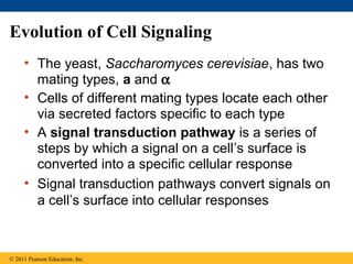
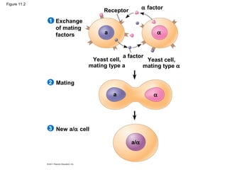
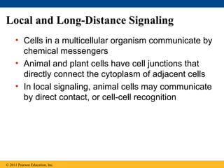
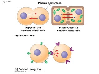
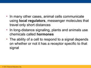
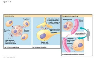
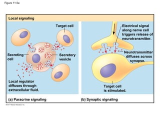
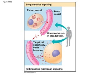
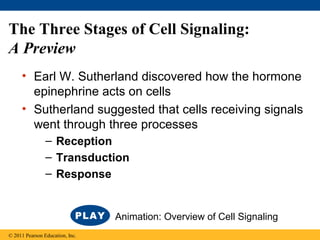
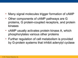
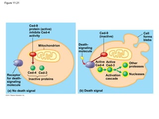
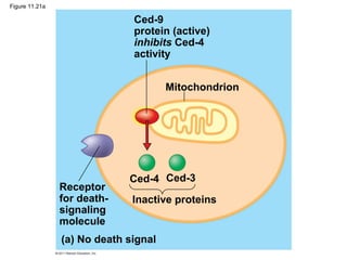
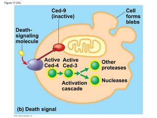
The document provides an overview of cell communication and signaling. It discusses:
- Cells communicate via chemical signals such as epinephrine.
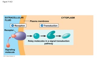
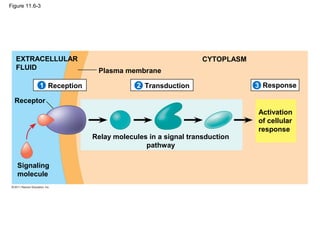
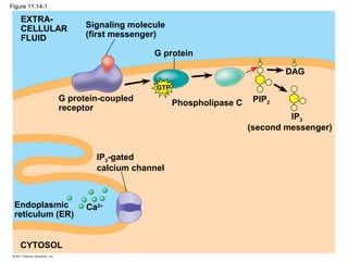
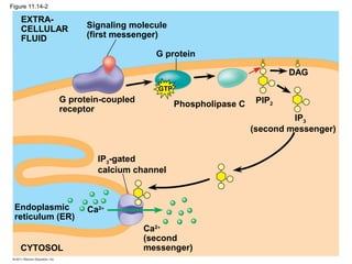
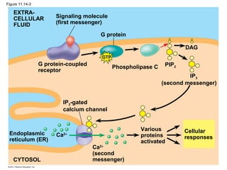
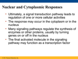
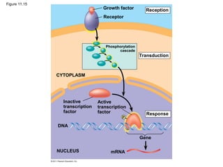
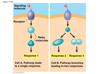
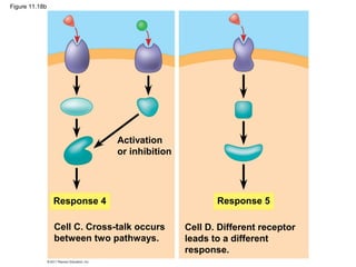
- Signal transduction pathways convert signals at the cell surface into responses.
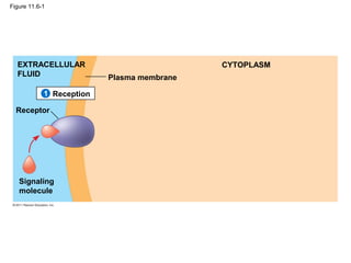
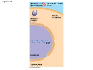
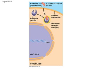
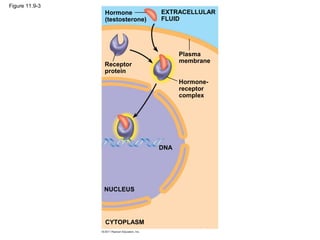
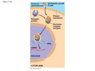
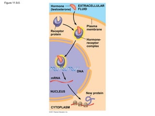
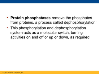
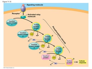
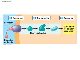
- Reception involves signal molecules binding receptors, transduction uses cascades of molecular interactions to relay signals from receptors to target molecules, and response leads to activation of cellular responses.
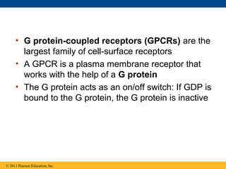
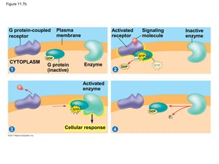
- Common mechanisms of signaling include G-protein coupled receptors, receptor tyrosine kinases, calcium ions, cyclic AMP, and inositol triphosphate.


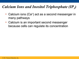
![Figure 11.13
Mitochondrion
EXTRACELLULAR
FLUID
Plasma
membrane
Ca2+
pump
Nucleus
CYTOSOL
Ca2+
pump
Ca2+
pump
Endoplasmic
reticulum
(ER)
ATP
ATP
Low [Ca2+
]High [Ca2+
]Key](https://image.slidesharecdn.com/11lecturecellcommunication-150106201808-conversion-gate02/85/Ch-11-Cell-Communication-53-320.jpg)


![Cell signaling -_introduction[1]](https://cdn.slidesharecdn.com/ss_thumbnails/cellsignaling-introduction1-160424161032-thumbnail.jpg?width=640&height=640&fit=bounds)