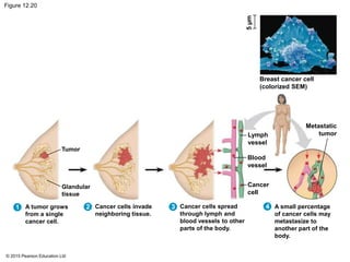
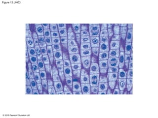
1) The document describes the process of cell division known as mitosis in eukaryotic cells.
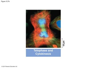
2) Mitosis consists of nuclear division (mitosis) and cytoplasmic division (cytokinesis). During interphase, the cell grows and DNA is replicated.
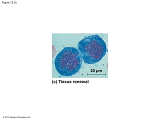
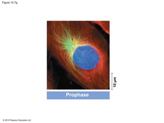
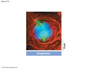
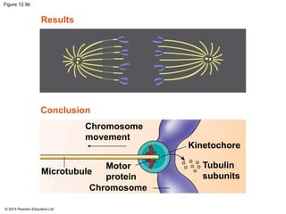
3) Mitosis involves five stages (prophase, prometaphase, metaphase, anaphase, telophase) where chromosomes align and separate, guided by the mitotic spindle. Cytokinesis then divides the cytoplasm, forming two daughter cells.