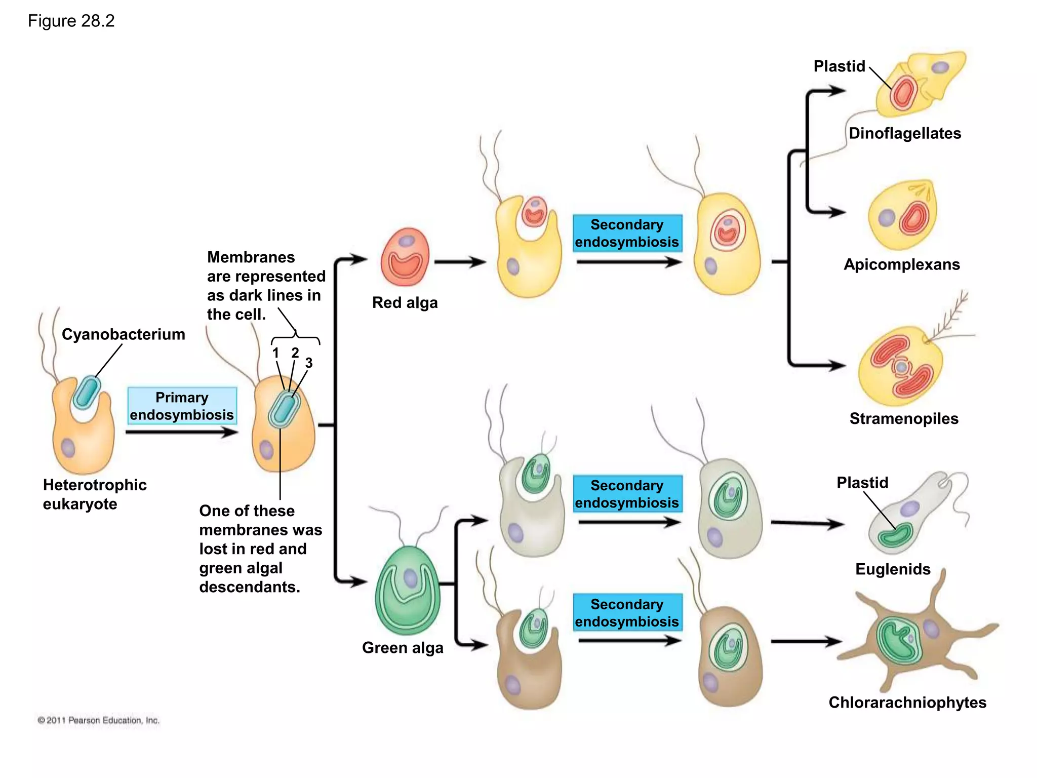
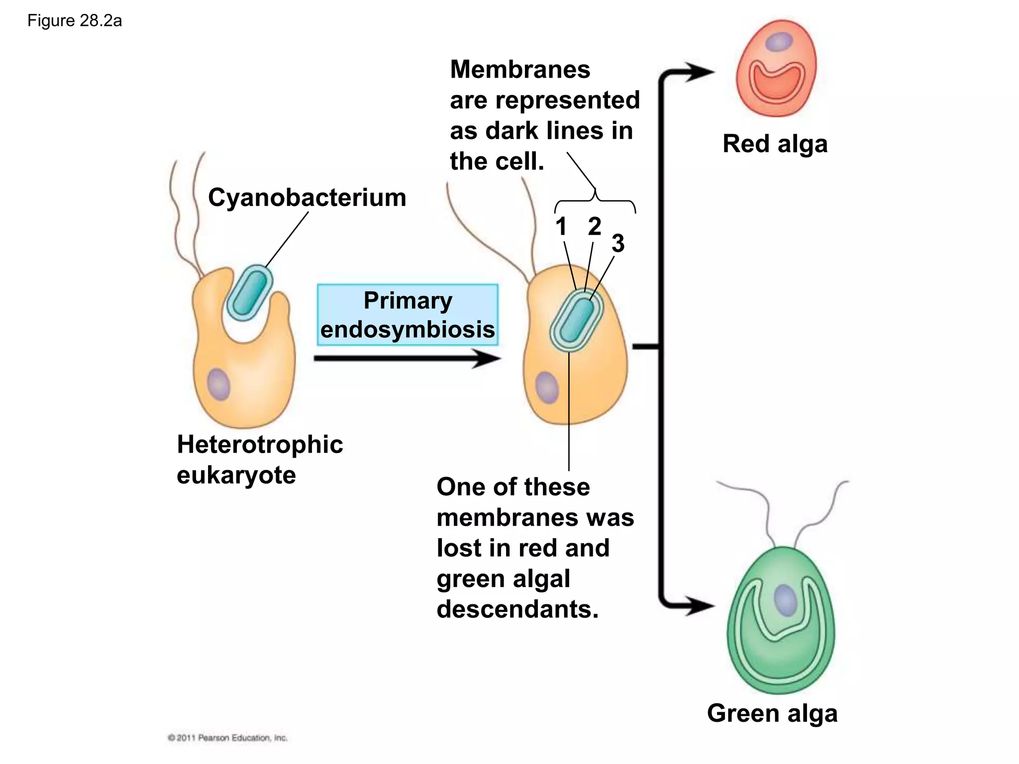
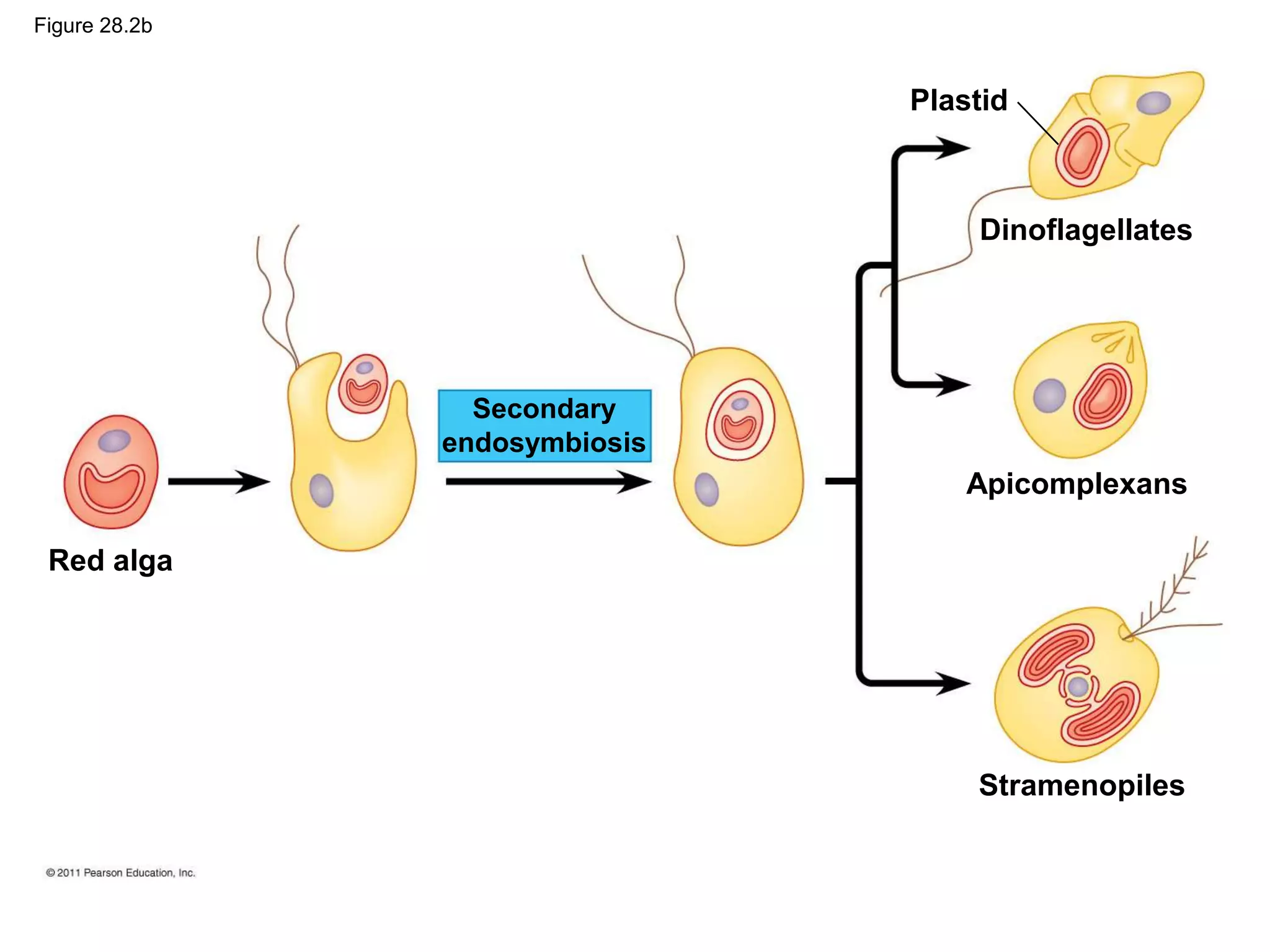
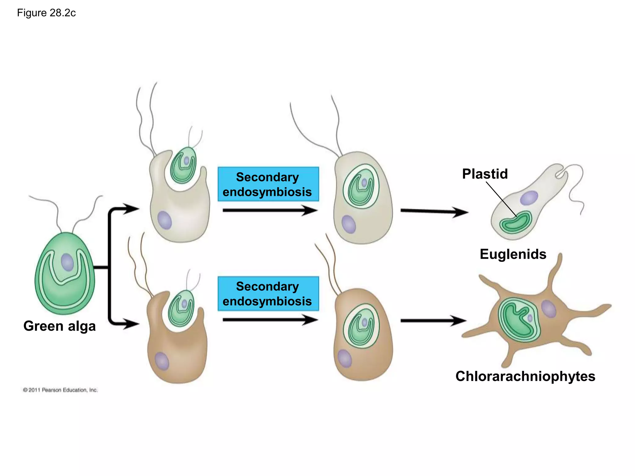
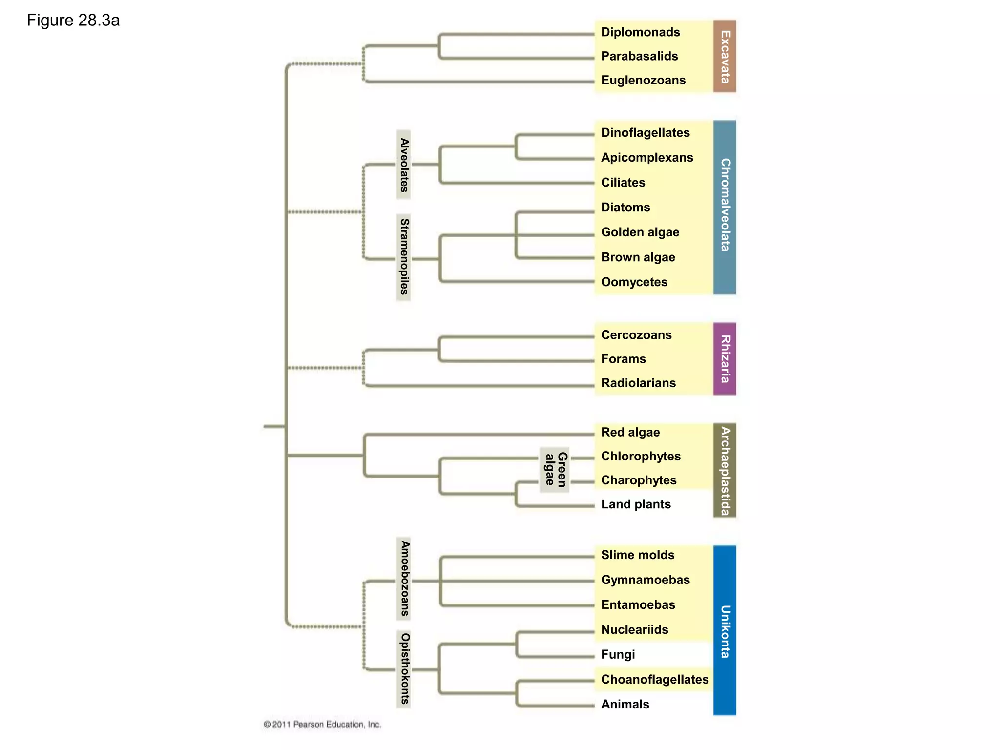
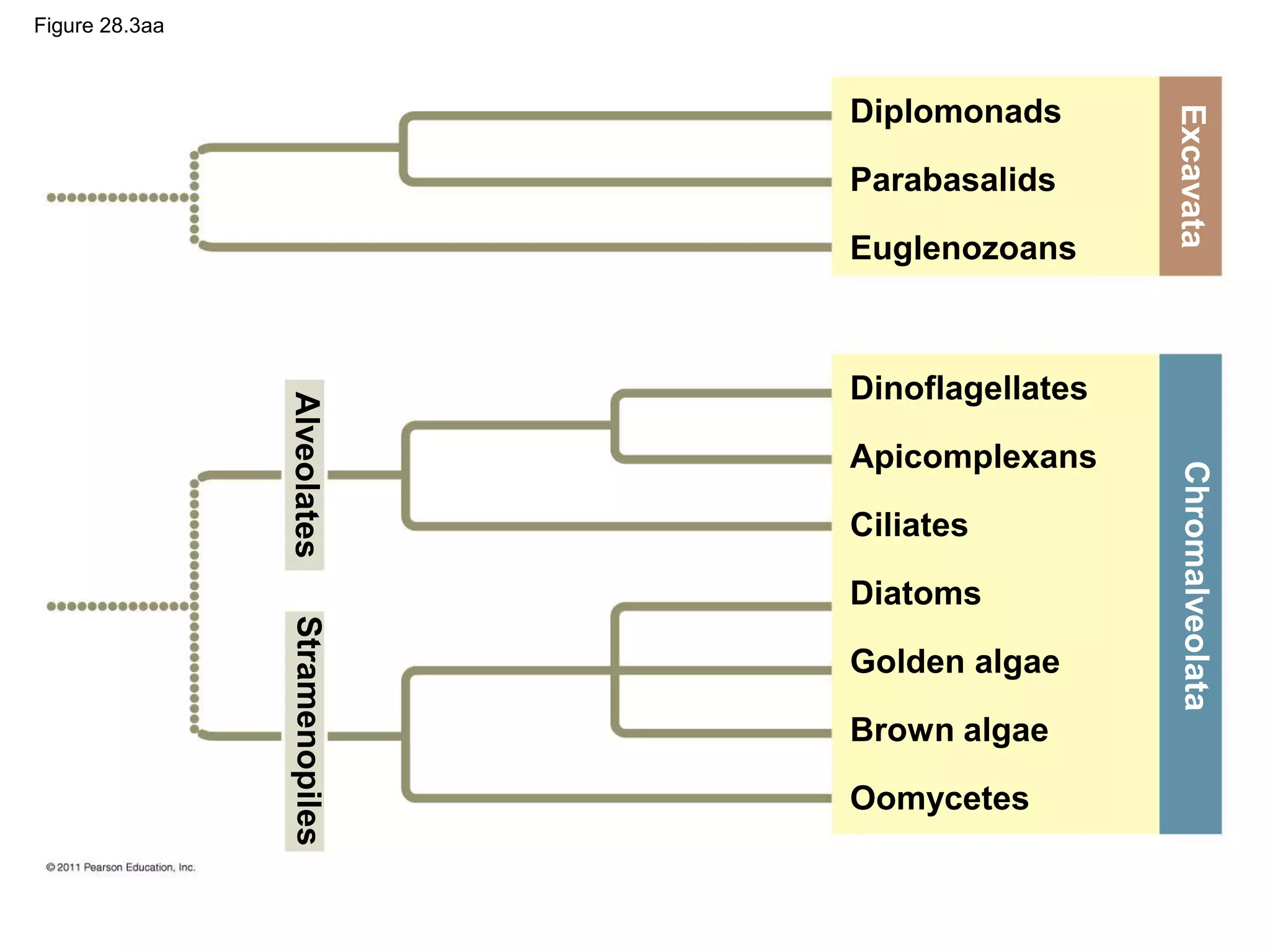
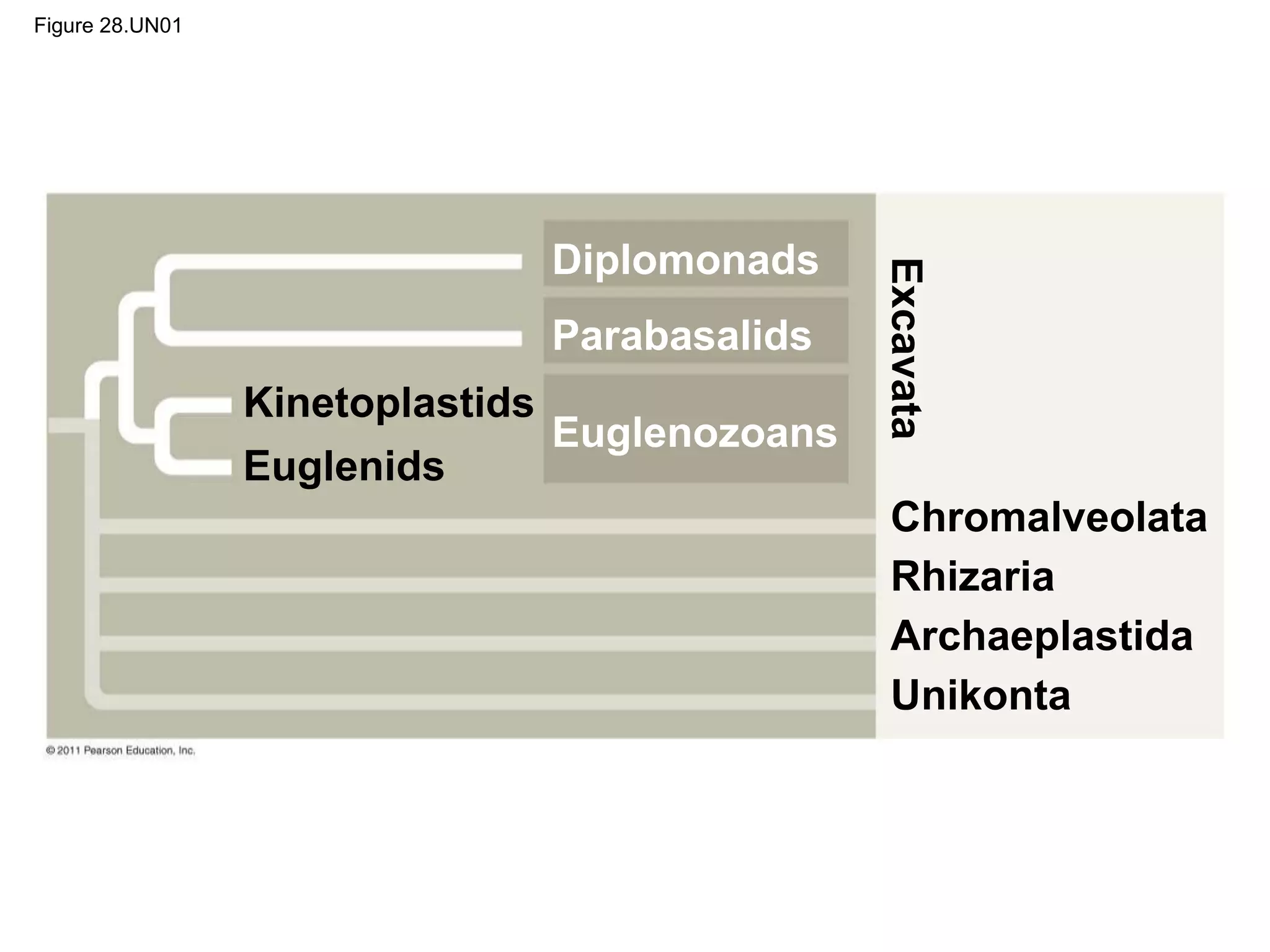
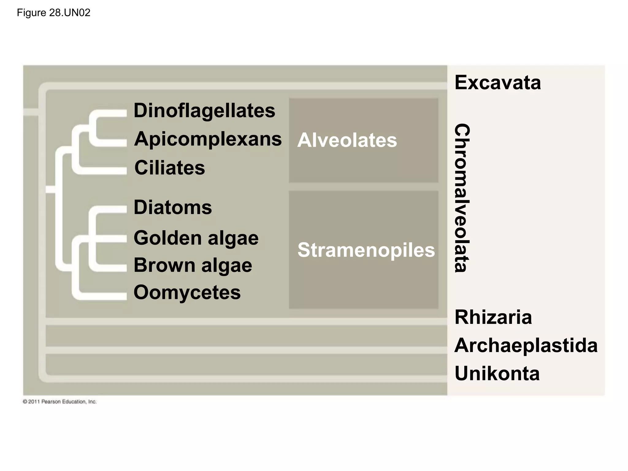
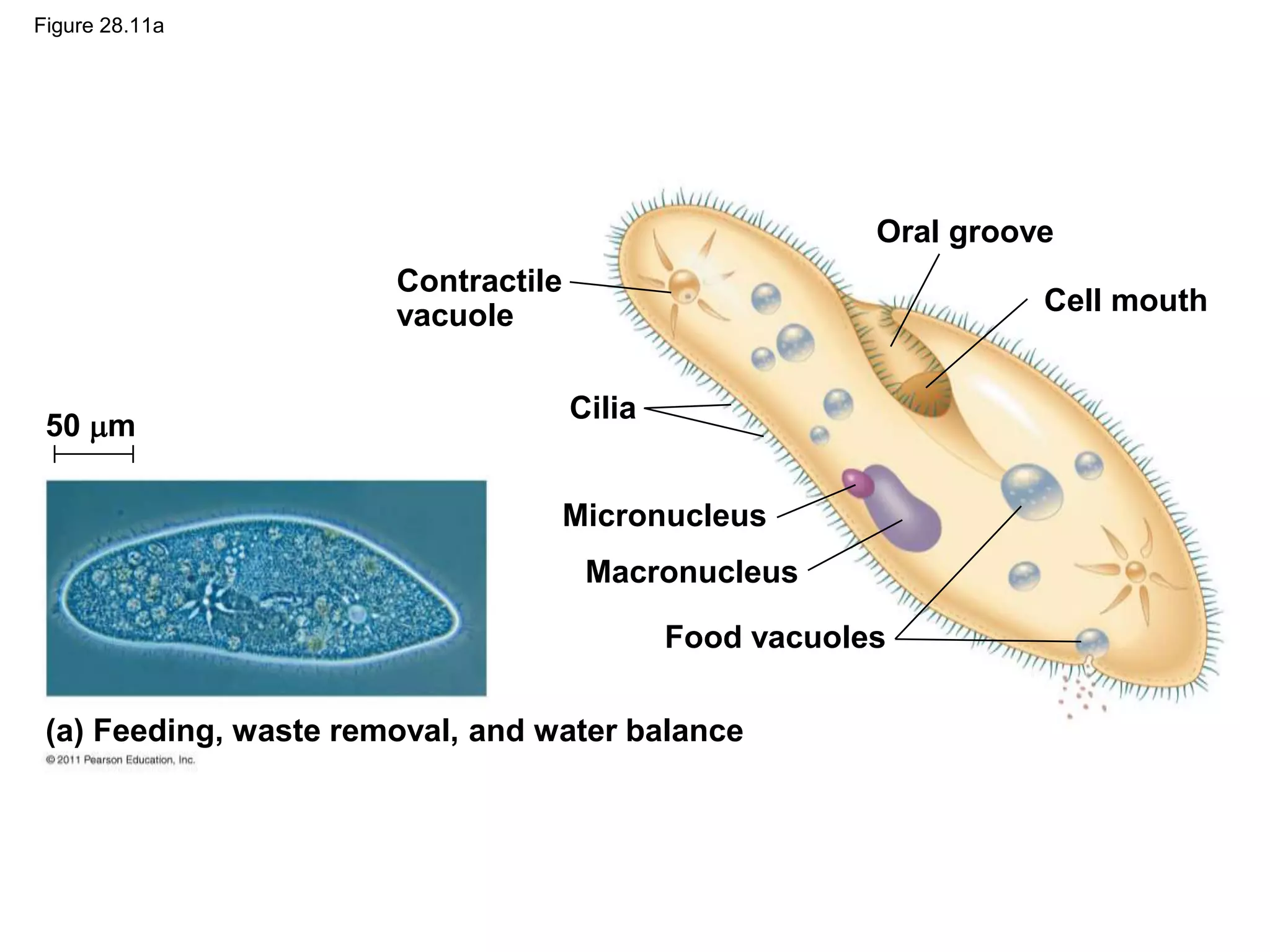
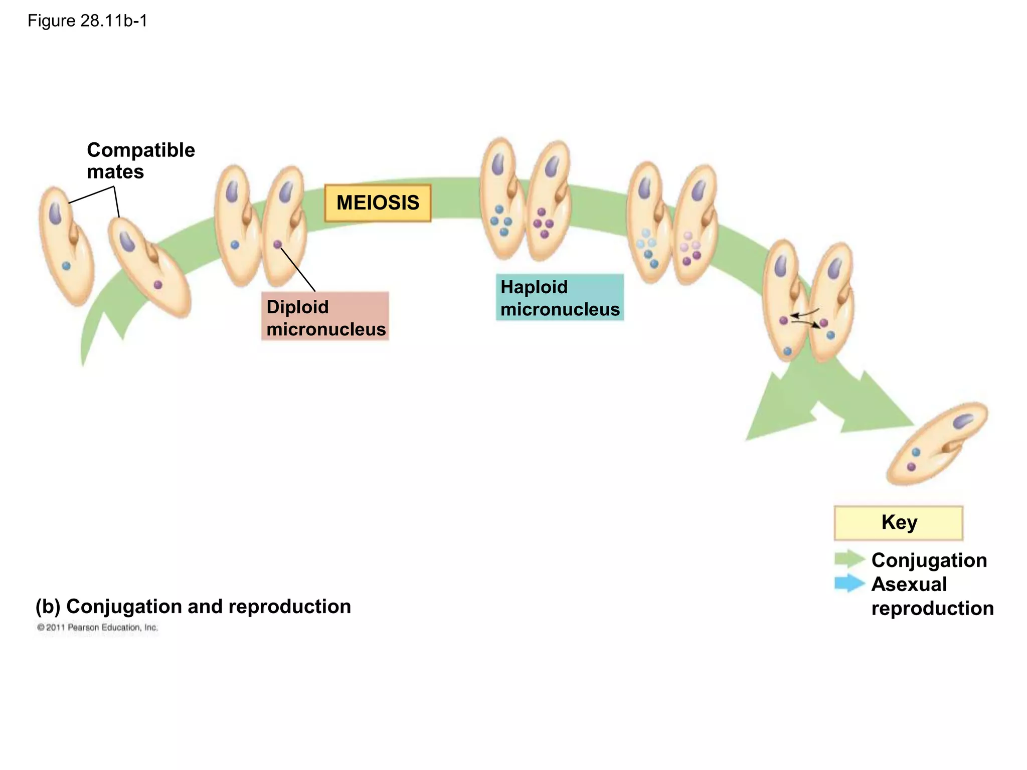
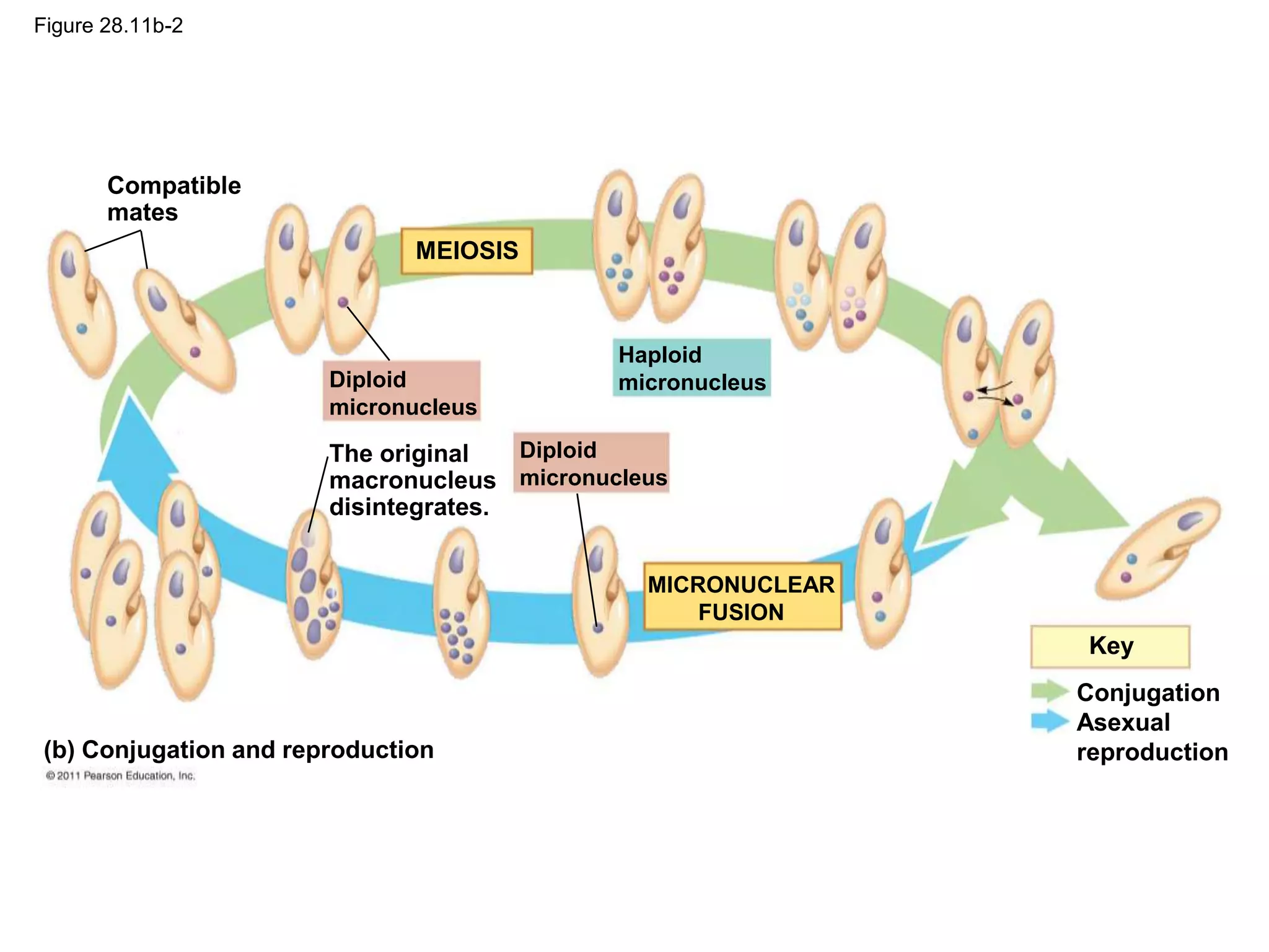
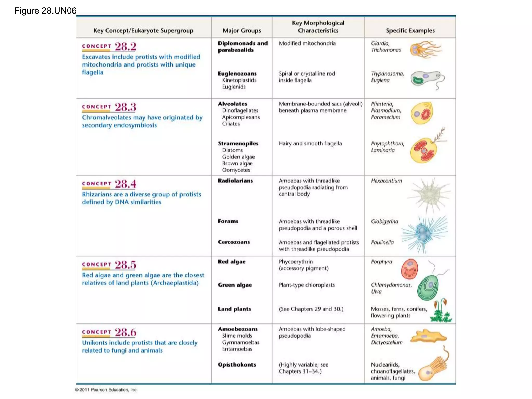
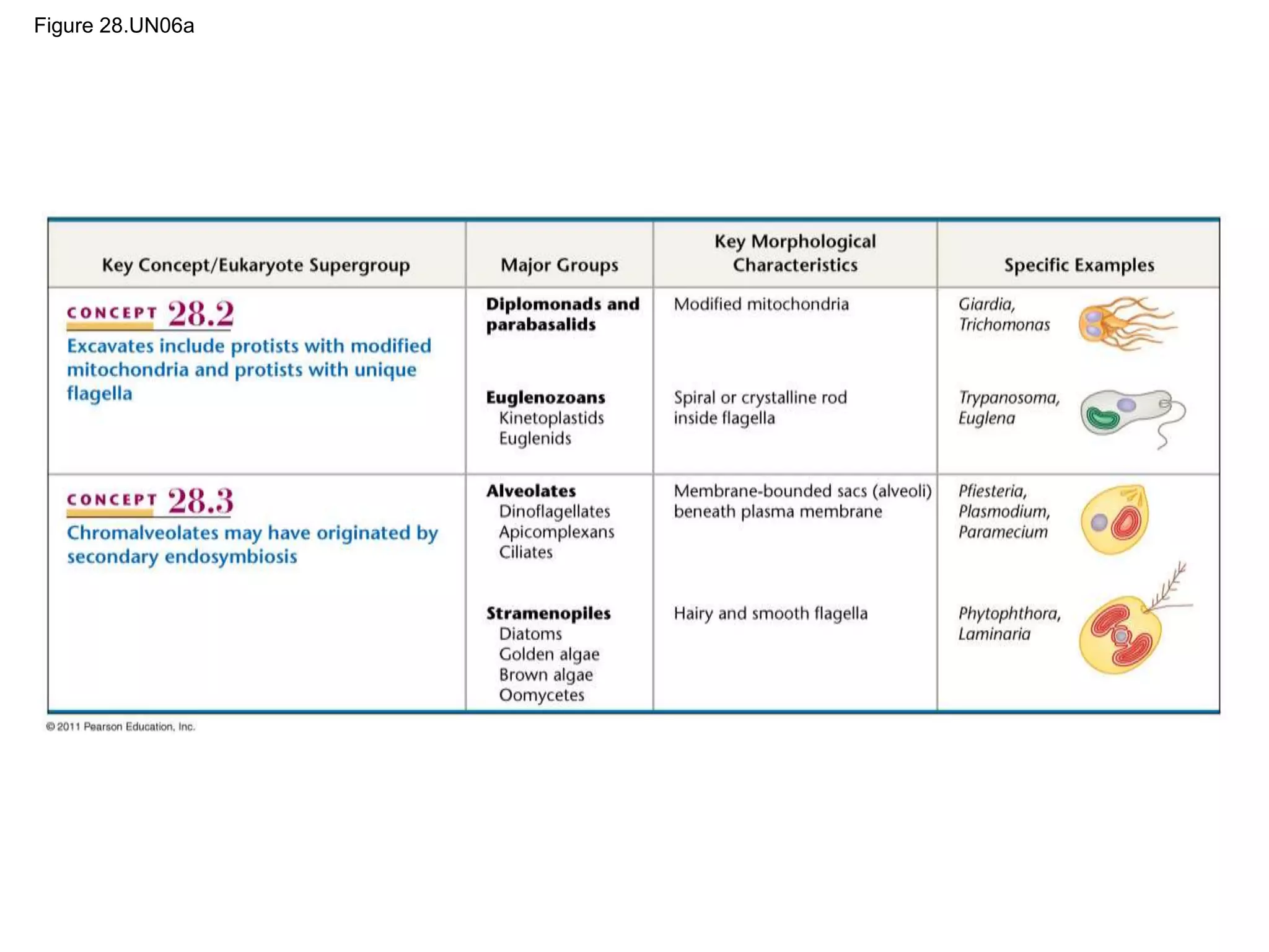
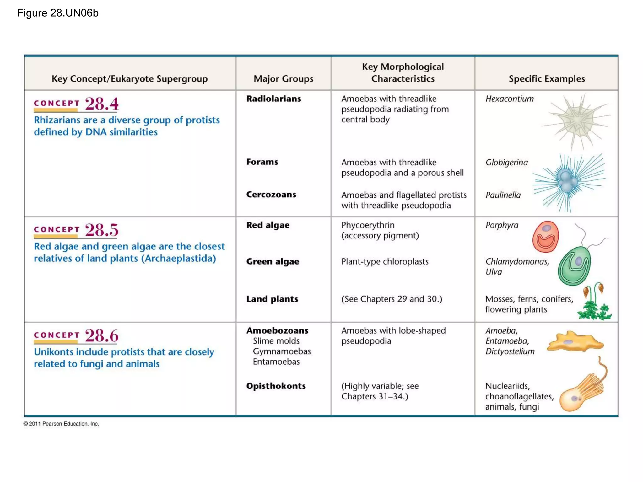
LECTURE PRESENTATIONS
For CAMPBELL BIOLOGY, NINTH EDITION
Jane B. Reece, Lisa A. Urry, Michael L. Cain, Steven A. Wasserman, Peter V. Minorsky, Robert B. Jackson
© 2011 Pearson Education, Inc.
Lectures by
Erin Barley
Kathleen Fitzpatrick
Protists
Chapter 28
Overview: Living Small
• Even a low-power microscope can reveal a great
variety of organisms in a drop of pond water
• Protist is the informal name of the group of mostly
unicellular eukaryotes
• Advances in eukaryotic systematics have caused
the classification of protists to change significantly
• Protists constitute a polyphyletic group, and
Protista is no longer valid as a kingdom
© 2011 Pearson Education, Inc.
1 m
Figure 28.1
Concept 28.1: Most eukaryotes are
single-celled organisms
• Protists are eukaryotes
• Eukaryotic cells have organelles and are more
complex than prokaryotic cells
• Most protists are unicellular, but there are some
colonial and multicellular species
© 2011 Pearson Education, Inc.
Structural and Functional Diversity in
Protists
• Protists exhibit more structural and functional
diversity than any other group of eukaryotes
• Single-celled protists can be very complex, as all
biological functions are carried out by organelles in
each individual cell
© 2011 Pearson Education, Inc.
• Protists, the most nutritionally diverse of all
eukaryotes, include
– Photoautotrophs, which contain chloroplasts
– Heterotrophs, which absorb organic molecules or
ingest larger food particles
– Mixotrophs, which combine photosynthesis and
heterotrophic nutrition
© 2011 Pearson Education, Inc.
• Some protists reproduce asexually, while
others reproduce sexually, or by the sexual
processes of meiosis and fertilization
© 2011 Pearson Education, Inc.
Endosymbiosis in Eukaryotic Evolution
• There is now considerable evidence that much
protist diversity has its origins in endosymbiosis
• Endosymbiosis is the process in which a
unicellular organism engulfs another cell, which
becomes an endosymbiont and then organelle in
the host cell
• Mitochondria evolved by endosymbiosis of an
aerobic prokaryote
• Plastids evolved by endosymbiosis of a
photosynthetic cyanobacterium
© 2011 Pearson Education, Inc.
Cyanobacterium
Heterotrophic
eukaryote
Primary
endosymbiosis
Membranes
are represented
as dark lines in
the cell.
1 2
3
One of these
membranes was
lost in red and
green algal
descendants.
Plastid
Red alga
Secondary
endosymbiosis
Secondary
endosymbiosis
Secondary
endosymbiosis
Green alga
Dinoflagellates
Apicomplexans
Stramenopiles
Plastid
Euglenids
Chlorarachniophytes
Figure 28.2
Figure 28.2a
Cyanobacterium
Heterotrophic
eukaryote
Primary
endosymbiosis
Membranes
are represented
as dark lines in
the cell.
1 2
3
One of these
membranes was
lost in red and
green algal
descendants.
Red alga
Green alga
Figure 28.2b
Plastid
Red alga
Secondary
endosymbiosis
Dinoflagellates
Apicomplexans
Stramenopiles
Figure 28.2c
Secondary
endosymbiosis
Green alga
Plastid
Euglenids
Chlorarachniophytes
Secondary
endosymbiosis
• The plastid-bearing lineage of protists evolved into
red and green algae
• The DNA of plastid genes in red algae and green
algae closely resemble the DNA of cyanobacteria
• On several occasions during eukaryotic evolution,
red and green algae underwent secondary
endosymbiosis, in which they were ingested by a
heterotrophic eukaryote
© 2011 Pearson Education, Inc.
Five Supergroups of Eukaryotes
• It is no longer thought that amitochondriates
(lacking mitochondria) are the oldest lineage of
eukaryotes
• Many have been shown to have mitochondria and
have been reclassified
• Our understanding of the relationships among
protist groups continues to change rapidly
• One hypothesis divides all eukaryotes (including
protists) into five supergroups
© 2011 Pearson Education, Inc.
Figure 28.3a
Diplomonads
Parabasalids
Euglenozoans
Dinoflagellates
Apicomplexans
Ciliates
Diatoms
Golden algae
Brown algae
Oomycetes
Cercozoans
Forams
Radiolarians
Red algae
Chlorophytes
Charophytes
Land plants
Slime molds
Gymnamoebas
Entamoebas
Nucleariids
Fungi
Choanoflagellates
Animals
Alveolates
Stramenopiles
Green
algae
Amoebozoans
Opisthokonts
Excavata
Chromalveolata
Rhizaria
Archaeplastida
Unikonta
Figure 28.3aa
Diplomonads
Parabasalids
Euglenozoans
Dinoflagellates
Apicomplexans
Ciliates
Diatoms
Golden algae
Brown algae
Oomycetes
Alveolates
Stramenopiles
Excavata
Chromalveolata
Figure 28.3ab
Cercozoans
Forams
Radiolarians
Red algae
Chlorophytes
Charophytes
Land plants
Green
algae
Rhizaria
Archaeplastida
Figure 28.3ac
Slime molds
Gymnamoebas
Entamoebas
Nucleariids
Fungi
Choanoflagellates
Animals
Amoebozoans
Opisthokonts
Unikonta
Figure 28.3b
Giardia intestinalis, a diplomonad
parasite
5 m
Figure 28.3c
Diatom diversity
50 m
Globigerina, a foram in the supergroup Rhizaria
100 m
Figure 28.3d
Figure 28.3da
Figure 28.3db
Globigerina, a foram in the supergroup Rhizaria
100 m
Figure 28.3e
Volvox, a colonial freshwater green alga
20 m
50 m
Figure 28.3ea
20 m
Figure 28.3eb
Volvox, a colonial freshwater green alga
50 m
Figure 28.3f
A unikont amoeba
100 m
• The clade Excavata is characterized by its
cytoskeleton
• Some members have a feeding groove
• This controversial group includes the
diplomonads, parabasalids, and euglenozoans
Concept 28.2: Excavates include protists
with modified mitochondria and protists
with unique flagella
© 2011 Pearson Education, Inc.
Figure 28.UN01
Kinetoplastids
Euglenids
Euglenozoans
Diplomonads
Parabasalids
Excavata
Chromalveolata
Rhizaria
Archaeplastida
Unikonta
Diplomonads and Parabasalids
• These two groups lack plastids, have modified
mitochondria, and most live in anaerobic
environments
• Diplomonads
– Have modified mitochondria called mitosomes
– Derive energy from anaerobic biochemical
pathways
– Have two equal-sized nuclei and multiple flagella
– Are often parasites, for example, Giardia
intestinalis (also known as Giardia lamblia)
© 2011 Pearson Education, Inc.
• Parabasalids
– Have reduced mitochondria called
hydrogenosomes that generate some energy
anaerobically
– Include Trichomonas vaginalis, the pathogen that
causes yeast infections in human females
© 2011 Pearson Education, Inc.
Figure 28.4
Flagella
5
m
Undulating
membrane
Euglenozoans
• Euglenozoa is a diverse clade that includes
predatory heterotrophs, photosynthetic autotrophs,
and parasites
• The main feature distinguishing them as a clade is
a spiral or crystalline rod of unknown function
inside their flagella
• This clade includes the kinetoplastids and
euglenids
© 2011 Pearson Education, Inc.
Figure 28.5
Crystalline rod
(cross section)
Flagella
Ring of microtubules
(cross section)
8 m
0.2 m
Figure 28.5a
Crystalline rod
(cross section)
Ring of microtubules
(cross section)
0.2 m
Kinetoplastids
• Kinetoplastids have a single mitochondrion with
an organized mass of DNA called a kinetoplast
• They include free-living consumers of prokaryotes
in freshwater, marine, and moist terrestrial
ecosystems
• This group includes Trypanosoma, which causes
sleeping sickness in humans
• Another pathogenic trypanosome causes Chagas’
disease
© 2011 Pearson Education, Inc.
Figure 28.6
9 m
• Trypanosomes evade immune responses by
switching surface proteins
• A cell produces millions of copies of a single
protein
• The new generation produces millions of copies of
a different protein
• These frequent changes prevent the host from
developing immunity
© 2011 Pearson Education, Inc.
Euglenids
• Euglenids have one or two flagella that emerge
from a pocket at one end of the cell
• Some species can be both autotrophic and
heterotrophic
© 2011 Pearson Education, Inc.
Video: Euglena Motion
Video: Euglena
Figure 28.7
Long flagellum
Eyespot
Short flagellum
Contractile vacuole
Nucleus
Chloroplast
Plasma membrane
Euglena (LM)
5 m
Pellicle
Light
detector
Figure 28.7a
Long flagellum
Eyespot
Contractile vacuole
Nucleus
Chloroplast
Plasma membrane
Euglena (LM) 5 m
Concept 28.3: Chromalveolates may have
originated by secondary endosymbiosis
• Some data suggest that the clade
Chromalveolata is monophyletic and originated
by a secondary endosymbiosis event
• The proposed endosymbiont is a red alga
• This clade is controversial and includes the
alveolates and the stramenopiles
© 2011 Pearson Education, Inc.
Figure 28.UN02
Apicomplexans
Ciliates
Diatoms
Stramenopiles
Alveolates
Chromalveolata
Rhizaria
Archaeplastida
Unikonta
Dinoflagellates
Golden algae
Brown algae
Oomycetes
Excavata
Alveolates
• Members of the clade Alveolata have
membrane-bounded sacs (alveoli) just under
the plasma membrane
• The function of the alveoli is unknown
• The alveolates include
– Dinoflagellates
– Apicomplexans
– Ciliates
© 2011 Pearson Education, Inc.
Figure 28.8
Flagellum Alveoli
Alveolate
0.2
m
Figure 28.8a
Flagellum Alveoli
0.2
m
Dinoflagellates
• Dinoflagellates have two flagella and each cell is
reinforced by cellulose plates
• They are abundant components of both marine
and freshwater phytoplankton
• They are a diverse group of aquatic phototrophs,
mixotrophs, and heterotrophs
• Toxic “red tides” are caused by dinoflagellate
blooms
© 2011 Pearson Education, Inc.
Video: Dinoflagellate
Figure 28.9
Flagella
3
m
Apicomplexans
• Apicomplexans are parasites of animals, and
some cause serious human diseases
• They spread through their host as infectious cells
called sporozoites
• One end, the apex, contains a complex of
organelles specialized for penetrating host cells
and tissues
• Most have sexual and asexual stages that require
two or more different host species for completion
© 2011 Pearson Education, Inc.
• The apicomplexan Plasmodium is the parasite that
causes malaria
• Plasmodium requires both mosquitoes and
humans to complete its life cycle
• Approximately 900,000 people die each year from
malaria
• Efforts are ongoing to develop vaccines that target
this pathogen
© 2011 Pearson Education, Inc.
Merozoite
(n)
Liver
Liver
cell
Red blood
cells
Gametocytes
(n)
Merozoite
Apex
0.5 m
Red blood
cell
Key
Haploid (n)
Diploid (2n)
Inside human
Figure 28.10-1
Zygote
(2n)
Merozoite
(n)
Gametes
Liver
Liver
cell
Red blood
cells
Gametocytes
(n)
Merozoite
Apex
0.5 m
Red blood
cell
Key
Haploid (n)
Diploid (2n)
Inside mosquito Inside human
FERTILIZATION
Figure 28.10-2
Sporozoites
(n)
Oocyst
Zygote
(2n)
Merozoite
(n)
Gametes
Liver
Liver
cell
Red blood
cells
Gametocytes
(n)
Merozoite
Apex
0.5 m
Red blood
cell
Key
Haploid (n)
Diploid (2n)
Inside mosquito Inside human
MEIOSIS
FERTILIZATION
Figure 28.10-3
Figure 28.10a
Merozoite
Apex
Red blood
cell
0.5 m
Ciliates
• Ciliates, a large varied group of protists, are
named for their use of cilia to move and feed
• They have large macronuclei and small
micronuclei
• Genetic variation results from conjugation, in
which two individuals exchange haploid
micronuclei
• Conjugation is a sexual process, and is separate
from reproduction, which generally occurs by
binary fission
© 2011 Pearson Education, Inc.
Contractile
vacuole Oral groove
Cell mouth
Food vacuoles
Cilia
Micronucleus
Macronucleus
50 m
(a) Feeding, waste removal, and water balance
Conjugation
Asexual
reproduction
Key
MEIOSIS
MICRONUCLEAR
FUSION
Diploid
micronucleus
Diploid
micronucleus
Haploid
micronucleus
Compatible
mates
The original
macronucleus
disintegrates.
(b) Conjugation and reproduction
Figure 28.11
Figure 28.11a
Contractile
vacuole
Oral groove
Cell mouth
Food vacuoles
Cilia
Micronucleus
Macronucleus
50 m
(a) Feeding, waste removal, and water balance
Figure 28.11b-1
Conjugation
Asexual
reproduction
Key
MEIOSIS
Diploid
micronucleus
Haploid
micronucleus
Compatible
mates
(b) Conjugation and reproduction
Figure 28.11b-2
Conjugation
Asexual
reproduction
Key
MEIOSIS
MICRONUCLEAR
FUSION
Diploid
micronucleus
Diploid
micronucleus
Haploid
micronucleus
Compatible
mates
The original
macronucleus
disintegrates.
(b) Conjugation and reproduction
Figure 28.11c
50 m
© 2011 Pearson Education, Inc.
Video: Vorticella Habitat
Video: Vorticella Detail
Video: Vorticella Cilia
Video: Paramecium Vacuole
Video: Paramecium Cilia
Stramenopiles
• The clade Stramenopila includes important
phototrophs as well as several clades of
heterotrophs
• Most have a “hairy” flagellum paired with a
“smooth” flagellum
• Stramenopiles include diatoms, golden algae,
brown algae, and oomycetes
© 2011 Pearson Education, Inc.
Figure 28.12
Smooth
flagellum
Hairy
flagellum
5 m
Diatoms
• Diatoms are unicellular algae with a unique two-
part, glass-like wall of hydrated silica
• Diatoms usually reproduce asexually, and
occasionally sexually
© 2011 Pearson Education, Inc.
Figure 28.13
40
m
• Diatoms are a major component of phytoplankton
and are highly diverse
• Fossilized diatom walls compose much of the
sediments known as diatomaceous earth
• After a diatom population has bloomed, many
dead individuals fall to the ocean floor
undecomposed
© 2011 Pearson Education, Inc.
• This removes carbon dioxide from the
atmosphere and “pumps” it to the ocean floor
© 2011 Pearson Education, Inc.
Video: Various Diatoms
Video: Diatoms Moving
Golden Algae
• Golden algae are named for their color, which
results from their yellow and brown carotenoids
• The cells of golden algae are typically
biflagellated, with both flagella near one end
• All golden algae are photosynthetic, and some are
mixotrophs
• Most are unicellular, but some are colonial
© 2011 Pearson Education, Inc.
Flagellum
Outer container
Living cell
25 m
Figure 28.14
Brown Algae
• Brown algae are the largest and most complex
algae
• All are multicellular, and most are marine
• Brown algae include many species commonly
called “seaweeds”
• Brown algae have the most complex multicellular
anatomy of all algae
© 2011 Pearson Education, Inc.
• Giant seaweeds called kelps live in deep parts of
the ocean
• The algal body is plantlike but lacks true roots,
stems, and leaves and is called a thallus
• The rootlike holdfast anchors the stemlike stipe,
which in turn supports the leaflike blades
© 2011 Pearson Education, Inc.
Figure 28.15
Blade
Stipe
Holdfast
Alternation of Generations
• A variety of life cycles have evolved among the
multicellular algae
• The most complex life cycles include an
alternation of generations, the alternation of
multicellular haploid and diploid forms
• Heteromorphic generations are structurally
different, while isomorphic generations look
similar
© 2011 Pearson Education, Inc.
• The diploid sporophyte produces haploid
flagellated spores called zoospores
• The zoospores develop into haploid male and
female gametophytes, which produce gametes
• Fertilization of gamates results in a diploid zygote,
which grows into a new sporophyte
© 2011 Pearson Education, Inc.
10 cm
Haploid (n)
Diploid (2n)
Key
Sporangia
Gametophytes
(n)
Zoospore
Female
Male
Sperm
MEIOSIS
Sporophyte
(2n)
Egg
Figure 28.16-1
10 cm
Mature female
gametophyte
(n)
Developing
sporophyte
Zygote
(2n)
Haploid (n)
Diploid (2n)
Key
Sporangia
Gametophytes
(n)
Zoospore
Female
Male
Sperm
MEIOSIS
FERTILIZATION
Sporophyte
(2n)
Egg
Figure 28.16-2
Figure 28.16a
10 cm
Oomycetes (Water Molds and Their Relatives)
• Oomycetes include water molds, white rusts, and
downy mildews
• They were once considered fungi based on
morphological studies
• Most oomycetes are decomposers or parasites
• They have filaments (hyphae) that facilitate
nutrient uptake
• Their ecological impact can be great, as in potato
blight caused by Phytophthora infestans
© 2011 Pearson Education, Inc.
Germ tube
Cyst
Hyphae
ASEXUAL
REPRODUCTION
Zoospore
(2n)
Zoosporangium
(2n)
Key
Haploid (n)
Diploid (2n)
Figure 28.17-1
Figure 28.17-2
Germ tube
Cyst
Hyphae
ASEXUAL
REPRODUCTION
Zoospore
(2n)
Zoosporangium
(2n)
Key
Haploid (n)
Diploid (2n)
MEIOSIS
Antheridial
hypha with
sperm nuclei
(n)
Egg nucleus
(n)
Oogonium
Figure 28.17-3
Germ tube
Cyst
Hyphae
ASEXUAL
REPRODUCTION
Zoospore
(2n)
Zoosporangium
(2n)
Zygote
germination SEXUAL
REPRODUCTION
Key
Haploid (n)
Diploid (2n)
MEIOSIS
FERTILIZATION
Zygotes
(oospores)
(2n)
Antheridial
hypha with
sperm nuclei
(n)
Egg nucleus
(n)
Oogonium
Figure 28.17a
© 2011 Pearson Education, Inc.
Video: Water Mold Oogonium
Video: Water Mold Zoospores
Concept 28.4: Rhizarians are a diverse
group of protists defined by DNA
similarities
• DNA evidence supports Rhizaria as a
monophyletic clade
• Amoebas move and feed by pseudopodia; some
but not all belong to the clade Rhizaria
• Rhizarians include radiolarians, forams, and
cercozoans
© 2011 Pearson Education, Inc.
Figure 28.UN03
Radiolarians
Foraminiferans
Cercozoans
Rhizaria
Excavata
Chromalveolata
Archaeplastida
Unikonta
Radiolarians
• Marine protists called radiolarians have tests
fused into one delicate piece, usually made of
silica
• Radiolarians use their pseudopodia to engulf
microorganisms through phagocytosis
• The pseudopodia of radiolarians radiate from the
central body
© 2011 Pearson Education, Inc.
Figure 28.18
Pseudopodia
200 m
Forams
• Foraminiferans, or forams, are named for
porous, generally multichambered shells, called
tests
• Pseudopodia extend through the pores in the test
• Foram tests in marine sediments form an
extensive fossil record
• Many forams have endosymbiotic algae
© 2011 Pearson Education, Inc.
Cercozoans
• Cercozoans include most amoeboid and
flagellated protists with threadlike pseudopodia
• They are common in marine, freshwater, and soil
ecosystems
• Most are heterotrophs, including parasites and
predators
© 2011 Pearson Education, Inc.
• Paulinella chromatophora is an autotroph with a
unique photosynthetic structure
• This structure evolved from a different
cyanobacterium than the plastids of other
photosynthetic eukaryotes
© 2011 Pearson Education, Inc.
Figure 28.19
Chromatophore
5 m
Concept 28.5: Red algae and green algae
are the closest relatives of land plants
• Over a billion years ago, a heterotrophic protist
acquired a cyanobacterial endosymbiont
• The photosynthetic descendants of this ancient
protist evolved into red algae and green algae
• Land plants are descended from the green algae
• Archaeplastida is the supergroup that includes
red algae, green algae, and land plants
© 2011 Pearson Education, Inc.
Figure 28.UN04
Chlorophytes
Charophytes
Archaeplastida
Excavata
Chromalveolata
Rhizaria
Unikonta
Red algae
Green algae
Land plants
Red Algae
• Red algae are reddish in color due to an
accessory pigment called phycoerythrin, which
masks the green of chlorophyll
• The color varies from greenish-red in shallow
water to dark red or almost black in deep water
• Red algae are usually multicellular; the largest are
seaweeds
• Red algae are the most abundant large algae in
coastal waters of the tropics
© 2011 Pearson Education, Inc.
Figure 28.20
20 cm
8 mm
Bonnemaisonia
hamifera
Dulse (Palmaria palmata)
Nori
Figure 28.20a
8 mm
Bonnemaisonia
hamifera
Figure 28.20b
20 cm
Dulse (Palmaria
palmata)
Figure 28.20c
Nori
Figure 28.20d
Nori
Figure 28.20e
Nori
Green Algae
• Green algae are named for their grass-green
chloroplasts
• Plants are descended from the green algae
• Green algae are a paraphyletic group
• The two main groups are chlorophytes and
charophyceans
• Charophytes are most closely related to land
plants
© 2011 Pearson Education, Inc.
• Most chlorophytes live in fresh water,
although many are marine
• Other chlorophytes live in damp soil, as
symbionts in lichens, or in snow
© 2011 Pearson Education, Inc.
• Larger size and greater complexity evolved in
chlorophytes by
1. The formation of colonies from individual cells
2. The formation of true multicellular bodies by
cell division and differentiation (e.g., Ulva)
3. The repeated division of nuclei with no
cytoplasmic division (e.g., Caulerpa)
© 2011 Pearson Education, Inc.
Figure 28.21
(a) Ulva, or sea lettuce
(b) Caulerpa, an
intertidal
chlorophyte
2 cm
Figure 28.21a
(a) Ulva, or sea lettuce
2 cm
Figure 28.21b
(b) Caulerpa, an intertidal chlorophyte
© 2011 Pearson Education, Inc.
Video: Volvox Sperm and Female
Video: Volvox Inversion 2
Video: Volvox Inversion 1
Video: Volvox Flagella
Video: Volvox Female Spheroid
Video: Volvox Daughter
Video: Volvox Colony
• Most chlorophytes have complex life cycles with
both sexual and asexual reproductive stages
© 2011 Pearson Education, Inc.
Video: Chlamydomonas
Figure 28.22
Flagella
Cell wall
Nucleus
Cross
section of
cup-shaped
chloroplast
1 m
(TEM)
Zoospore
ASEXUAL
REPRODUCTION
SEXUAL
REPRODUCTION
Gamete
(n)
Mature cell
(n)
Zygote
(2n)
FERTILIZATION
MEIOSIS
Key
Haploid (n)
Diploid (2n)








Zoospore
ASEXUAL
REPRODUCTION
Mature cell
(n)
Key
Haploid (n)
Diploid (2n)
Figure 28.22a-1
Zoospore
ASEXUAL
REPRODUCTION
SEXUAL
REPRODUCTION
Gamete
(n)
Mature cell
(n)
Zygote
(2n)
FERTILIZATION
MEIOSIS
Key
Haploid (n)
Diploid (2n)








Figure 28.22a-2
Figure 28.22b
Flagella
Cell wall
Nucleus
Cross
section of
cup-shaped
chloroplast
1 m
(TEM)
Concept 28.6: Unikonts include protists that
are closely related to fungi and animals
• The supergroup Unikonta includes animals, fungi,
and some protists
• This group includes two clades: the amoebozoans
and the opisthokonts (animals, fungi, and related
protists)
• The root of the eukaryotic tree remains
controversial
• It is unclear whether unikonts separated from
other eukaryotes relatively early or late
© 2011 Pearson Education, Inc.
Figure 28.UN05
Choanoflagellates
Animals
Unikonta
Excavata
Chromalveolata
Rhizaria
Archaeplastida
Amoebozoans
Nucleariids
Fungi
Unikonta
Excavata
Chromalveolata
Rhizaria
Archaeplastida
RESULTS
Common
ancestor
of all
eukaryotes
DHFR-TS
gene
fusion
Choanoflagellates
Animals
Fungl
Amoebozoans
Euglenozoans
Alveolates
Stramenopiles
Rhizarians
Diplomonads
Red algae
Green algae
Plants
Figure 28.23
Amoebozoans
• Amoebozoans are amoeba that have lobe- or
tube-shaped, rather than threadlike, pseudopodia
• They include slime molds, gymnamoebas, and
entamoebas
© 2011 Pearson Education, Inc.
Slime Molds
• Slime molds, or mycetozoans, were once thought
to be fungi
• Molecular systematics places slime molds in the
clade Amoebozoa
© 2011 Pearson Education, Inc.
Plasmodial Slime Molds
• Many species of plasmodial slime molds are
brightly pigmented, usually yellow or orange
© 2011 Pearson Education, Inc.
Video: Plasmodial Slime Mold Streaming
Video: Plasmodial Slime Mold
Stalk
Zygote
(2n)
FERTILIZATION
MEIOSIS
Amoeboid cells
(n)
Germinating
spore
Spores
(n)
Flagellated
cells
(n)
Mature
sporangium
Young
sporangium
Feeding
plasmodium
Mature
plasmodium
(preparing to fruit)
4 cm
1 mm
Key
Haploid (n)
Diploid (2n)
Figure 28.24
Stalk
Mature
sporangium
Young
sporangium
Feeding
plasmodium
Mature
plasmodium
(preparing to fruit)
Key
Haploid (n)
Diploid (2n)
Figure 28.24a-1
Stalk
MEIOSIS
Amoeboid cells
(n)
Germinating
spore
Spores
(n)
Flagellated
cells
(n)
Mature
sporangium
Young
sporangium
Feeding
plasmodium
Mature
plasmodium
(preparing to fruit)
Key
Haploid (n)
Diploid (2n)
Figure 28.24a-2
Stalk
Zygote
(2n)
FERTILIZATION
MEIOSIS
Amoeboid cells
(n)
Germinating
spore
Spores
(n)
Flagellated
cells
(n)
Mature
sporangium
Young
sporangium
Feeding
plasmodium
Mature
plasmodium
(preparing to fruit)
Key
Haploid (n)
Diploid (2n)
Figure 28.24a-3
Figure 28.24b
4 cm
Figure 28.24c
1 mm
• At one point in the life cycle, plasmodial slime
molds form a mass called a plasmodium (not to
be confused with malarial Plasmodium)
• The plasmodium is not multicellular
• It is undivided by plasma membranes and contains
many diploid nuclei
• It extends pseudopodia through decomposing
material, engulfing food by phagocytosis
© 2011 Pearson Education, Inc.
Cellular Slime Molds
• Cellular slime molds form multicellular
aggregates in which cells are separated by their
membranes
• Cells feed individually, but can aggregate to form a
fruiting body
• Dictyostelium discoideum is an experimental
model for studying the evolution of multicellularity
© 2011 Pearson Education, Inc.
Spores
(n)
600 m
200 m
Solitary
amoebas (n)
Emerging
amoeba
(n)
Fruiting
bodies
(n)
Aggregated
amoebas
ASEXUAL
REPRODUCTION
Migrating
aggregate
Key
Haploid (n)
Diploid (2n)
Figure 28.25-1
Spores
(n)
600 m
200 m
Solitary
amoebas (n)
Emerging
amoeba
(n)
Fruiting
bodies
(n)
Aggregated
amoebas
Amoebas (n)
ASEXUAL
REPRODUCTION
SEXUAL
REPRODUCTION
Zygote
(2n)
Migrating
aggregate
Key
Haploid (n)
Diploid (2n)
FERTILIZATION
MEIOSIS
Figure 28.25-2
Figure 28.25a
600 m
Figure 28.25b
200 m
Gymnamoebas
• Gymnamoebas are common unicellular
amoebozoans in soil as well as freshwater and
marine environments
• Most gymnamoebas are heterotrophic and actively
seek and consume bacteria and other protists
© 2011 Pearson Education, Inc.
Video: Amoeba Pseudopodia
Video: Amoeba
Entamoebas
• Entamoebas are parasites of vertebrates and
some invertebrates
• Entamoeba histolytica causes amebic
dysentery, the third-leading cause of human
death due to eukaryotic parasites
© 2011 Pearson Education, Inc.
Opisthokonts
• Opisthokonts include animals, fungi, and several
groups of protists
© 2011 Pearson Education, Inc.
Concept 28.7: Protists play key roles in
ecological communities
• Protists are found in diverse aquatic environments
• Protists often play the role of symbiont or producer
© 2011 Pearson Education, Inc.
Symbiotic Protists
• Some protist symbionts benefit their hosts
– Dinoflagellates nourish coral polyps that build
reefs
– Wood-digesting protists digest cellulose in the gut
of termites
© 2011 Pearson Education, Inc.
Figure 28.26
10
m
• Some protists are parasitic
– Plasmodium causes malaria
– Pfiesteria shumwayae is a dinoflagellate that
causes fish kills
– Phytophthora ramorum causes sudden oak death
© 2011 Pearson Education, Inc.
Photosynthetic Protists
• Many protists are important producers that obtain
energy from the sun
• In aquatic environments, photosynthetic protists
and prokaryotes are the main producers
• In aquatic environments, photosynthetic protists
are limited by nutrients
• These populations can explode when limiting
nutrients are added
© 2011 Pearson Education, Inc.
Figure 28.27
Herbivorous
plankton
Other
consumers
Carnivorous
plankton
Protistan
producers
Prokaryotic
producers
• Biomass of photosynthetic protists has declined as
sea surface temperature has increased
• If sea surface temperature continues to warm due
to global warming, this could have large effects on
– Marine ecosystems
– Fishery yields
– The global carbon cycle
© 2011 Pearson Education, Inc.
Figure 28.28
Higher
Lower
SST
SST
Growth
Growth
In regions between the
black lines, a layer of warm water
rests on top of colder waters.
In the yellow regions, high SSTs increase the
temperature differences between warm and cold
waters, which reduces upwelling.
Figure 28.UN06
Figure 28.UN06a
Figure 28.UN06b
Figure 28.UN07
Figure 28.UN08

28_Lecture_Presentation.ppt