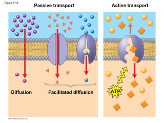
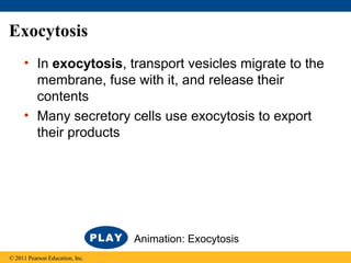
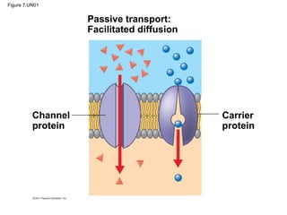
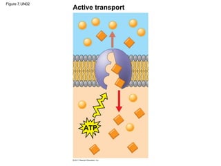
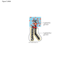
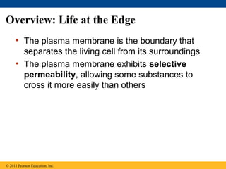
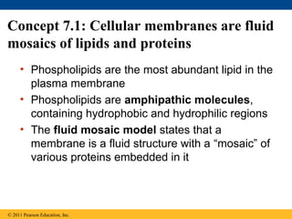
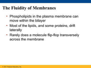
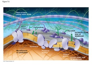
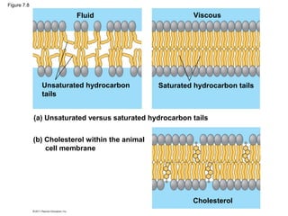
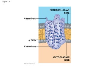
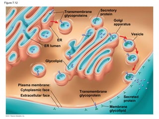
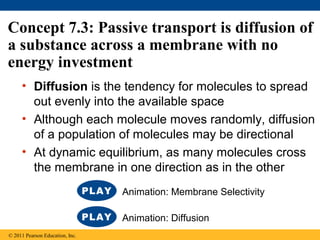
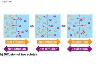
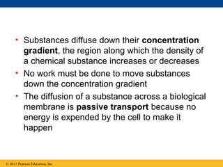
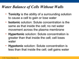
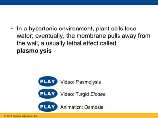
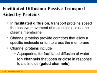
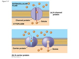
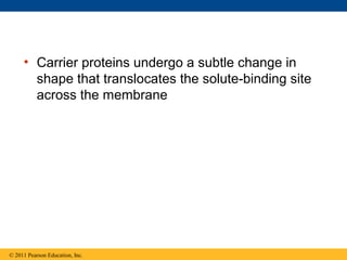
SlideShare a Scribd company logo
© 2011 Pearson Education, Inc.
LECTURE PRESENTATIONS
For CAMPBELL BIOLOGY, NINTH EDITION
Jane B. Reece, Lisa A. Urry, Michael L. Cain, Steven A. Wasserman, Peter V. Minorsky, Robert B. Jackson
Lectures by
Erin Barley
Kathleen Fitzpatrick
Membrane Structure and
Function
Chapter 7
Overview: Life at the Edge
• The plasma membrane is the boundary that
separates the living cell from its surroundings
• The plasma membrane exhibits selective
permeability, allowing some substances to
cross it more easily than others
© 2011 Pearson Education, Inc.
Figure 7.1
Concept 7.1: Cellular membranes are fluid
mosaics of lipids and proteins
• Phospholipids are the most abundant lipid in the
plasma membrane
• Phospholipids are amphipathic molecules,
containing hydrophobic and hydrophilic regions
• The fluid mosaic model states that a
membrane is a fluid structure with a “mosaic” of
various proteins embedded in it
© 2011 Pearson Education, Inc.
Membrane Models: Scientific Inquiry
• Membranes have been chemically analyzed and
found to be made of proteins and lipids
• Scientists studying the plasma membrane
reasoned that it must be a phospholipid bilayer
© 2011 Pearson Education, Inc.
Figure 7.2
Hydrophilic
head
Hydrophobic
tail
WATER
WATER
© 2011 Pearson Education, Inc.
• In 1935, Hugh Davson and James Danielli
proposed a sandwich model in which the
phospholipid bilayer lies between two layers of
globular proteins
• Later studies found problems with this model,
particularly the placement of membrane proteins,
which have hydrophilic and hydrophobic regions
• In 1972, S. J. Singer and G. Nicolson proposed
that the membrane is a mosaic of proteins
dispersed within the bilayer, with only the
hydrophilic regions exposed to water
Figure 7.3
Phospholipid
bilayer
Hydrophobic regions
of protein
Hydrophilic
regions of protein
• Freeze-fracture studies of the plasma
membrane supported the fluid mosaic model
• Freeze-fracture is a specialized preparation
technique that splits a membrane along the
middle of the phospholipid bilayer
© 2011 Pearson Education, Inc.
Figure 7.4
Knife
Plasma membrane Cytoplasmic layer
Proteins
Extracellular
layer
Inside of extracellular layer Inside of cytoplasmic layer
TECHNIQUE
RESULTS
Figure 7.4a
Inside of extracellular layer
Figure 7.4b
Inside of cytoplasmic layer
The Fluidity of Membranes
• Phospholipids in the plasma membrane can
move within the bilayer
• Most of the lipids, and some proteins, drift
laterally
• Rarely does a molecule flip-flop transversely
across the membrane
© 2011 Pearson Education, Inc.
Figure 7.5
Glyco-
protein
Carbohydrate
Glycolipid
Microfilaments
of cytoskeleton
EXTRACELLULAR
SIDE OF
MEMBRANE
CYTOPLASMIC SIDE
OF MEMBRANE
Integral
protein
Peripheral
proteins
Cholesterol
Fibers of extra-
cellular matrix (ECM)
Figure 7.6
Lateral movement occurs
∼107
times per second.
Flip-flopping across the membrane
is rare (∼ once per month).
Figure 7.7
Membrane proteins
Mouse cell
Human cell
Hybrid cell
Mixed proteins
after 1 hour
RESULTS
• As temperatures cool, membranes switch
from a fluid state to a solid state
• The temperature at which a membrane
solidifies depends on the types of lipids
• Membranes rich in unsaturated fatty acids
are more fluid than those rich in saturated
fatty acids
• Membranes must be fluid to work properly;
they are usually about as fluid as salad oil
© 2011 Pearson Education, Inc.
• The steroid cholesterol has different effects on
membrane fluidity at different temperatures
• At warm temperatures (such as 37°C),
cholesterol restrains movement of
phospholipids
• At cool temperatures, it maintains fluidity by
preventing tight packing
© 2011 Pearson Education, Inc.
Figure 7.8
Fluid
Unsaturated hydrocarbon
tails
Viscous
Saturated hydrocarbon tails
(a) Unsaturated versus saturated hydrocarbon tails
(b) Cholesterol within the animal
cell membrane
Cholesterol
Evolution of Differences in Membrane Lipid
Composition
• Variations in lipid composition of cell
membranes of many species appear to be
adaptations to specific environmental
conditions
• Ability to change the lipid compositions in
response to temperature changes has evolved
in organisms that live where temperatures vary
© 2011 Pearson Education, Inc.
Membrane Proteins and Their Functions
• A membrane is a collage of different proteins,
often grouped together, embedded in the fluid
matrix of the lipid bilayer
• Proteins determine most of the membrane’s
specific functions
© 2011 Pearson Education, Inc.
• Peripheral proteins are bound to the surface
of the membrane
• Integral proteins penetrate the hydrophobic
core
• Integral proteins that span the membrane are
called transmembrane proteins
• The hydrophobic regions of an integral protein
consist of one or more stretches of nonpolar
amino acids, often coiled into alpha helices
© 2011 Pearson Education, Inc.
Figure 7.9
N-terminus
α helix
C-terminus
EXTRACELLULAR
SIDE
CYTOPLASMIC
SIDE
• Six major functions of membrane proteins
– Transport
– Enzymatic activity
– Signal transduction
– Cell-cell recognition
– Intercellular joining
– Attachment to the cytoskeleton and
extracellular matrix (ECM)
© 2011 Pearson Education, Inc.
Figure 7.10
Enzymes
Signaling molecule
Receptor
Signal transduction
Glyco-
protein
ATP
(a) Transport (b) Enzymatic activity (c) Signal transduction
(d) Cell-cell recognition (e) Intercellular joining (f) Attachment to
the cytoskeleton
and extracellular
matrix (ECM)
Figure 7.10a
Enzymes
Signaling molecule
Receptor
Signal transduction
ATP
(a) Transport (b) Enzymatic activity (c) Signal transduction
Figure 7.10b
Glyco-
protein
(d) Cell-cell recognition(e) Intercellular joining (f) Attachment to
the cytoskeleton
and extracellular
matrix (ECM)
The Role of Membrane Carbohydrates in
Cell-Cell Recognition
• Cells recognize each other by binding to surface
molecules, often containing carbohydrates, on
the extracellular surface of the plasma
membrane
• Membrane carbohydrates may be covalently
bonded to lipids (forming glycolipids) or more
commonly to proteins (forming glycoproteins)
• Carbohydrates on the external side of the
plasma membrane vary among species,
individuals, and even cell types in an individual
© 2011 Pearson Education, Inc.
Figure 7.11
Receptor
(CD4)
Co-receptor
(CCR5)
HIV
Receptor (CD4)
but no CCR5 Plasma
membrane
HIV can infect a cell that
has CCR5 on its surface,
as in most people.
HIV cannot infect a cell lacking
CCR5 on its surface, as in
resistant individuals.
Synthesis and Sidedness of Membranes
• Membranes have distinct inside and outside
faces
• The asymmetrical distribution of proteins,
lipids, and associated carbohydrates in the
plasma membrane is determined when the
membrane is built by the ER and Golgi
apparatus
© 2011 Pearson Education, Inc.
Figure 7.12
Transmembrane
glycoproteins
ER
ER lumen
Glycolipid
Plasma membrane:
Cytoplasmic face
Extracellular face
Secretory
protein
Golgi
apparatus
Vesicle
Transmembrane
glycoprotein Secreted
protein
Membrane
glycolipid
Concept 7.2: Membrane structure results
in selective permeability
• A cell must exchange materials with its
surroundings, a process controlled by the
plasma membrane
• Plasma membranes are selectively permeable,
regulating the cell’s molecular traffic
© 2011 Pearson Education, Inc.
The Permeability of the Lipid Bilayer
• Hydrophobic (nonpolar) molecules, such as
hydrocarbons, can dissolve in the lipid bilayer
and pass through the membrane rapidly
• Polar molecules, such as sugars, do not cross
the membrane easily
© 2011 Pearson Education, Inc.
Transport Proteins
• Transport proteins allow passage of
hydrophilic substances across the membrane
• Some transport proteins, called channel
proteins, have a hydrophilic channel that
certain molecules or ions can use as a tunnel
• Channel proteins called aquaporins facilitate
the passage of water
© 2011 Pearson Education, Inc.
• Other transport proteins, called carrier proteins,
bind to molecules and change shape to shuttle
them across the membrane
• A transport protein is specific for the substance
it moves
© 2011 Pearson Education, Inc.
Concept 7.3: Passive transport is diffusion of
a substance across a membrane with no
energy investment
• Diffusion is the tendency for molecules to spread
out evenly into the available space
• Although each molecule moves randomly, diffusion
of a population of molecules may be directional
• At dynamic equilibrium, as many molecules cross
the membrane in one direction as in the other
© 2011 Pearson Education, Inc.
Animation: Membrane Selectivity
Animation: Diffusion
Figure 7.13
Molecules of dye
Membrane (cross section)
WATER
(a) Diffusion of one solute
(b) Diffusion of two solutes
Net diffusion Net diffusion
Equilibrium
Equilibrium
Equilibrium
Figure 7.13a
Molecules of dye
Membrane (cross section)
WATER
(a) Diffusion of one solute
Net diffusion Net diffusion Equilibrium
Figure 7.13b
(b) Diffusion of two solutes
Net diffusion Net diffusion
Equilibrium
Equilibrium
• Substances diffuse down their concentration
gradient, the region along which the density of
a chemical substance increases or decreases
• No work must be done to move substances
down the concentration gradient
• The diffusion of a substance across a biological
membrane is passive transport because no
energy is expended by the cell to make it
happen
© 2011 Pearson Education, Inc.
Effects of Osmosis on Water Balance
• Osmosis is the diffusion of water across a
selectively permeable membrane
• Water diffuses across a membrane from the
region of lower solute concentration to the
region of higher solute concentration until the
solute concentration is equal on both sides
© 2011 Pearson Education, Inc.
Figure 7.14
Lower
concentration
of solute (sugar)
Higher
concentration
of solute
Sugar
molecule
H2O
Same concentration
of solute
Selectively
permeable
membrane
Osmosis
Water Balance of Cells Without Walls
• Tonicity is the ability of a surrounding solution
to cause a cell to gain or lose water
• Isotonic solution: Solute concentration is the
same as that inside the cell; no net water
movement across the plasma membrane
• Hypertonic solution: Solute concentration is
greater than that inside the cell; cell loses
water
• Hypotonic solution: Solute concentration is
less than that inside the cell; cell gains water
© 2011 Pearson Education, Inc.
Figure 7.15
Hypotonic
solution
Osmosis
Isotonic
solution
Hypertonic
solution
(a) Animal cell
(b) Plant cell
H2O H2O H2O H2O
H2O H2O H2O H2OCell wall
Lysed Normal Shriveled
Turgid (normal) Flaccid Plasmolyzed
• Hypertonic or hypotonic environments create
osmotic problems for organisms
• Osmoregulation, the control of solute
concentrations and water balance, is a necessary
adaptation for life in such environments
• The protist Paramecium, which is hypertonic to its
pond water environment, has a contractile
vacuole that acts as a pump
© 2011 Pearson Education, Inc.
Video: Chlamydomonas
Video: Paramecium Vacuole
Figure 7.16
Contractile vacuole
50 µm
Water Balance of Cells with Walls
• Cell walls help maintain water balance
• A plant cell in a hypotonic solution swells until
the wall opposes uptake; the cell is now turgid
(firm)
• If a plant cell and its surroundings are isotonic,
there is no net movement of water into the cell;
the cell becomes flaccid (limp), and the plant
may wilt
© 2011 Pearson Education, Inc.
• In a hypertonic environment, plant cells lose
water; eventually, the membrane pulls away from
the wall, a usually lethal effect called
plasmolysis
© 2011 Pearson Education, Inc.
Video: Turgid Elodea
Animation: Osmosis
Video: Plasmolysis
Facilitated Diffusion: Passive Transport
Aided by Proteins
• In facilitated diffusion, transport proteins speed
the passive movement of molecules across the
plasma membrane
• Channel proteins provide corridors that allow a
specific molecule or ion to cross the membrane
• Channel proteins include
– Aquaporins, for facilitated diffusion of water
– Ion channels that open or close in response
to a stimulus (gated channels)
© 2011 Pearson Education, Inc.
Figure 7.17
EXTRACELLULAR
FLUID
CYTOPLASM
Channel protein
Solute
SoluteCarrier protein
(a) A channel
protein
(b) A carrier protein
• Carrier proteins undergo a subtle change in
shape that translocates the solute-binding site
across the membrane
© 2011 Pearson Education, Inc.
• Some diseases are caused by malfunctions in
specific transport systems, for example the
kidney disease cystinuria
© 2011 Pearson Education, Inc.
Concept 7.4: Active transport uses energy to
move solutes against their gradients
• Facilitated diffusion is still passive because the
solute moves down its concentration gradient,
and the transport requires no energy
• Some transport proteins, however, can move
solutes against their concentration gradients
© 2011 Pearson Education, Inc.
The Need for Energy in Active Transport
• Active transport moves substances against
their concentration gradients
• Active transport requires energy, usually in the
form of ATP
• Active transport is performed by specific
proteins embedded in the membranes
© 2011 Pearson Education, Inc.
Animation: Active Transport
• Active transport allows cells to maintain
concentration gradients that differ from their
surroundings
• The sodium-potassium pump is one type of
active transport system
© 2011 Pearson Education, Inc.
Figure 7.18-1
EXTRACELLULAR
FLUID
[Na+
] high
[K+
] low
[Na+
] low
[K+
] high
CYTOPLASM
Na+
Na+
Na+
1
Figure 7.18-2
EXTRACELLULAR
FLUID
[Na+
] high
[K+
] low
[Na+
] low
[K+
] high
CYTOPLASM
Na+
Na+
Na+
1 2
Na+
Na+
Na+
P
ATP
ADP
Figure 7.18-3
EXTRACELLULAR
FLUID
[Na+
] high
[K+
] low
[Na+
] low
[K+
] high
CYTOPLASM
Na+
Na+
Na+
1 2 3
Na+
Na+
Na+
Na+
Na+
Na+
P
P
ATP
ADP
Figure 7.18-4
EXTRACELLULAR
FLUID
[Na+
] high
[K+
] low
[Na+
] low
[K+
] high
CYTOPLASM
Na+
Na+
Na+
1 2 3
4
Na+
Na+
Na+
Na+
Na+
Na+
K+
K+
P
P
P
P i
ATP
ADP
Figure 7.18-5
EXTRACELLULAR
FLUID
[Na+
] high
[K+
] low
[Na+
] low
[K+
] high
CYTOPLASM
Na+
Na+
Na+
1 2 3
45
Na+
Na+
Na+
Na+
Na+
Na+
K+
K+
K+
K+
P
P
P
P i
ATP
ADP
Figure 7.18-6
EXTRACELLULAR
FLUID
[Na+
] high
[K+
] low
[Na+
] low
[K+
] high
CYTOPLASM
Na+
Na+
Na+
1 2 3
456
Na+
Na+
Na+
Na+
Na+
Na+
K+
K+
K+
K+
K+
K+
P
P
P
P i
ATP
ADP
Figure 7.19
Passive transport Active transport
Diffusion Facilitated diffusion ATP
How Ion Pumps Maintain Membrane
Potential
• Membrane potential is the voltage difference
across a membrane
• Voltage is created by differences in the
distribution of positive and negative ions across
a membrane
© 2011 Pearson Education, Inc.
• Two combined forces, collectively called the
electrochemical gradient, drive the diffusion
of ions across a membrane
– A chemical force (the ion’s concentration
gradient)
– An electrical force (the effect of the membrane
potential on the ion’s movement)
© 2011 Pearson Education, Inc.
• An electrogenic pump is a transport protein
that generates voltage across a membrane
• The sodium-potassium pump is the major
electrogenic pump of animal cells
• The main electrogenic pump of plants, fungi,
and bacteria is a proton pump
• Electrogenic pumps help store energy that can
be used for cellular work
© 2011 Pearson Education, Inc.
Figure 7.20
CYTOPLASM
ATP EXTRACELLULAR
FLUID
Proton pump
H+
H+
H+
H+
H+
H+
−
−
−
−
+
+
+
+
Cotransport: Coupled Transport by a
Membrane Protein
• Cotransport occurs when active transport of a
solute indirectly drives transport of other
solutes
• Plants commonly use the gradient of hydrogen
ions generated by proton pumps to drive
active transport of nutrients into the cell
© 2011 Pearson Education, Inc.
Figure 7.21
ATP
H+
H+
H+
H+
H+
H+
H+
H+
Proton pump
Sucrose-H+
cotransporter
Sucrose
Sucrose
Diffusion of H+
−
−
−
−
+
+
+
+
Concept 7.5: Bulk transport across the
plasma membrane occurs by exocytosis
and endocytosis
• Small molecules and water enter or leave the
cell through the lipid bilayer or via transport
proteins
• Large molecules, such as polysaccharides and
proteins, cross the membrane in bulk via
vesicles
• Bulk transport requires energy
© 2011 Pearson Education, Inc.
Exocytosis
• In exocytosis, transport vesicles migrate to the
membrane, fuse with it, and release their
contents
• Many secretory cells use exocytosis to export
their products
© 2011 Pearson Education, Inc.
Animation: Exocytosis
Endocytosis
• In endocytosis, the cell takes in macromolecules
by forming vesicles from the plasma membrane
• Endocytosis is a reversal of exocytosis, involving
different proteins
• There are three types of endocytosis
– Phagocytosis (“cellular eating”)
– Pinocytosis (“cellular drinking”)
– Receptor-mediated endocytosis
© 2011 Pearson Education, Inc.
Animation: Exocytosis and Endocytosis Introduction
• In phagocytosis a cell engulfs a particle in a
vacuole
• The vacuole fuses with a lysosome to digest
the particle
© 2011 Pearson Education, Inc.
Animation: Phagocytosis
• In pinocytosis, molecules are taken up when
extracellular fluid is “gulped” into tiny vesicles
© 2011 Pearson Education, Inc.
Animation: Pinocytosis
• In receptor-mediated endocytosis, binding of
ligands to receptors triggers vesicle formation
• A ligand is any molecule that binds specifically
to a receptor site of another molecule
© 2011 Pearson Education, Inc.
Animation: Receptor-Mediated Endocytosis
Figure 7.22
Solutes
Pseudopodium
“Food” or
other particle
Food
vacuole
CYTOPLASM
Plasma
membrane
Vesicle
Receptor
Ligand
Coat proteins
Coated
pit
Coated
vesicle
EXTRACELLULAR
FLUID
Phagocytosis Pinocytosis Receptor-Mediated Endocytosis
Figure 7.22a
Pseudopodium
Solutes
“Food”
or other
particle
Food
vacuole
CYTOPLASM
EXTRACELLULAR
FLUID
Pseudopodium
of amoeba
Bacterium
Food vacuole
An amoeba engulfing a bacterium
via phagocytosis (TEM).
Phagocytosis
1µm
Figure 7.22b
Pinocytosis vesicles forming
in a cell lining a small blood
vessel (TEM).
Plasma
membrane
Vesicle
0.5µm
Pinocytosis
Figure 7.22c
Top: A coated pit. Bottom: A
coated vesicle forming during
receptor-mediated endocytosis
(TEMs).
Receptor
0.25µm
Receptor-Mediated Endocytosis
Ligand
Coat proteins
Coated
pit
Coated
vesicle
Coat
proteins
Plasma
membrane
Figure 7.22d
Bacterium
Food vacuole
Pseudopodium
of amoeba
An amoeba engulfing a bacterium via
phagocytosis (TEM).
1µm
Figure 7.22e
Pinocytosis vesicles forming (indicated by arrows)
in a cell lining a small blood vessel (TEM).
0.5µm
Figure 7.22f
Top: A coated pit. Bottom: A coated
vesicle forming during receptor-mediated
endocytosis (TEMs).
Plasma
membrane
Coat
proteins
0.25µm
Figure 7.UN01
Passive transport:
Facilitated diffusion
Channel
protein
Carrier
protein
Figure 7.UN02
Active transport
ATP
Figure 7.UN03
0.03 M sucrose
0.02 M glucose
“Cell” “Environment”
0.01 M sucrose
0.01 M glucose
0.01 M fructose
Figure 7.UN04

More Related Content

What's hot

AP Biology-Ch.6 A Tour of the Cell
AP Biology-Ch.6 A Tour of the CellAP Biology-Ch.6 A Tour of the Cell
AP Biology-Ch.6 A Tour of the Cell
sciencewithsuresh
 
18 Regulation of Gene Expression
18 Regulation of Gene Expression18 Regulation of Gene Expression
18 Regulation of Gene Expressionkindarspirit
 
Ch 3: Water and Life
Ch 3: Water and LifeCh 3: Water and Life
Ch 3: Water and Life
veneethmathew
 
25 lecture history_of_life
25 lecture history_of_life25 lecture history_of_life
25 lecture history_of_lifeveneethmathew
 
Ch 15: The Chromosomal Basis of Inheritance
Ch 15: The Chromosomal Basis of InheritanceCh 15: The Chromosomal Basis of Inheritance
Ch 15: The Chromosomal Basis of Inheritanceveneethmathew
 
Ch 11: Cell Communication
Ch 11: Cell CommunicationCh 11: Cell Communication
Ch 11: Cell Communication
veneethmathew
 
Ch 9: Cell Respiration and Fermentation
Ch 9: Cell Respiration and FermentationCh 9: Cell Respiration and Fermentation
Ch 9: Cell Respiration and Fermentation
veneethmathew
 
Ch 2: The Chemical Context of Life
Ch 2: The Chemical Context of LifeCh 2: The Chemical Context of Life
Ch 2: The Chemical Context of Life
veneethmathew
 
06 a tour of the cell
06 a tour of the cell06 a tour of the cell
06 a tour of the cellkindarspirit
 
02 the chemical context of life
02 the chemical context of life02 the chemical context of life
02 the chemical context of lifekindarspirit
 
Ch 16: The Molecular Basis of Inheritance
Ch 16: The Molecular Basis of Inheritance Ch 16: The Molecular Basis of Inheritance
Ch 16: The Molecular Basis of Inheritance veneethmathew
 
14 mendel and the gene idea
14 mendel and the gene idea14 mendel and the gene idea
14 mendel and the gene ideakindarspirit
 
01 themes in the study of life
01 themes in the study of life01 themes in the study of life
01 themes in the study of lifekindarspirit
 
26 lecture phylogeny
26 lecture phylogeny26 lecture phylogeny
26 lecture phylogenyveneethmathew
 
Ch 17: From Gene to Protein
Ch 17: From Gene to Protein Ch 17: From Gene to Protein
Ch 17: From Gene to Protein veneethmathew
 
17 - From Gene to Protein
17 - From Gene to Protein17 - From Gene to Protein
17 - From Gene to Proteinkindarspirit
 

What's hot (20)

AP Biology-Ch.6 A Tour of the Cell
AP Biology-Ch.6 A Tour of the CellAP Biology-Ch.6 A Tour of the Cell
AP Biology-Ch.6 A Tour of the Cell
 
18 Regulation of Gene Expression
18 Regulation of Gene Expression18 Regulation of Gene Expression
18 Regulation of Gene Expression
 
Ch 3: Water and Life
Ch 3: Water and LifeCh 3: Water and Life
Ch 3: Water and Life
 
25 lecture history_of_life
25 lecture history_of_life25 lecture history_of_life
25 lecture history_of_life
 
27 lecture bacteria
27 lecture bacteria27 lecture bacteria
27 lecture bacteria
 
Ch 15: The Chromosomal Basis of Inheritance
Ch 15: The Chromosomal Basis of InheritanceCh 15: The Chromosomal Basis of Inheritance
Ch 15: The Chromosomal Basis of Inheritance
 
Ch 11: Cell Communication
Ch 11: Cell CommunicationCh 11: Cell Communication
Ch 11: Cell Communication
 
Ch 9: Cell Respiration and Fermentation
Ch 9: Cell Respiration and FermentationCh 9: Cell Respiration and Fermentation
Ch 9: Cell Respiration and Fermentation
 
Ch 2: The Chemical Context of Life
Ch 2: The Chemical Context of LifeCh 2: The Chemical Context of Life
Ch 2: The Chemical Context of Life
 
06 a tour of the cell
06 a tour of the cell06 a tour of the cell
06 a tour of the cell
 
02 the chemical context of life
02 the chemical context of life02 the chemical context of life
02 the chemical context of life
 
Ch 16: The Molecular Basis of Inheritance
Ch 16: The Molecular Basis of Inheritance Ch 16: The Molecular Basis of Inheritance
Ch 16: The Molecular Basis of Inheritance
 
14 mendel and the gene idea
14 mendel and the gene idea14 mendel and the gene idea
14 mendel and the gene idea
 
01 themes in the study of life
01 themes in the study of life01 themes in the study of life
01 themes in the study of life
 
26 lecture phylogeny
26 lecture phylogeny26 lecture phylogeny
26 lecture phylogeny
 
Ch 17: From Gene to Protein
Ch 17: From Gene to Protein Ch 17: From Gene to Protein
Ch 17: From Gene to Protein
 
17 - From Gene to Protein
17 - From Gene to Protein17 - From Gene to Protein
17 - From Gene to Protein
 
54 lectures ppt
54 lectures ppt54 lectures ppt
54 lectures ppt
 
12 the cell cycle
12 the cell cycle12 the cell cycle
12 the cell cycle
 
19 - Viruses
19 - Viruses19 - Viruses
19 - Viruses
 

Viewers also liked

Membrane Structure and Function
Membrane Structure and FunctionMembrane Structure and Function
Membrane Structure and Function
Wesley McCammon
 
Ch 14: Mendel and the Gene Idea
Ch 14: Mendel and the Gene IdeaCh 14: Mendel and the Gene Idea
Ch 14: Mendel and the Gene Ideaveneethmathew
 
Ch 1: Themes in the Study of Life
Ch 1: Themes in the Study of LifeCh 1: Themes in the Study of Life
Ch 1: Themes in the Study of Life
veneethmathew
 
21 lecture genome_and_evolution
21 lecture genome_and_evolution21 lecture genome_and_evolution
21 lecture genome_and_evolutionveneethmathew
 
History and overview of immunology
History and overview of immunologyHistory and overview of immunology
History and overview of immunologyBruno Mmassy
 
Biology in Focus - Chapter 7
Biology in Focus - Chapter 7Biology in Focus - Chapter 7
Biology in Focus - Chapter 7
mpattani
 
Bio 2 - Lab 2 Photos
Bio 2 - Lab 2 PhotosBio 2 - Lab 2 Photos
Bio 2 - Lab 2 Photos
kindarspirit
 
33. introduction to virology
33. introduction to virology33. introduction to virology
33. introduction to virologysulochan_lohani
 
15 the chromosomal basis of inheritance
15 the chromosomal basis of inheritance15 the chromosomal basis of inheritance
15 the chromosomal basis of inheritancekindarspirit
 
An introduction to virology
An introduction to virologyAn introduction to virology
An introduction to virology
Kaveh Haratian
 
16 the molecular basis of inheritance
16 the molecular basis of inheritance16 the molecular basis of inheritance
16 the molecular basis of inheritancekindarspirit
 

Viewers also liked (14)

19 lecture viruses
19 lecture viruses19 lecture viruses
19 lecture viruses
 
AP Bio Ch 7 ppt
AP Bio Ch 7 pptAP Bio Ch 7 ppt
AP Bio Ch 7 ppt
 
Membrane Structure and Function
Membrane Structure and FunctionMembrane Structure and Function
Membrane Structure and Function
 
Bio Ch 7 Pwpt
Bio  Ch 7 PwptBio  Ch 7 Pwpt
Bio Ch 7 Pwpt
 
Ch 14: Mendel and the Gene Idea
Ch 14: Mendel and the Gene IdeaCh 14: Mendel and the Gene Idea
Ch 14: Mendel and the Gene Idea
 
Ch 1: Themes in the Study of Life
Ch 1: Themes in the Study of LifeCh 1: Themes in the Study of Life
Ch 1: Themes in the Study of Life
 
21 lecture genome_and_evolution
21 lecture genome_and_evolution21 lecture genome_and_evolution
21 lecture genome_and_evolution
 
History and overview of immunology
History and overview of immunologyHistory and overview of immunology
History and overview of immunology
 
Biology in Focus - Chapter 7
Biology in Focus - Chapter 7Biology in Focus - Chapter 7
Biology in Focus - Chapter 7
 
Bio 2 - Lab 2 Photos
Bio 2 - Lab 2 PhotosBio 2 - Lab 2 Photos
Bio 2 - Lab 2 Photos
 
33. introduction to virology
33. introduction to virology33. introduction to virology
33. introduction to virology
 
15 the chromosomal basis of inheritance
15 the chromosomal basis of inheritance15 the chromosomal basis of inheritance
15 the chromosomal basis of inheritance
 
An introduction to virology
An introduction to virologyAn introduction to virology
An introduction to virology
 
16 the molecular basis of inheritance
16 the molecular basis of inheritance16 the molecular basis of inheritance
16 the molecular basis of inheritance
 

Similar to Ch 7: Membrane Structure and Function

membrana celular y sus componentes estructurarles
membrana celular y sus componentes estructurarlesmembrana celular y sus componentes estructurarles
membrana celular y sus componentes estructurarles
brayancriollo6
 
chapter7.ppt for mechanical engineering gtu
chapter7.ppt for mechanical engineering gtuchapter7.ppt for mechanical engineering gtu
chapter7.ppt for mechanical engineering gtu
abhishagi22
 
2-Cell Membranes.ppt
2-Cell Membranes.ppt2-Cell Membranes.ppt
2-Cell Membranes.ppt
VamsikrishnaVaidyula
 
07membranestructureandfunction 130311053334-phpapp01
07membranestructureandfunction 130311053334-phpapp0107membranestructureandfunction 130311053334-phpapp01
07membranestructureandfunction 130311053334-phpapp01
Cleophas Rwemera
 
chapter 7.ppt
chapter 7.pptchapter 7.ppt
chapter 7.ppt
Walled1
 
Cell membrane structure an function.pptx
Cell membrane structure an function.pptxCell membrane structure an function.pptx
Cell membrane structure an function.pptx
drn00r
 
Membrane Structure and Function
Membrane Structure and FunctionMembrane Structure and Function
Membrane Structure and Function
John Canuel
 
07 membrane structure and function
07 membrane structure and function07 membrane structure and function
07 membrane structure and functionkindarspirit
 
07 lecture presentation_171-187_1
07 lecture presentation_171-187_107 lecture presentation_171-187_1
07 lecture presentation_171-187_1omar_1994
 
cell membrane structure and function
cell membrane structure and functioncell membrane structure and function
cell membrane structure and function
msarwa
 
07 lecture BIOL 1010-30 Gillette College
07 lecture BIOL 1010-30 Gillette College07 lecture BIOL 1010-30 Gillette College
07 lecture BIOL 1010-30 Gillette College
deskam2
 
chapter7_membrane_structure_and_function.ppt
chapter7_membrane_structure_and_function.pptchapter7_membrane_structure_and_function.ppt
chapter7_membrane_structure_and_function.ppt
somethingspecial16
 
Cell Membrane & ion transport.pptx
Cell Membrane & ion transport.pptxCell Membrane & ion transport.pptx
Cell Membrane & ion transport.pptx
Mira426412
 
Membrane structure and membrane chemistry.pptx
Membrane structure and membrane chemistry.pptxMembrane structure and membrane chemistry.pptx
Membrane structure and membrane chemistry.pptx
rajashri101
 
membrane function and structure and Cell transport
membrane function and structure and Cell transportmembrane function and structure and Cell transport
membrane function and structure and Cell transportColine Angela Guce
 
CELL-MEMBRANE-STRUCTURE-FUNCTION-WEEK-5.ppt
CELL-MEMBRANE-STRUCTURE-FUNCTION-WEEK-5.pptCELL-MEMBRANE-STRUCTURE-FUNCTION-WEEK-5.ppt
CELL-MEMBRANE-STRUCTURE-FUNCTION-WEEK-5.ppt
MichelleGAntoque
 
Membrane structure
Membrane structureMembrane structure
Membrane structure
Subham Preetam
 
membrane_structure.pptgy hj hh buhh nijjb
membrane_structure.pptgy hj hh buhh nijjbmembrane_structure.pptgy hj hh buhh nijjb
membrane_structure.pptgy hj hh buhh nijjb
SanjeevKumar11th1291
 
membrane_structure.ppt
membrane_structure.pptmembrane_structure.ppt
membrane_structure.ppt
somethingspecial16
 

Similar to Ch 7: Membrane Structure and Function (20)

membrana celular y sus componentes estructurarles
membrana celular y sus componentes estructurarlesmembrana celular y sus componentes estructurarles
membrana celular y sus componentes estructurarles
 
chapter7.ppt for mechanical engineering gtu
chapter7.ppt for mechanical engineering gtuchapter7.ppt for mechanical engineering gtu
chapter7.ppt for mechanical engineering gtu
 
2-Cell Membranes.ppt
2-Cell Membranes.ppt2-Cell Membranes.ppt
2-Cell Membranes.ppt
 
07membranestructureandfunction 130311053334-phpapp01
07membranestructureandfunction 130311053334-phpapp0107membranestructureandfunction 130311053334-phpapp01
07membranestructureandfunction 130311053334-phpapp01
 
chapter 7.ppt
chapter 7.pptchapter 7.ppt
chapter 7.ppt
 
Cell membrane structure an function.pptx
Cell membrane structure an function.pptxCell membrane structure an function.pptx
Cell membrane structure an function.pptx
 
Membrane Structure and Function
Membrane Structure and FunctionMembrane Structure and Function
Membrane Structure and Function
 
07 membrane structure and function
07 membrane structure and function07 membrane structure and function
07 membrane structure and function
 
07 lecture presentation_171-187_1
07 lecture presentation_171-187_107 lecture presentation_171-187_1
07 lecture presentation_171-187_1
 
cell membrane structure and function
cell membrane structure and functioncell membrane structure and function
cell membrane structure and function
 
07 lecture BIOL 1010-30 Gillette College
07 lecture BIOL 1010-30 Gillette College07 lecture BIOL 1010-30 Gillette College
07 lecture BIOL 1010-30 Gillette College
 
chapter7_membrane_structure_and_function.ppt
chapter7_membrane_structure_and_function.pptchapter7_membrane_structure_and_function.ppt
chapter7_membrane_structure_and_function.ppt
 
Cell Membrane & ion transport.pptx
Cell Membrane & ion transport.pptxCell Membrane & ion transport.pptx
Cell Membrane & ion transport.pptx
 
Membrane structure
Membrane structureMembrane structure
Membrane structure
 
Membrane structure and membrane chemistry.pptx
Membrane structure and membrane chemistry.pptxMembrane structure and membrane chemistry.pptx
Membrane structure and membrane chemistry.pptx
 
membrane function and structure and Cell transport
membrane function and structure and Cell transportmembrane function and structure and Cell transport
membrane function and structure and Cell transport
 
CELL-MEMBRANE-STRUCTURE-FUNCTION-WEEK-5.ppt
CELL-MEMBRANE-STRUCTURE-FUNCTION-WEEK-5.pptCELL-MEMBRANE-STRUCTURE-FUNCTION-WEEK-5.ppt
CELL-MEMBRANE-STRUCTURE-FUNCTION-WEEK-5.ppt
 
Membrane structure
Membrane structureMembrane structure
Membrane structure
 
membrane_structure.pptgy hj hh buhh nijjb
membrane_structure.pptgy hj hh buhh nijjbmembrane_structure.pptgy hj hh buhh nijjb
membrane_structure.pptgy hj hh buhh nijjb
 
membrane_structure.ppt
membrane_structure.pptmembrane_structure.ppt
membrane_structure.ppt
 

Recently uploaded

June 3, 2024 Anti-Semitism Letter Sent to MIT President Kornbluth and MIT Cor...
June 3, 2024 Anti-Semitism Letter Sent to MIT President Kornbluth and MIT Cor...June 3, 2024 Anti-Semitism Letter Sent to MIT President Kornbluth and MIT Cor...
June 3, 2024 Anti-Semitism Letter Sent to MIT President Kornbluth and MIT Cor...
Levi Shapiro
 
A Strategic Approach: GenAI in Education
A Strategic Approach: GenAI in EducationA Strategic Approach: GenAI in Education
A Strategic Approach: GenAI in Education
Peter Windle
 
CLASS 11 CBSE B.St Project AIDS TO TRADE - INSURANCE
CLASS 11 CBSE B.St Project AIDS TO TRADE - INSURANCECLASS 11 CBSE B.St Project AIDS TO TRADE - INSURANCE
CLASS 11 CBSE B.St Project AIDS TO TRADE - INSURANCE
BhavyaRajput3
 
BÀI TẬP BỔ TRỢ TIẾNG ANH GLOBAL SUCCESS LỚP 3 - CẢ NĂM (CÓ FILE NGHE VÀ ĐÁP Á...
BÀI TẬP BỔ TRỢ TIẾNG ANH GLOBAL SUCCESS LỚP 3 - CẢ NĂM (CÓ FILE NGHE VÀ ĐÁP Á...BÀI TẬP BỔ TRỢ TIẾNG ANH GLOBAL SUCCESS LỚP 3 - CẢ NĂM (CÓ FILE NGHE VÀ ĐÁP Á...
BÀI TẬP BỔ TRỢ TIẾNG ANH GLOBAL SUCCESS LỚP 3 - CẢ NĂM (CÓ FILE NGHE VÀ ĐÁP Á...
Nguyen Thanh Tu Collection
 
1.4 modern child centered education - mahatma gandhi-2.pptx
1.4 modern child centered education - mahatma gandhi-2.pptx1.4 modern child centered education - mahatma gandhi-2.pptx
1.4 modern child centered education - mahatma gandhi-2.pptx
JosvitaDsouza2
 
Additional Benefits for Employee Website.pdf
Additional Benefits for Employee Website.pdfAdditional Benefits for Employee Website.pdf
Additional Benefits for Employee Website.pdf
joachimlavalley1
 
Unit 2- Research Aptitude (UGC NET Paper I).pdf
Unit 2- Research Aptitude (UGC NET Paper I).pdfUnit 2- Research Aptitude (UGC NET Paper I).pdf
Unit 2- Research Aptitude (UGC NET Paper I).pdf
Thiyagu K
 
Palestine last event orientationfvgnh .pptx
Palestine last event orientationfvgnh .pptxPalestine last event orientationfvgnh .pptx
Palestine last event orientationfvgnh .pptx
RaedMohamed3
 
The approach at University of Liverpool.pptx
The approach at University of Liverpool.pptxThe approach at University of Liverpool.pptx
The approach at University of Liverpool.pptx
Jisc
 
special B.ed 2nd year old paper_20240531.pdf
special B.ed 2nd year old paper_20240531.pdfspecial B.ed 2nd year old paper_20240531.pdf
special B.ed 2nd year old paper_20240531.pdf
Special education needs
 
How to Make a Field invisible in Odoo 17
How to Make a Field invisible in Odoo 17How to Make a Field invisible in Odoo 17
How to Make a Field invisible in Odoo 17
Celine George
 
Instructions for Submissions thorugh G- Classroom.pptx
Instructions for Submissions thorugh G- Classroom.pptxInstructions for Submissions thorugh G- Classroom.pptx
Instructions for Submissions thorugh G- Classroom.pptx
Jheel Barad
 
Phrasal Verbs.XXXXXXXXXXXXXXXXXXXXXXXXXX
Phrasal Verbs.XXXXXXXXXXXXXXXXXXXXXXXXXXPhrasal Verbs.XXXXXXXXXXXXXXXXXXXXXXXXXX
Phrasal Verbs.XXXXXXXXXXXXXXXXXXXXXXXXXX
MIRIAMSALINAS13
 
The Accursed House by Émile Gaboriau.pptx
The Accursed House by Émile Gaboriau.pptxThe Accursed House by Émile Gaboriau.pptx
The Accursed House by Émile Gaboriau.pptx
DhatriParmar
 
Language Across the Curriculm LAC B.Ed.
Language Across the  Curriculm LAC B.Ed.Language Across the  Curriculm LAC B.Ed.
Language Across the Curriculm LAC B.Ed.
Atul Kumar Singh
 
"Protectable subject matters, Protection in biotechnology, Protection of othe...
"Protectable subject matters, Protection in biotechnology, Protection of othe..."Protectable subject matters, Protection in biotechnology, Protection of othe...
"Protectable subject matters, Protection in biotechnology, Protection of othe...
SACHIN R KONDAGURI
 
Chapter 3 - Islamic Banking Products and Services.pptx
Chapter 3 - Islamic Banking Products and Services.pptxChapter 3 - Islamic Banking Products and Services.pptx
Chapter 3 - Islamic Banking Products and Services.pptx
Mohd Adib Abd Muin, Senior Lecturer at Universiti Utara Malaysia
 
The Roman Empire A Historical Colossus.pdf
The Roman Empire A Historical Colossus.pdfThe Roman Empire A Historical Colossus.pdf
The Roman Empire A Historical Colossus.pdf
kaushalkr1407
 
Biological Screening of Herbal Drugs in detailed.
Biological Screening of Herbal Drugs in detailed.Biological Screening of Herbal Drugs in detailed.
Biological Screening of Herbal Drugs in detailed.
Ashokrao Mane college of Pharmacy Peth-Vadgaon
 
Overview on Edible Vaccine: Pros & Cons with Mechanism
Overview on Edible Vaccine: Pros & Cons with MechanismOverview on Edible Vaccine: Pros & Cons with Mechanism
Overview on Edible Vaccine: Pros & Cons with Mechanism
DeeptiGupta154
 

Recently uploaded (20)

June 3, 2024 Anti-Semitism Letter Sent to MIT President Kornbluth and MIT Cor...
June 3, 2024 Anti-Semitism Letter Sent to MIT President Kornbluth and MIT Cor...June 3, 2024 Anti-Semitism Letter Sent to MIT President Kornbluth and MIT Cor...
June 3, 2024 Anti-Semitism Letter Sent to MIT President Kornbluth and MIT Cor...
 
A Strategic Approach: GenAI in Education
A Strategic Approach: GenAI in EducationA Strategic Approach: GenAI in Education
A Strategic Approach: GenAI in Education
 
CLASS 11 CBSE B.St Project AIDS TO TRADE - INSURANCE
CLASS 11 CBSE B.St Project AIDS TO TRADE - INSURANCECLASS 11 CBSE B.St Project AIDS TO TRADE - INSURANCE
CLASS 11 CBSE B.St Project AIDS TO TRADE - INSURANCE
 
BÀI TẬP BỔ TRỢ TIẾNG ANH GLOBAL SUCCESS LỚP 3 - CẢ NĂM (CÓ FILE NGHE VÀ ĐÁP Á...
BÀI TẬP BỔ TRỢ TIẾNG ANH GLOBAL SUCCESS LỚP 3 - CẢ NĂM (CÓ FILE NGHE VÀ ĐÁP Á...BÀI TẬP BỔ TRỢ TIẾNG ANH GLOBAL SUCCESS LỚP 3 - CẢ NĂM (CÓ FILE NGHE VÀ ĐÁP Á...
BÀI TẬP BỔ TRỢ TIẾNG ANH GLOBAL SUCCESS LỚP 3 - CẢ NĂM (CÓ FILE NGHE VÀ ĐÁP Á...
 
1.4 modern child centered education - mahatma gandhi-2.pptx
1.4 modern child centered education - mahatma gandhi-2.pptx1.4 modern child centered education - mahatma gandhi-2.pptx
1.4 modern child centered education - mahatma gandhi-2.pptx
 
Additional Benefits for Employee Website.pdf
Additional Benefits for Employee Website.pdfAdditional Benefits for Employee Website.pdf
Additional Benefits for Employee Website.pdf
 
Unit 2- Research Aptitude (UGC NET Paper I).pdf
Unit 2- Research Aptitude (UGC NET Paper I).pdfUnit 2- Research Aptitude (UGC NET Paper I).pdf
Unit 2- Research Aptitude (UGC NET Paper I).pdf
 
Palestine last event orientationfvgnh .pptx
Palestine last event orientationfvgnh .pptxPalestine last event orientationfvgnh .pptx
Palestine last event orientationfvgnh .pptx
 
The approach at University of Liverpool.pptx
The approach at University of Liverpool.pptxThe approach at University of Liverpool.pptx
The approach at University of Liverpool.pptx
 
special B.ed 2nd year old paper_20240531.pdf
special B.ed 2nd year old paper_20240531.pdfspecial B.ed 2nd year old paper_20240531.pdf
special B.ed 2nd year old paper_20240531.pdf
 
How to Make a Field invisible in Odoo 17
How to Make a Field invisible in Odoo 17How to Make a Field invisible in Odoo 17
How to Make a Field invisible in Odoo 17
 
Instructions for Submissions thorugh G- Classroom.pptx
Instructions for Submissions thorugh G- Classroom.pptxInstructions for Submissions thorugh G- Classroom.pptx
Instructions for Submissions thorugh G- Classroom.pptx
 
Phrasal Verbs.XXXXXXXXXXXXXXXXXXXXXXXXXX
Phrasal Verbs.XXXXXXXXXXXXXXXXXXXXXXXXXXPhrasal Verbs.XXXXXXXXXXXXXXXXXXXXXXXXXX
Phrasal Verbs.XXXXXXXXXXXXXXXXXXXXXXXXXX
 
The Accursed House by Émile Gaboriau.pptx
The Accursed House by Émile Gaboriau.pptxThe Accursed House by Émile Gaboriau.pptx
The Accursed House by Émile Gaboriau.pptx
 
Language Across the Curriculm LAC B.Ed.
Language Across the  Curriculm LAC B.Ed.Language Across the  Curriculm LAC B.Ed.
Language Across the Curriculm LAC B.Ed.
 
"Protectable subject matters, Protection in biotechnology, Protection of othe...
"Protectable subject matters, Protection in biotechnology, Protection of othe..."Protectable subject matters, Protection in biotechnology, Protection of othe...
"Protectable subject matters, Protection in biotechnology, Protection of othe...
 
Chapter 3 - Islamic Banking Products and Services.pptx
Chapter 3 - Islamic Banking Products and Services.pptxChapter 3 - Islamic Banking Products and Services.pptx
Chapter 3 - Islamic Banking Products and Services.pptx
 
The Roman Empire A Historical Colossus.pdf
The Roman Empire A Historical Colossus.pdfThe Roman Empire A Historical Colossus.pdf
The Roman Empire A Historical Colossus.pdf
 
Biological Screening of Herbal Drugs in detailed.
Biological Screening of Herbal Drugs in detailed.Biological Screening of Herbal Drugs in detailed.
Biological Screening of Herbal Drugs in detailed.
 
Overview on Edible Vaccine: Pros & Cons with Mechanism
Overview on Edible Vaccine: Pros & Cons with MechanismOverview on Edible Vaccine: Pros & Cons with Mechanism
Overview on Edible Vaccine: Pros & Cons with Mechanism
 

Ch 7: Membrane Structure and Function

  • 1. © 2011 Pearson Education, Inc. LECTURE PRESENTATIONS For CAMPBELL BIOLOGY, NINTH EDITION Jane B. Reece, Lisa A. Urry, Michael L. Cain, Steven A. Wasserman, Peter V. Minorsky, Robert B. Jackson Lectures by Erin Barley Kathleen Fitzpatrick Membrane Structure and Function Chapter 7
  • 2. Overview: Life at the Edge • The plasma membrane is the boundary that separates the living cell from its surroundings • The plasma membrane exhibits selective permeability, allowing some substances to cross it more easily than others © 2011 Pearson Education, Inc.
  • 4. Concept 7.1: Cellular membranes are fluid mosaics of lipids and proteins • Phospholipids are the most abundant lipid in the plasma membrane • Phospholipids are amphipathic molecules, containing hydrophobic and hydrophilic regions • The fluid mosaic model states that a membrane is a fluid structure with a “mosaic” of various proteins embedded in it © 2011 Pearson Education, Inc.
  • 5. Membrane Models: Scientific Inquiry • Membranes have been chemically analyzed and found to be made of proteins and lipids • Scientists studying the plasma membrane reasoned that it must be a phospholipid bilayer © 2011 Pearson Education, Inc.
  • 7. © 2011 Pearson Education, Inc. • In 1935, Hugh Davson and James Danielli proposed a sandwich model in which the phospholipid bilayer lies between two layers of globular proteins • Later studies found problems with this model, particularly the placement of membrane proteins, which have hydrophilic and hydrophobic regions • In 1972, S. J. Singer and G. Nicolson proposed that the membrane is a mosaic of proteins dispersed within the bilayer, with only the hydrophilic regions exposed to water
  • 8. Figure 7.3 Phospholipid bilayer Hydrophobic regions of protein Hydrophilic regions of protein
  • 9. • Freeze-fracture studies of the plasma membrane supported the fluid mosaic model • Freeze-fracture is a specialized preparation technique that splits a membrane along the middle of the phospholipid bilayer © 2011 Pearson Education, Inc.
  • 10. Figure 7.4 Knife Plasma membrane Cytoplasmic layer Proteins Extracellular layer Inside of extracellular layer Inside of cytoplasmic layer TECHNIQUE RESULTS
  • 11. Figure 7.4a Inside of extracellular layer
  • 12. Figure 7.4b Inside of cytoplasmic layer
  • 13. The Fluidity of Membranes • Phospholipids in the plasma membrane can move within the bilayer • Most of the lipids, and some proteins, drift laterally • Rarely does a molecule flip-flop transversely across the membrane © 2011 Pearson Education, Inc.
  • 14. Figure 7.5 Glyco- protein Carbohydrate Glycolipid Microfilaments of cytoskeleton EXTRACELLULAR SIDE OF MEMBRANE CYTOPLASMIC SIDE OF MEMBRANE Integral protein Peripheral proteins Cholesterol Fibers of extra- cellular matrix (ECM)
  • 15. Figure 7.6 Lateral movement occurs ∼107 times per second. Flip-flopping across the membrane is rare (∼ once per month).
  • 16. Figure 7.7 Membrane proteins Mouse cell Human cell Hybrid cell Mixed proteins after 1 hour RESULTS
  • 17. • As temperatures cool, membranes switch from a fluid state to a solid state • The temperature at which a membrane solidifies depends on the types of lipids • Membranes rich in unsaturated fatty acids are more fluid than those rich in saturated fatty acids • Membranes must be fluid to work properly; they are usually about as fluid as salad oil © 2011 Pearson Education, Inc.
  • 18. • The steroid cholesterol has different effects on membrane fluidity at different temperatures • At warm temperatures (such as 37°C), cholesterol restrains movement of phospholipids • At cool temperatures, it maintains fluidity by preventing tight packing © 2011 Pearson Education, Inc.
  • 19. Figure 7.8 Fluid Unsaturated hydrocarbon tails Viscous Saturated hydrocarbon tails (a) Unsaturated versus saturated hydrocarbon tails (b) Cholesterol within the animal cell membrane Cholesterol
  • 20. Evolution of Differences in Membrane Lipid Composition • Variations in lipid composition of cell membranes of many species appear to be adaptations to specific environmental conditions • Ability to change the lipid compositions in response to temperature changes has evolved in organisms that live where temperatures vary © 2011 Pearson Education, Inc.
  • 21. Membrane Proteins and Their Functions • A membrane is a collage of different proteins, often grouped together, embedded in the fluid matrix of the lipid bilayer • Proteins determine most of the membrane’s specific functions © 2011 Pearson Education, Inc.
  • 22. • Peripheral proteins are bound to the surface of the membrane • Integral proteins penetrate the hydrophobic core • Integral proteins that span the membrane are called transmembrane proteins • The hydrophobic regions of an integral protein consist of one or more stretches of nonpolar amino acids, often coiled into alpha helices © 2011 Pearson Education, Inc.
  • 24. • Six major functions of membrane proteins – Transport – Enzymatic activity – Signal transduction – Cell-cell recognition – Intercellular joining – Attachment to the cytoskeleton and extracellular matrix (ECM) © 2011 Pearson Education, Inc.
  • 25. Figure 7.10 Enzymes Signaling molecule Receptor Signal transduction Glyco- protein ATP (a) Transport (b) Enzymatic activity (c) Signal transduction (d) Cell-cell recognition (e) Intercellular joining (f) Attachment to the cytoskeleton and extracellular matrix (ECM)
  • 26. Figure 7.10a Enzymes Signaling molecule Receptor Signal transduction ATP (a) Transport (b) Enzymatic activity (c) Signal transduction
  • 27. Figure 7.10b Glyco- protein (d) Cell-cell recognition(e) Intercellular joining (f) Attachment to the cytoskeleton and extracellular matrix (ECM)
  • 28. The Role of Membrane Carbohydrates in Cell-Cell Recognition • Cells recognize each other by binding to surface molecules, often containing carbohydrates, on the extracellular surface of the plasma membrane • Membrane carbohydrates may be covalently bonded to lipids (forming glycolipids) or more commonly to proteins (forming glycoproteins) • Carbohydrates on the external side of the plasma membrane vary among species, individuals, and even cell types in an individual © 2011 Pearson Education, Inc.
  • 29. Figure 7.11 Receptor (CD4) Co-receptor (CCR5) HIV Receptor (CD4) but no CCR5 Plasma membrane HIV can infect a cell that has CCR5 on its surface, as in most people. HIV cannot infect a cell lacking CCR5 on its surface, as in resistant individuals.
  • 30. Synthesis and Sidedness of Membranes • Membranes have distinct inside and outside faces • The asymmetrical distribution of proteins, lipids, and associated carbohydrates in the plasma membrane is determined when the membrane is built by the ER and Golgi apparatus © 2011 Pearson Education, Inc.
  • 31. Figure 7.12 Transmembrane glycoproteins ER ER lumen Glycolipid Plasma membrane: Cytoplasmic face Extracellular face Secretory protein Golgi apparatus Vesicle Transmembrane glycoprotein Secreted protein Membrane glycolipid
  • 32. Concept 7.2: Membrane structure results in selective permeability • A cell must exchange materials with its surroundings, a process controlled by the plasma membrane • Plasma membranes are selectively permeable, regulating the cell’s molecular traffic © 2011 Pearson Education, Inc.
  • 33. The Permeability of the Lipid Bilayer • Hydrophobic (nonpolar) molecules, such as hydrocarbons, can dissolve in the lipid bilayer and pass through the membrane rapidly • Polar molecules, such as sugars, do not cross the membrane easily © 2011 Pearson Education, Inc.
  • 34. Transport Proteins • Transport proteins allow passage of hydrophilic substances across the membrane • Some transport proteins, called channel proteins, have a hydrophilic channel that certain molecules or ions can use as a tunnel • Channel proteins called aquaporins facilitate the passage of water © 2011 Pearson Education, Inc.
  • 35. • Other transport proteins, called carrier proteins, bind to molecules and change shape to shuttle them across the membrane • A transport protein is specific for the substance it moves © 2011 Pearson Education, Inc.
  • 36. Concept 7.3: Passive transport is diffusion of a substance across a membrane with no energy investment • Diffusion is the tendency for molecules to spread out evenly into the available space • Although each molecule moves randomly, diffusion of a population of molecules may be directional • At dynamic equilibrium, as many molecules cross the membrane in one direction as in the other © 2011 Pearson Education, Inc. Animation: Membrane Selectivity Animation: Diffusion
  • 37. Figure 7.13 Molecules of dye Membrane (cross section) WATER (a) Diffusion of one solute (b) Diffusion of two solutes Net diffusion Net diffusion Net diffusion Net diffusion Net diffusion Net diffusion Equilibrium Equilibrium Equilibrium
  • 38. Figure 7.13a Molecules of dye Membrane (cross section) WATER (a) Diffusion of one solute Net diffusion Net diffusion Equilibrium
  • 39. Figure 7.13b (b) Diffusion of two solutes Net diffusion Net diffusion Net diffusion Net diffusion Equilibrium Equilibrium
  • 40. • Substances diffuse down their concentration gradient, the region along which the density of a chemical substance increases or decreases • No work must be done to move substances down the concentration gradient • The diffusion of a substance across a biological membrane is passive transport because no energy is expended by the cell to make it happen © 2011 Pearson Education, Inc.
  • 41. Effects of Osmosis on Water Balance • Osmosis is the diffusion of water across a selectively permeable membrane • Water diffuses across a membrane from the region of lower solute concentration to the region of higher solute concentration until the solute concentration is equal on both sides © 2011 Pearson Education, Inc.
  • 42. Figure 7.14 Lower concentration of solute (sugar) Higher concentration of solute Sugar molecule H2O Same concentration of solute Selectively permeable membrane Osmosis
  • 43. Water Balance of Cells Without Walls • Tonicity is the ability of a surrounding solution to cause a cell to gain or lose water • Isotonic solution: Solute concentration is the same as that inside the cell; no net water movement across the plasma membrane • Hypertonic solution: Solute concentration is greater than that inside the cell; cell loses water • Hypotonic solution: Solute concentration is less than that inside the cell; cell gains water © 2011 Pearson Education, Inc.
  • 44. Figure 7.15 Hypotonic solution Osmosis Isotonic solution Hypertonic solution (a) Animal cell (b) Plant cell H2O H2O H2O H2O H2O H2O H2O H2OCell wall Lysed Normal Shriveled Turgid (normal) Flaccid Plasmolyzed
  • 45. • Hypertonic or hypotonic environments create osmotic problems for organisms • Osmoregulation, the control of solute concentrations and water balance, is a necessary adaptation for life in such environments • The protist Paramecium, which is hypertonic to its pond water environment, has a contractile vacuole that acts as a pump © 2011 Pearson Education, Inc. Video: Chlamydomonas Video: Paramecium Vacuole
  • 47. Water Balance of Cells with Walls • Cell walls help maintain water balance • A plant cell in a hypotonic solution swells until the wall opposes uptake; the cell is now turgid (firm) • If a plant cell and its surroundings are isotonic, there is no net movement of water into the cell; the cell becomes flaccid (limp), and the plant may wilt © 2011 Pearson Education, Inc.
  • 48. • In a hypertonic environment, plant cells lose water; eventually, the membrane pulls away from the wall, a usually lethal effect called plasmolysis © 2011 Pearson Education, Inc. Video: Turgid Elodea Animation: Osmosis Video: Plasmolysis
  • 49. Facilitated Diffusion: Passive Transport Aided by Proteins • In facilitated diffusion, transport proteins speed the passive movement of molecules across the plasma membrane • Channel proteins provide corridors that allow a specific molecule or ion to cross the membrane • Channel proteins include – Aquaporins, for facilitated diffusion of water – Ion channels that open or close in response to a stimulus (gated channels) © 2011 Pearson Education, Inc.
  • 50. Figure 7.17 EXTRACELLULAR FLUID CYTOPLASM Channel protein Solute SoluteCarrier protein (a) A channel protein (b) A carrier protein
  • 51. • Carrier proteins undergo a subtle change in shape that translocates the solute-binding site across the membrane © 2011 Pearson Education, Inc.
  • 52. • Some diseases are caused by malfunctions in specific transport systems, for example the kidney disease cystinuria © 2011 Pearson Education, Inc.
  • 53. Concept 7.4: Active transport uses energy to move solutes against their gradients • Facilitated diffusion is still passive because the solute moves down its concentration gradient, and the transport requires no energy • Some transport proteins, however, can move solutes against their concentration gradients © 2011 Pearson Education, Inc.
  • 54. The Need for Energy in Active Transport • Active transport moves substances against their concentration gradients • Active transport requires energy, usually in the form of ATP • Active transport is performed by specific proteins embedded in the membranes © 2011 Pearson Education, Inc. Animation: Active Transport
  • 55. • Active transport allows cells to maintain concentration gradients that differ from their surroundings • The sodium-potassium pump is one type of active transport system © 2011 Pearson Education, Inc.
  • 56. Figure 7.18-1 EXTRACELLULAR FLUID [Na+ ] high [K+ ] low [Na+ ] low [K+ ] high CYTOPLASM Na+ Na+ Na+ 1
  • 57. Figure 7.18-2 EXTRACELLULAR FLUID [Na+ ] high [K+ ] low [Na+ ] low [K+ ] high CYTOPLASM Na+ Na+ Na+ 1 2 Na+ Na+ Na+ P ATP ADP
  • 58. Figure 7.18-3 EXTRACELLULAR FLUID [Na+ ] high [K+ ] low [Na+ ] low [K+ ] high CYTOPLASM Na+ Na+ Na+ 1 2 3 Na+ Na+ Na+ Na+ Na+ Na+ P P ATP ADP
  • 59. Figure 7.18-4 EXTRACELLULAR FLUID [Na+ ] high [K+ ] low [Na+ ] low [K+ ] high CYTOPLASM Na+ Na+ Na+ 1 2 3 4 Na+ Na+ Na+ Na+ Na+ Na+ K+ K+ P P P P i ATP ADP
  • 60. Figure 7.18-5 EXTRACELLULAR FLUID [Na+ ] high [K+ ] low [Na+ ] low [K+ ] high CYTOPLASM Na+ Na+ Na+ 1 2 3 45 Na+ Na+ Na+ Na+ Na+ Na+ K+ K+ K+ K+ P P P P i ATP ADP
  • 61. Figure 7.18-6 EXTRACELLULAR FLUID [Na+ ] high [K+ ] low [Na+ ] low [K+ ] high CYTOPLASM Na+ Na+ Na+ 1 2 3 456 Na+ Na+ Na+ Na+ Na+ Na+ K+ K+ K+ K+ K+ K+ P P P P i ATP ADP
  • 62. Figure 7.19 Passive transport Active transport Diffusion Facilitated diffusion ATP
  • 63. How Ion Pumps Maintain Membrane Potential • Membrane potential is the voltage difference across a membrane • Voltage is created by differences in the distribution of positive and negative ions across a membrane © 2011 Pearson Education, Inc.
  • 64. • Two combined forces, collectively called the electrochemical gradient, drive the diffusion of ions across a membrane – A chemical force (the ion’s concentration gradient) – An electrical force (the effect of the membrane potential on the ion’s movement) © 2011 Pearson Education, Inc.
  • 65. • An electrogenic pump is a transport protein that generates voltage across a membrane • The sodium-potassium pump is the major electrogenic pump of animal cells • The main electrogenic pump of plants, fungi, and bacteria is a proton pump • Electrogenic pumps help store energy that can be used for cellular work © 2011 Pearson Education, Inc.
  • 66. Figure 7.20 CYTOPLASM ATP EXTRACELLULAR FLUID Proton pump H+ H+ H+ H+ H+ H+ − − − − + + + +
  • 67. Cotransport: Coupled Transport by a Membrane Protein • Cotransport occurs when active transport of a solute indirectly drives transport of other solutes • Plants commonly use the gradient of hydrogen ions generated by proton pumps to drive active transport of nutrients into the cell © 2011 Pearson Education, Inc.
  • 69. Concept 7.5: Bulk transport across the plasma membrane occurs by exocytosis and endocytosis • Small molecules and water enter or leave the cell through the lipid bilayer or via transport proteins • Large molecules, such as polysaccharides and proteins, cross the membrane in bulk via vesicles • Bulk transport requires energy © 2011 Pearson Education, Inc.
  • 70. Exocytosis • In exocytosis, transport vesicles migrate to the membrane, fuse with it, and release their contents • Many secretory cells use exocytosis to export their products © 2011 Pearson Education, Inc. Animation: Exocytosis
  • 71. Endocytosis • In endocytosis, the cell takes in macromolecules by forming vesicles from the plasma membrane • Endocytosis is a reversal of exocytosis, involving different proteins • There are three types of endocytosis – Phagocytosis (“cellular eating”) – Pinocytosis (“cellular drinking”) – Receptor-mediated endocytosis © 2011 Pearson Education, Inc. Animation: Exocytosis and Endocytosis Introduction
  • 72. • In phagocytosis a cell engulfs a particle in a vacuole • The vacuole fuses with a lysosome to digest the particle © 2011 Pearson Education, Inc. Animation: Phagocytosis
  • 73. • In pinocytosis, molecules are taken up when extracellular fluid is “gulped” into tiny vesicles © 2011 Pearson Education, Inc. Animation: Pinocytosis
  • 74. • In receptor-mediated endocytosis, binding of ligands to receptors triggers vesicle formation • A ligand is any molecule that binds specifically to a receptor site of another molecule © 2011 Pearson Education, Inc. Animation: Receptor-Mediated Endocytosis
  • 75. Figure 7.22 Solutes Pseudopodium “Food” or other particle Food vacuole CYTOPLASM Plasma membrane Vesicle Receptor Ligand Coat proteins Coated pit Coated vesicle EXTRACELLULAR FLUID Phagocytosis Pinocytosis Receptor-Mediated Endocytosis
  • 76. Figure 7.22a Pseudopodium Solutes “Food” or other particle Food vacuole CYTOPLASM EXTRACELLULAR FLUID Pseudopodium of amoeba Bacterium Food vacuole An amoeba engulfing a bacterium via phagocytosis (TEM). Phagocytosis 1µm
  • 77. Figure 7.22b Pinocytosis vesicles forming in a cell lining a small blood vessel (TEM). Plasma membrane Vesicle 0.5µm Pinocytosis
  • 78. Figure 7.22c Top: A coated pit. Bottom: A coated vesicle forming during receptor-mediated endocytosis (TEMs). Receptor 0.25µm Receptor-Mediated Endocytosis Ligand Coat proteins Coated pit Coated vesicle Coat proteins Plasma membrane
  • 79. Figure 7.22d Bacterium Food vacuole Pseudopodium of amoeba An amoeba engulfing a bacterium via phagocytosis (TEM). 1µm
  • 80. Figure 7.22e Pinocytosis vesicles forming (indicated by arrows) in a cell lining a small blood vessel (TEM). 0.5µm
  • 81. Figure 7.22f Top: A coated pit. Bottom: A coated vesicle forming during receptor-mediated endocytosis (TEMs). Plasma membrane Coat proteins 0.25µm
  • 82. Figure 7.UN01 Passive transport: Facilitated diffusion Channel protein Carrier protein
  • 84. Figure 7.UN03 0.03 M sucrose 0.02 M glucose “Cell” “Environment” 0.01 M sucrose 0.01 M glucose 0.01 M fructose

Editor's Notes

  1. Figure 7.1 How do cell membrane proteins help regulate chemical traffic?
  2. For the Cell Biology Video Structure of the Cell Membrane, go to Animation and Video Files.
  3. Figure 7.2 Phospholipid bilayer (cross section).
  4. Figure 7.3 The original fluid mosaic model for membranes.
  5. Figure 7.4 Research Method: Freeze-fracture
  6. Figure 7.4 Research Method: Freeze-fracture
  7. Figure 7.4 Research Method: Freeze-fracture
  8. Figure 7.5 Updated model of an animal cell’s plasma membrane (cutaway view).
  9. Figure 7.6 The movement of phospholipids.
  10. Figure 7.7 Inquiry: Do membrane proteins move?
  11. Figure 7.8 Factors that affect membrane fluidity.
  12. Figure 7.9 The structure of a transmembrane protein.
  13. Figure 7.10 Some functions of membrane proteins.
  14. Figure 7.10 Some functions of membrane proteins.
  15. Figure 7.10 Some functions of membrane proteins.
  16. Figure 7.11 Impact: Blocking HIV Entry into Cells as a Treatment for HIV Infections
  17. Figure 7.12 Synthesis of membrane components and their orientation in the membrane.
  18. Figure 7.13 The diffusion of solutes across a synthetic membrane.
  19. Figure 7.13 The diffusion of solutes across a synthetic membrane.
  20. Figure 7.13 The diffusion of solutes across a synthetic membrane.
  21. Figure 7.14 Osmosis.
  22. Figure 7.15 The water balance of living cells.
  23. Figure 7.16 The contractile vacuole of Paramecium caudatum.
  24. For the Cell Biology Video Water Movement through an Aquaporin, go to Animation and Video Files.
  25. Figure 7.17 Two types of transport proteins that carry out facilitated diffusion.
  26. For the Cell Biology Video Na+/K+ATPase Cycle, go to Animation and Video Files.
  27. Figure 7.18 The sodium-potassium pump: a specific case of active transport.
  28. Figure 7.18 The sodium-potassium pump: a specific case of active transport.
  29. Figure 7.18 The sodium-potassium pump: a specific case of active transport.
  30. Figure 7.18 The sodium-potassium pump: a specific case of active transport.
  31. Figure 7.18 The sodium-potassium pump: a specific case of active transport.
  32. Figure 7.18 The sodium-potassium pump: a specific case of active transport.
  33. Figure 7.19 Review: passive and active transport.
  34. Figure 7.20 A proton pump.
  35. Figure 7.21 Cotransport: active transport driven by a concentration gradient.
  36. For the Cell Biology Video Phagocytosis in Action, go to Animation and Video Files.
  37. Figure 7.22 Exploring: Endocytosis in Animal Cells
  38. Figure 7.22 Exploring: Endocytosis in Animal Cells
  39. Figure 7.22 Exploring: Endocytosis in Animal Cells
  40. Figure 7.22 Exploring: Endocytosis in Animal Cells
  41. Figure 7.22 Exploring: Endocytosis in Animal Cells
  42. Figure 7.22 Exploring: Endocytosis in Animal Cells
  43. Figure 7.22 Exploring: Endocytosis in Animal Cells
  44. Figure 7.UN01 Summary figure, Concept 7.3
  45. Figure 7.UN02 Summary figure, Concept 7.4
  46. Figure 7.UN03 Test Your Understanding, question 6
  47. Figure 7.UN04 Appendix A: answer to Figure 7.2 legend question