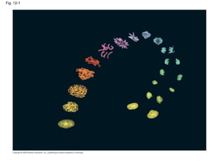
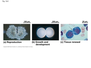
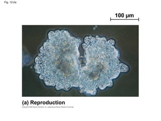
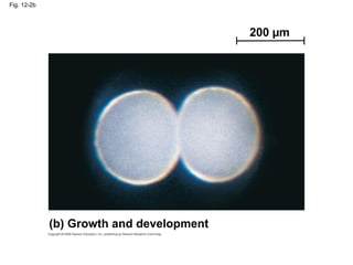
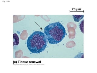
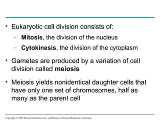
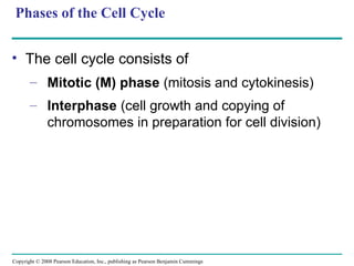
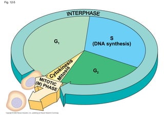
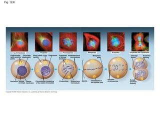
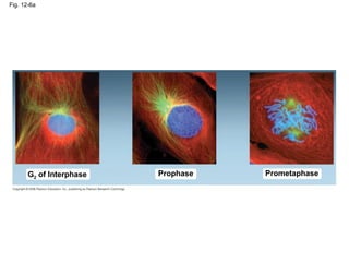
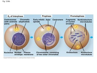
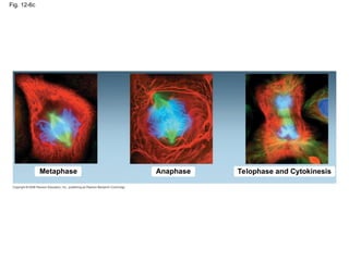
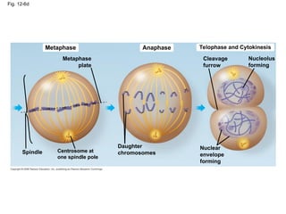
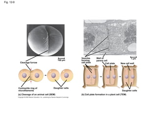
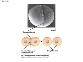
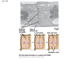
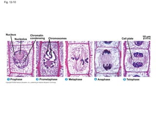
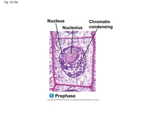
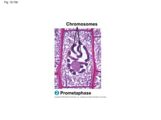
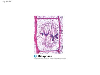
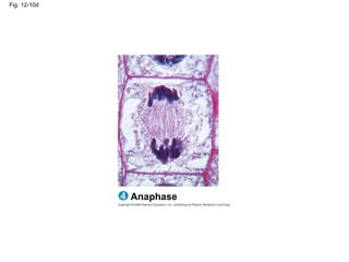
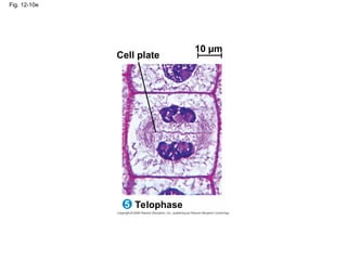
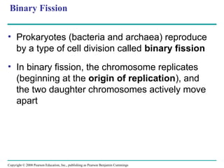
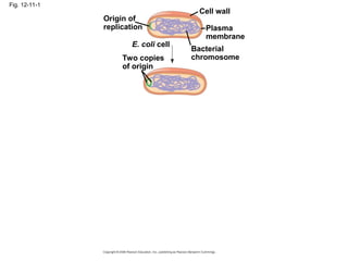
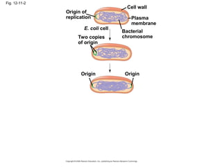
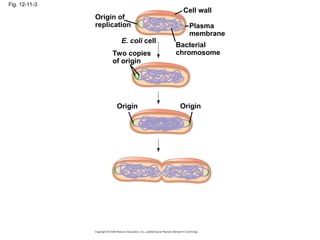
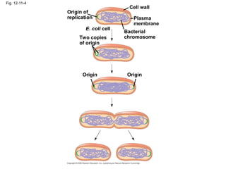
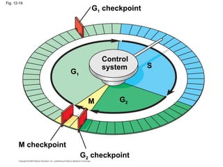
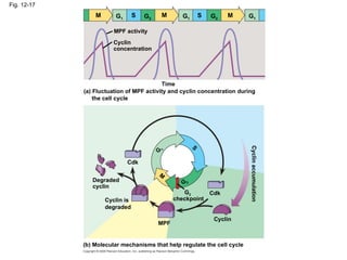
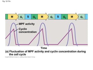
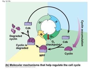
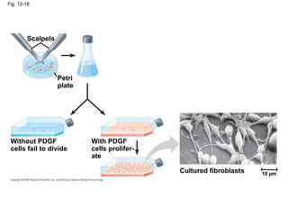
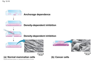
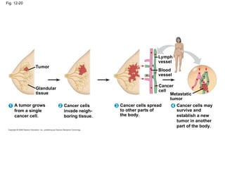
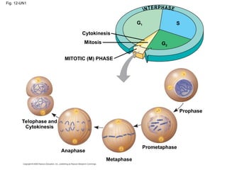
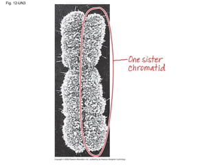
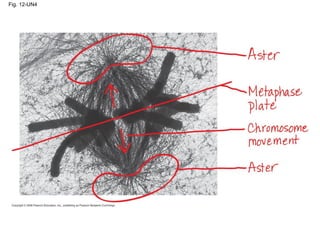
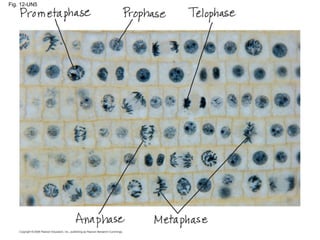
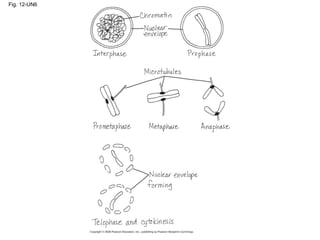
Copyright © 2008 Pearson Education, Inc., publishing as Pearson Benjamin Cummings
PowerPoint®
Lecture Presentations for
Biology
Eighth Edition
Neil Campbell and Jane Reece
Lectures by Chris Romero, updated by Erin Barley with contributions from Joan Sharp
Chapter 12
The Cell Cycle
Overview: The Key Roles of Cell Division
• The ability of organisms to reproduce best
distinguishes living things from nonliving matter
• The continuity of life is based on the
reproduction of cells, or cell division
Copyright © 2008 Pearson Education, Inc., publishing as Pearson Benjamin Cummings
Fig. 12-1
• In unicellular organisms, division of one cell
reproduces the entire organism
• Multicellular organisms depend on cell division
for:
– Development from a fertilized cell
– Growth
– Repair
• Cell division is an integral part of the cell cycle,
the life of a cell from formation to its own
division
Copyright © 2008 Pearson Education, Inc., publishing as Pearson Benjamin Cummings
Fig. 12-2
100 µm 200 µm 20 µm
(a) Reproduction (b) Growth and
development
(c) Tissue renewal
Fig. 12-2a
100 µm
(a) Reproduction
Fig. 12-2b
200 µm
(b) Growth and development
Fig. 12-2c
20 µm
(c) Tissue renewal
Concept 12.1: Cell division results in genetically
identical daughter cells
• Most cell division results in daughter cells with
identical genetic information, DNA
• A special type of division produces nonidentical
daughter cells (gametes, or sperm and egg
cells)
Copyright © 2008 Pearson Education, Inc., publishing as Pearson Benjamin Cummings
Cellular Organization of the Genetic Material
• All the DNA in a cell constitutes the cell’s
genome
• A genome can consist of a single DNA
molecule (common in prokaryotic cells) or a
number of DNA molecules (common in
eukaryotic cells)
• DNA molecules in a cell are packaged into
chromosomes
Copyright © 2008 Pearson Education, Inc., publishing as Pearson Benjamin Cummings
Fig. 12-3
20 µm
• Every eukaryotic species has a characteristic
number of chromosomes in each cell nucleus
• Somatic cells (nonreproductive cells) have
two sets of chromosomes
• Gametes (reproductive cells: sperm and eggs)
have half as many chromosomes as somatic
cells
• Eukaryotic chromosomes consist of
chromatin, a complex of DNA and protein that
condenses during cell division
Copyright © 2008 Pearson Education, Inc., publishing as Pearson Benjamin Cummings
Distribution of Chromosomes During Eukaryotic
Cell Division
• In preparation for cell division, DNA is
replicated and the chromosomes condense
• Each duplicated chromosome has two sister
chromatids, which separate during cell
division
• The centromere is the narrow “waist” of the
duplicated chromosome, where the two
chromatids are most closely attached
Copyright © 2008 Pearson Education, Inc., publishing as Pearson Benjamin Cummings
Fig. 12-4
0.5 µm Chromosomes
Chromosome
duplication
(including DNA
synthesis)
Chromo-
some arm
Centromere
Sister
chromatids
DNA molecules
Separation of
sister chromatids
Centromere
Sister chromatids
• Eukaryotic cell division consists of:
– Mitosis, the division of the nucleus
– Cytokinesis, the division of the cytoplasm
• Gametes are produced by a variation of cell
division called meiosis
• Meiosis yields nonidentical daughter cells that
have only one set of chromosomes, half as
many as the parent cell
Copyright © 2008 Pearson Education, Inc., publishing as Pearson Benjamin Cummings
Concept 12.2: The mitotic phase alternates with
interphase in the cell cycle
• In 1882, the German anatomist Walther
Flemming developed dyes to observe
chromosomes during mitosis and cytokinesis
Copyright © 2008 Pearson Education, Inc., publishing as Pearson Benjamin Cummings
Phases of the Cell Cycle
• The cell cycle consists of
– Mitotic (M) phase (mitosis and cytokinesis)
– Interphase (cell growth and copying of
chromosomes in preparation for cell division)
Copyright © 2008 Pearson Education, Inc., publishing as Pearson Benjamin Cummings
• Interphase (about 90% of the cell cycle) can be
divided into subphases:
– G1 phase (“first gap”)
– S phase (“synthesis”)
– G2 phase (“second gap”)
• The cell grows during all three phases, but
chromosomes are duplicated only during the S
phase
Copyright © 2008 Pearson Education, Inc., publishing as Pearson Benjamin Cummings
Fig. 12-5
S
(DNA synthesis)
MITOTIC
(M) PHASE
M
i
t
o
s
i
s
Cytokinesis
G1
G2
• Mitosis is conventionally divided into five
phases:
– Prophase
– Prometaphase
– Metaphase
– Anaphase
– Telophase
• Cytokinesis is well underway by late telophase
BioFlix: Mitosis
BioFlix: Mitosis
Copyright © 2008 Pearson Education, Inc., publishing as Pearson Benjamin Cummings
Fig. 12-6
G2 of Interphase
Centrosomes
(with centriole
pairs)
Chromatin
(duplicated)
Nucleolus Nuclear
envelope
Plasma
membrane
Early mitotic
spindle
Aster Centromere
Chromosome, consisting
of two sister chromatids
Prophase Prometaphase
Fragments
of nuclear
envelope
Nonkinetochore
microtubules
Kinetochore Kinetochore
microtubule
Metaphase
Metaphase
plate
Spindle Centrosome at
one spindle pole
Anaphase
Daughter
chromosomes
Telophase and Cytokinesis
Cleavage
furrow
Nucleolus
forming
Nuclear
envelope
forming
Prophase
Fig. 12-6a
Prometaphase
G2 of Interphase
Fig. 12-6b
Prometaphase
Prophase
G2 of Interphase
Nonkinetochore
microtubules
Fragments
of nuclear
envelope
Aster Centromere
Early mitotic
spindle
Chromatin
(duplicated)
Centrosomes
(with centriole
pairs)
Nucleolus Nuclear
envelope
Plasma
membrane
Chromosome, consisting
of two sister chromatids
Kinetochore Kinetochore
microtubule
Fig. 12-6c
Metaphase Anaphase Telophase and Cytokinesis
Fig. 12-6d
Metaphase Anaphase Telophase and Cytokinesis
Cleavage
furrow
Nucleolus
forming
Metaphase
plate
Centrosome at
one spindle pole
Spindle
Daughter
chromosomes
Nuclear
envelope
forming
The Mitotic Spindle: A Closer Look
• The mitotic spindle is an apparatus of
microtubules that controls chromosome
movement during mitosis
• During prophase, assembly of spindle
microtubules begins in the centrosome, the
microtubule organizing center
• The centrosome replicates, forming two
centrosomes that migrate to opposite ends of
the cell, as spindle microtubules grow out from
them
Copyright © 2008 Pearson Education, Inc., publishing as Pearson Benjamin Cummings
• An aster (a radial array of short microtubules)
extends from each centrosome
• The spindle includes the centrosomes, the
spindle microtubules, and the asters
Copyright © 2008 Pearson Education, Inc., publishing as Pearson Benjamin Cummings
• During prometaphase, some spindle
microtubules attach to the kinetochores of
chromosomes and begin to move the
chromosomes
• At metaphase, the chromosomes are all lined
up at the metaphase plate, the midway point
between the spindle’s two poles
Copyright © 2008 Pearson Education, Inc., publishing as Pearson Benjamin Cummings
Fig. 12-7
Microtubules Chromosomes
Sister
chromatids
Aster
Metaphase
plate
Centrosome
Kineto-
chores
Kinetochore
microtubules
Overlapping
nonkinetochore
microtubules
Centrosome 1 µm
0.5 µm
• In anaphase, sister chromatids separate and
move along the kinetochore microtubules
toward opposite ends of the cell
• The microtubules shorten by depolymerizing at
their kinetochore ends
Copyright © 2008 Pearson Education, Inc., publishing as Pearson Benjamin Cummings
Fig. 12-8
EXPERIMENT
Kinetochore
RESULTS
CONCLUSION
Spindle
pole
Mark
Chromosome
movement
Kinetochore
Microtubule
Motor
protein
Chromosome
Tubulin
subunits
Fig. 12-8a
Kinetochore
Spindle
pole
Mark
EXPERIMENT
RESULTS
Fig. 12-8b
Kinetochore
Microtubule
Tubulin
Subunits
Chromosome
Chromosome
movement
Motor
protein
CONCLUSION
• Nonkinetochore microtubules from opposite
poles overlap and push against each other,
elongating the cell
• In telophase, genetically identical daughter
nuclei form at opposite ends of the cell
Copyright © 2008 Pearson Education, Inc., publishing as Pearson Benjamin Cummings
Cytokinesis: A Closer Look
• In animal cells, cytokinesis occurs by a process
known as cleavage, forming a cleavage
furrow
• In plant cells, a cell plate forms during
cytokinesis
Animation: Cytokinesis
Copyright © 2008 Pearson Education, Inc., publishing as Pearson Benjamin Cummings
Video: Sea Urchin (Time Lapse)
Video: Animal Mitosis
Copyright © 2008 Pearson Education, Inc., publishing as Pearson Benjamin Cummings
Fig. 12-9
Cleavage furrow
100 µm
Contractile ring of
microfilaments
Daughter cells
(a) Cleavage of an animal cell (SEM) (b) Cell plate formation in a plant cell (TEM)
Vesicles
forming
cell plate
Wall of
parent cell
Cell plate
Daughter cells
New cell wall
1 µm
Cleavage furrow
Fig. 12-9a
100 µm
Daughter cells
(a) Cleavage of an animal cell (SEM)
Contractile ring of
microfilaments
Fig. 12-9b
Daughter cells
(b) Cell plate formation in a plant cell (TEM)
Vesicles
forming
cell plate
Wall of
parent cell
New cell wall
Cell plate
1 µm
Fig. 12-10
Chromatin
condensing
Metaphase Anaphase Telophase
Prometaphase
Nucleus
Prophase
1 2 3 5
4
Nucleolus Chromosomes Cell plate
10 µm
Fig. 12-10a
Nucleus
Prophase
1
Nucleolus
Chromatin
condensing
Fig. 12-10b
Prometaphase
2
Chromosomes
Fig. 12-10c
Metaphase
3
Fig. 12-10d
Anaphase
4
Fig. 12-10e
Telophase
5
Cell plate
10 µm
Binary Fission
• Prokaryotes (bacteria and archaea) reproduce
by a type of cell division called binary fission
• In binary fission, the chromosome replicates
(beginning at the origin of replication), and
the two daughter chromosomes actively move
apart
Copyright © 2008 Pearson Education, Inc., publishing as Pearson Benjamin Cummings
Fig. 12-11-1
Origin of
replication
Two copies
of origin
E. coli cell
Bacterial
chromosome
Plasma
membrane
Cell wall
Fig. 12-11-2
Origin of
replication
Two copies
of origin
E. coli cell
Bacterial
chromosome
Plasma
membrane
Cell wall
Origin Origin
Fig. 12-11-3
Origin of
replication
Two copies
of origin
E. coli cell
Bacterial
chromosome
Plasma
membrane
Cell wall
Origin Origin
Fig. 12-11-4
Origin of
replication
Two copies
of origin
E. coli cell
Bacterial
chromosome
Plasma
membrane
Cell wall
Origin Origin
The Evolution of Mitosis
• Since prokaryotes evolved before eukaryotes,
mitosis probably evolved from binary fission
• Certain protists exhibit types of cell division that
seem intermediate between binary fission and
mitosis
Copyright © 2008 Pearson Education, Inc., publishing as Pearson Benjamin Cummings
Fig. 12-12
(a) Bacteria
Bacterial
chromosome
Chromosomes
Microtubules
Intact nuclear
envelope
(b) Dinoflagellates
Kinetochore
microtubule
Intact nuclear
envelope
(c) Diatoms and yeasts
Kinetochore
microtubule
Fragments of
nuclear envelope
(d) Most eukaryotes
Fig. 12-12ab
Bacterial
chromosome
Chromosomes
Microtubules
(a) Bacteria
(b) Dinoflagellates
Intact nuclear
envelope
Fig. 12-12cd
Kinetochore
microtubule
(c) Diatoms and yeasts
Kinetochore
microtubule
(d) Most eukaryotes
Fragments of
nuclear envelope
Intact nuclear
envelope
Concept 12.3: The eukaryotic cell cycle is regulated
by a molecular control system
• The frequency of cell division varies with the
type of cell
• These cell cycle differences result from
regulation at the molecular level
Copyright © 2008 Pearson Education, Inc., publishing as Pearson Benjamin Cummings
Evidence for Cytoplasmic Signals
• The cell cycle appears to be driven by specific
chemical signals present in the cytoplasm
• Some evidence for this hypothesis comes from
experiments in which cultured mammalian cells
at different phases of the cell cycle were fused
to form a single cell with two nuclei
Copyright © 2008 Pearson Education, Inc., publishing as Pearson Benjamin Cummings
Fig. 12-13
Experiment 1 Experiment 2
EXPERIMENT
RESULTS
S G1
M G1
M M
S
S
When a cell in the
S phase was fused
with a cell in G1, the G1
nucleus immediately
entered the S
phase—DNA was
synthesized.
When a cell in the
M phase was fused with
a cell in G1, the G1
nucleus immediately
began mitosis—a
spindle formed and
chromatin condensed,
even though the
chromosome had not
been duplicated.
The Cell Cycle Control System
• The sequential events of the cell cycle are
directed by a distinct cell cycle control
system, which is similar to a clock
• The cell cycle control system is regulated by
both internal and external controls
• The clock has specific checkpoints where the
cell cycle stops until a go-ahead signal is
received
Copyright © 2008 Pearson Education, Inc., publishing as Pearson Benjamin Cummings
Fig. 12-14
S
G1
M checkpoint
G2
M
Control
system
G1 checkpoint
G2 checkpoint
• For many cells, the G1 checkpoint seems to be
the most important one
• If a cell receives a go-ahead signal at the G1
checkpoint, it will usually complete the S, G2,
and M phases and divide
• If the cell does not receive the go-ahead signal,
it will exit the cycle, switching into a nondividing
state called the G0 phase
Copyright © 2008 Pearson Education, Inc., publishing as Pearson Benjamin Cummings
Fig. 12-15
G1
G0
G1 checkpoint
(a) Cell receives a go-ahead
signal
G1
(b) Cell does not receive a
go-ahead signal
The Cell Cycle Clock: Cyclins and
Cyclin-Dependent Kinases
• Two types of regulatory proteins are involved in
cell cycle control: cyclins and cyclin-
dependent kinases (Cdks)
• The activity of cyclins and Cdks fluctuates
during the cell cycle
• MPF (maturation-promoting factor) is a cyclin-
Cdk complex that triggers a cell’s passage past
the G2 checkpoint into the M phase
Copyright © 2008 Pearson Education, Inc., publishing as Pearson Benjamin Cummings
Fig. 12-16
Protein
kinase
activity
(–
)
%
of
dividing
cells
(–
)
Time (min)
300
200 400
100
0
1
2
3
4
5 30
500
0
20
10
RESULTS
Fig. 12-17
M G1
S G2
M G1
S G2
M G1
MPF activity
Cyclin
concentration
Time
(a) Fluctuation of MPF activity and cyclin concentration during
the cell cycle
Degraded
cyclin
Cdk
G 1
S
G2
M
Cdk
G2
checkpoint
Cyclin is
degraded
Cyclin
MPF
(b) Molecular mechanisms that help regulate the cell cycle
Cyclin
accumulation
Fig. 12-17a
Time
(a) Fluctuation of MPF activity and cyclin concentration during
the cell cycle
Cyclin
concentration
MPF activity
M M M
S
S
G1 G1 G1
G2 G2
Fig. 12-17b
Cyclin is
degraded
Cdk
MPF
Cdk
M
S
G
1
G2
checkpoint
Degraded
cyclin
Cyclin
(b) Molecular mechanisms that help regulate the cell cycle
G2
Cyclin
accumulation
Stop and Go Signs: Internal and External Signals at
the Checkpoints
• An example of an internal signal is that
kinetochores not attached to spindle
microtubules send a molecular signal that
delays anaphase
• Some external signals are growth factors,
proteins released by certain cells that stimulate
other cells to divide
• For example, platelet-derived growth factor
(PDGF) stimulates the division of human
fibroblast cells in culture
Copyright © 2008 Pearson Education, Inc., publishing as Pearson Benjamin Cummings
Fig. 12-18
Petri
plate
Scalpels
Cultured fibroblasts
Without PDGF
cells fail to divide
With PDGF
cells prolifer-
ate
10 µm
• Another example of external signals is density-
dependent inhibition, in which crowded cells
stop dividing
• Most animal cells also exhibit anchorage
dependence, in which they must be attached
to a substratum in order to divide
Copyright © 2008 Pearson Education, Inc., publishing as Pearson Benjamin Cummings
Fig. 12-19
Anchorage dependence
Density-dependent inhibition
(a) Normal mammalian cells (b) Cancer cells
25 µm
25 µm
• Cancer cells exhibit neither density-dependent
inhibition nor anchorage dependence
Copyright © 2008 Pearson Education, Inc., publishing as Pearson Benjamin Cummings
Loss of Cell Cycle Controls in Cancer Cells
• Cancer cells do not respond normally to the
body’s control mechanisms
• Cancer cells may not need growth factors to
grow and divide:
– They may make their own growth factor
– They may convey a growth factor’s signal
without the presence of the growth factor
– They may have an abnormal cell cycle control
system
Copyright © 2008 Pearson Education, Inc., publishing as Pearson Benjamin Cummings
• A normal cell is converted to a cancerous cell
by a process called transformation
• Cancer cells form tumors, masses of abnormal
cells within otherwise normal tissue
• If abnormal cells remain at the original site, the
lump is called a benign tumor
• Malignant tumors invade surrounding tissues
and can metastasize, exporting cancer cells to
other parts of the body, where they may form
secondary tumors
Copyright © 2008 Pearson Education, Inc., publishing as Pearson Benjamin Cummings
Fig. 12-20
Tumor
A tumor grows
from a single
cancer cell.
Glandular
tissue
Lymph
vessel
Blood
vessel
Metastatic
tumor
Cancer
cell
Cancer cells
invade neigh-
boring tissue.
Cancer cells spread
to other parts of
the body.
Cancer cells may
survive and
establish a new
tumor in another
part of the body.
1 2 3 4
Fig. 12-UN1
Telophase and
Cytokinesis
Anaphase
Metaphase
Prometaphase
Prophase
MITOTIC (M) PHASE
Cytokinesis
Mitosis
S
G1
G2
Fig. 12-UN2
Fig. 12-UN3
Fig. 12-UN4
Fig. 12-UN5
Fig. 12-UN6
You should now be able to:
1. Describe the structural organization of the
prokaryotic genome and the eukaryotic
genome
2. List the phases of the cell cycle; describe the
sequence of events during each phase
3. List the phases of mitosis and describe the
events characteristic of each phase
4. Draw or describe the mitotic spindle, including
centrosomes, kinetochore microtubules,
nonkinetochore microtubules, and asters
Copyright © 2008 Pearson Education, Inc., publishing as Pearson Benjamin Cummings
5. Compare cytokinesis in animals and plants
6. Describe the process of binary fission in
bacteria and explain how eukaryotic mitosis
may have evolved from binary fission
7. Explain how the abnormal cell division of
cancerous cells escapes normal cell cycle
controls
8. Distinguish between benign, malignant, and
metastatic tumors
Copyright © 2008 Pearson Education, Inc., publishing as Pearson Benjamin Cummings

Chapter 12 the cell cycle campbell 7th edition

  • 1.
    Copyright © 2008Pearson Education, Inc., publishing as Pearson Benjamin Cummings PowerPoint® Lecture Presentations for Biology Eighth Edition Neil Campbell and Jane Reece Lectures by Chris Romero, updated by Erin Barley with contributions from Joan Sharp Chapter 12 The Cell Cycle
  • 2.
    Overview: The KeyRoles of Cell Division • The ability of organisms to reproduce best distinguishes living things from nonliving matter • The continuity of life is based on the reproduction of cells, or cell division Copyright © 2008 Pearson Education, Inc., publishing as Pearson Benjamin Cummings
  • 3.
  • 4.
    • In unicellularorganisms, division of one cell reproduces the entire organism • Multicellular organisms depend on cell division for: – Development from a fertilized cell – Growth – Repair • Cell division is an integral part of the cell cycle, the life of a cell from formation to its own division Copyright © 2008 Pearson Education, Inc., publishing as Pearson Benjamin Cummings
  • 5.
    Fig. 12-2 100 µm200 µm 20 µm (a) Reproduction (b) Growth and development (c) Tissue renewal
  • 6.
  • 7.
    Fig. 12-2b 200 µm (b)Growth and development
  • 8.
    Fig. 12-2c 20 µm (c)Tissue renewal
  • 9.
    Concept 12.1: Celldivision results in genetically identical daughter cells • Most cell division results in daughter cells with identical genetic information, DNA • A special type of division produces nonidentical daughter cells (gametes, or sperm and egg cells) Copyright © 2008 Pearson Education, Inc., publishing as Pearson Benjamin Cummings
  • 10.
    Cellular Organization ofthe Genetic Material • All the DNA in a cell constitutes the cell’s genome • A genome can consist of a single DNA molecule (common in prokaryotic cells) or a number of DNA molecules (common in eukaryotic cells) • DNA molecules in a cell are packaged into chromosomes Copyright © 2008 Pearson Education, Inc., publishing as Pearson Benjamin Cummings
  • 11.
  • 12.
    • Every eukaryoticspecies has a characteristic number of chromosomes in each cell nucleus • Somatic cells (nonreproductive cells) have two sets of chromosomes • Gametes (reproductive cells: sperm and eggs) have half as many chromosomes as somatic cells • Eukaryotic chromosomes consist of chromatin, a complex of DNA and protein that condenses during cell division Copyright © 2008 Pearson Education, Inc., publishing as Pearson Benjamin Cummings
  • 13.
    Distribution of ChromosomesDuring Eukaryotic Cell Division • In preparation for cell division, DNA is replicated and the chromosomes condense • Each duplicated chromosome has two sister chromatids, which separate during cell division • The centromere is the narrow “waist” of the duplicated chromosome, where the two chromatids are most closely attached Copyright © 2008 Pearson Education, Inc., publishing as Pearson Benjamin Cummings
  • 14.
    Fig. 12-4 0.5 µmChromosomes Chromosome duplication (including DNA synthesis) Chromo- some arm Centromere Sister chromatids DNA molecules Separation of sister chromatids Centromere Sister chromatids
  • 15.
    • Eukaryotic celldivision consists of: – Mitosis, the division of the nucleus – Cytokinesis, the division of the cytoplasm • Gametes are produced by a variation of cell division called meiosis • Meiosis yields nonidentical daughter cells that have only one set of chromosomes, half as many as the parent cell Copyright © 2008 Pearson Education, Inc., publishing as Pearson Benjamin Cummings
  • 16.
    Concept 12.2: Themitotic phase alternates with interphase in the cell cycle • In 1882, the German anatomist Walther Flemming developed dyes to observe chromosomes during mitosis and cytokinesis Copyright © 2008 Pearson Education, Inc., publishing as Pearson Benjamin Cummings
  • 17.
    Phases of theCell Cycle • The cell cycle consists of – Mitotic (M) phase (mitosis and cytokinesis) – Interphase (cell growth and copying of chromosomes in preparation for cell division) Copyright © 2008 Pearson Education, Inc., publishing as Pearson Benjamin Cummings
  • 18.
    • Interphase (about90% of the cell cycle) can be divided into subphases: – G1 phase (“first gap”) – S phase (“synthesis”) – G2 phase (“second gap”) • The cell grows during all three phases, but chromosomes are duplicated only during the S phase Copyright © 2008 Pearson Education, Inc., publishing as Pearson Benjamin Cummings
  • 19.
    Fig. 12-5 S (DNA synthesis) MITOTIC (M)PHASE M i t o s i s Cytokinesis G1 G2
  • 20.
    • Mitosis isconventionally divided into five phases: – Prophase – Prometaphase – Metaphase – Anaphase – Telophase • Cytokinesis is well underway by late telophase BioFlix: Mitosis BioFlix: Mitosis Copyright © 2008 Pearson Education, Inc., publishing as Pearson Benjamin Cummings
  • 21.
    Fig. 12-6 G2 ofInterphase Centrosomes (with centriole pairs) Chromatin (duplicated) Nucleolus Nuclear envelope Plasma membrane Early mitotic spindle Aster Centromere Chromosome, consisting of two sister chromatids Prophase Prometaphase Fragments of nuclear envelope Nonkinetochore microtubules Kinetochore Kinetochore microtubule Metaphase Metaphase plate Spindle Centrosome at one spindle pole Anaphase Daughter chromosomes Telophase and Cytokinesis Cleavage furrow Nucleolus forming Nuclear envelope forming
  • 22.
  • 23.
    Fig. 12-6b Prometaphase Prophase G2 ofInterphase Nonkinetochore microtubules Fragments of nuclear envelope Aster Centromere Early mitotic spindle Chromatin (duplicated) Centrosomes (with centriole pairs) Nucleolus Nuclear envelope Plasma membrane Chromosome, consisting of two sister chromatids Kinetochore Kinetochore microtubule
  • 24.
    Fig. 12-6c Metaphase AnaphaseTelophase and Cytokinesis
  • 25.
    Fig. 12-6d Metaphase AnaphaseTelophase and Cytokinesis Cleavage furrow Nucleolus forming Metaphase plate Centrosome at one spindle pole Spindle Daughter chromosomes Nuclear envelope forming
  • 26.
    The Mitotic Spindle:A Closer Look • The mitotic spindle is an apparatus of microtubules that controls chromosome movement during mitosis • During prophase, assembly of spindle microtubules begins in the centrosome, the microtubule organizing center • The centrosome replicates, forming two centrosomes that migrate to opposite ends of the cell, as spindle microtubules grow out from them Copyright © 2008 Pearson Education, Inc., publishing as Pearson Benjamin Cummings
  • 27.
    • An aster(a radial array of short microtubules) extends from each centrosome • The spindle includes the centrosomes, the spindle microtubules, and the asters Copyright © 2008 Pearson Education, Inc., publishing as Pearson Benjamin Cummings
  • 28.
    • During prometaphase,some spindle microtubules attach to the kinetochores of chromosomes and begin to move the chromosomes • At metaphase, the chromosomes are all lined up at the metaphase plate, the midway point between the spindle’s two poles Copyright © 2008 Pearson Education, Inc., publishing as Pearson Benjamin Cummings
  • 29.
  • 30.
    • In anaphase,sister chromatids separate and move along the kinetochore microtubules toward opposite ends of the cell • The microtubules shorten by depolymerizing at their kinetochore ends Copyright © 2008 Pearson Education, Inc., publishing as Pearson Benjamin Cummings
  • 31.
  • 32.
  • 33.
  • 34.
    • Nonkinetochore microtubulesfrom opposite poles overlap and push against each other, elongating the cell • In telophase, genetically identical daughter nuclei form at opposite ends of the cell Copyright © 2008 Pearson Education, Inc., publishing as Pearson Benjamin Cummings
  • 35.
    Cytokinesis: A CloserLook • In animal cells, cytokinesis occurs by a process known as cleavage, forming a cleavage furrow • In plant cells, a cell plate forms during cytokinesis Animation: Cytokinesis Animation: Cytokinesis Copyright © 2008 Pearson Education, Inc., publishing as Pearson Benjamin Cummings
  • 36.
    Video: Sea Urchin(Time Lapse) Video: Sea Urchin (Time Lapse) Video: Animal Mitosis Video: Animal Mitosis Copyright © 2008 Pearson Education, Inc., publishing as Pearson Benjamin Cummings
  • 37.
    Fig. 12-9 Cleavage furrow 100µm Contractile ring of microfilaments Daughter cells (a) Cleavage of an animal cell (SEM) (b) Cell plate formation in a plant cell (TEM) Vesicles forming cell plate Wall of parent cell Cell plate Daughter cells New cell wall 1 µm
  • 38.
    Cleavage furrow Fig. 12-9a 100µm Daughter cells (a) Cleavage of an animal cell (SEM) Contractile ring of microfilaments
  • 39.
    Fig. 12-9b Daughter cells (b)Cell plate formation in a plant cell (TEM) Vesicles forming cell plate Wall of parent cell New cell wall Cell plate 1 µm
  • 40.
    Fig. 12-10 Chromatin condensing Metaphase AnaphaseTelophase Prometaphase Nucleus Prophase 1 2 3 5 4 Nucleolus Chromosomes Cell plate 10 µm
  • 41.
  • 42.
  • 43.
  • 44.
  • 45.
  • 46.
    Binary Fission • Prokaryotes(bacteria and archaea) reproduce by a type of cell division called binary fission • In binary fission, the chromosome replicates (beginning at the origin of replication), and the two daughter chromosomes actively move apart Copyright © 2008 Pearson Education, Inc., publishing as Pearson Benjamin Cummings
  • 47.
    Fig. 12-11-1 Origin of replication Twocopies of origin E. coli cell Bacterial chromosome Plasma membrane Cell wall
  • 48.
    Fig. 12-11-2 Origin of replication Twocopies of origin E. coli cell Bacterial chromosome Plasma membrane Cell wall Origin Origin
  • 49.
    Fig. 12-11-3 Origin of replication Twocopies of origin E. coli cell Bacterial chromosome Plasma membrane Cell wall Origin Origin
  • 50.
    Fig. 12-11-4 Origin of replication Twocopies of origin E. coli cell Bacterial chromosome Plasma membrane Cell wall Origin Origin
  • 51.
    The Evolution ofMitosis • Since prokaryotes evolved before eukaryotes, mitosis probably evolved from binary fission • Certain protists exhibit types of cell division that seem intermediate between binary fission and mitosis Copyright © 2008 Pearson Education, Inc., publishing as Pearson Benjamin Cummings
  • 52.
    Fig. 12-12 (a) Bacteria Bacterial chromosome Chromosomes Microtubules Intactnuclear envelope (b) Dinoflagellates Kinetochore microtubule Intact nuclear envelope (c) Diatoms and yeasts Kinetochore microtubule Fragments of nuclear envelope (d) Most eukaryotes
  • 53.
  • 54.
    Fig. 12-12cd Kinetochore microtubule (c) Diatomsand yeasts Kinetochore microtubule (d) Most eukaryotes Fragments of nuclear envelope Intact nuclear envelope
  • 55.
    Concept 12.3: Theeukaryotic cell cycle is regulated by a molecular control system • The frequency of cell division varies with the type of cell • These cell cycle differences result from regulation at the molecular level Copyright © 2008 Pearson Education, Inc., publishing as Pearson Benjamin Cummings
  • 56.
    Evidence for CytoplasmicSignals • The cell cycle appears to be driven by specific chemical signals present in the cytoplasm • Some evidence for this hypothesis comes from experiments in which cultured mammalian cells at different phases of the cell cycle were fused to form a single cell with two nuclei Copyright © 2008 Pearson Education, Inc., publishing as Pearson Benjamin Cummings
  • 57.
    Fig. 12-13 Experiment 1Experiment 2 EXPERIMENT RESULTS S G1 M G1 M M S S When a cell in the S phase was fused with a cell in G1, the G1 nucleus immediately entered the S phase—DNA was synthesized. When a cell in the M phase was fused with a cell in G1, the G1 nucleus immediately began mitosis—a spindle formed and chromatin condensed, even though the chromosome had not been duplicated.
  • 58.
    The Cell CycleControl System • The sequential events of the cell cycle are directed by a distinct cell cycle control system, which is similar to a clock • The cell cycle control system is regulated by both internal and external controls • The clock has specific checkpoints where the cell cycle stops until a go-ahead signal is received Copyright © 2008 Pearson Education, Inc., publishing as Pearson Benjamin Cummings
  • 59.
  • 60.
    • For manycells, the G1 checkpoint seems to be the most important one • If a cell receives a go-ahead signal at the G1 checkpoint, it will usually complete the S, G2, and M phases and divide • If the cell does not receive the go-ahead signal, it will exit the cycle, switching into a nondividing state called the G0 phase Copyright © 2008 Pearson Education, Inc., publishing as Pearson Benjamin Cummings
  • 61.
    Fig. 12-15 G1 G0 G1 checkpoint (a)Cell receives a go-ahead signal G1 (b) Cell does not receive a go-ahead signal
  • 62.
    The Cell CycleClock: Cyclins and Cyclin-Dependent Kinases • Two types of regulatory proteins are involved in cell cycle control: cyclins and cyclin- dependent kinases (Cdks) • The activity of cyclins and Cdks fluctuates during the cell cycle • MPF (maturation-promoting factor) is a cyclin- Cdk complex that triggers a cell’s passage past the G2 checkpoint into the M phase Copyright © 2008 Pearson Education, Inc., publishing as Pearson Benjamin Cummings
  • 63.
  • 64.
    Fig. 12-17 M G1 SG2 M G1 S G2 M G1 MPF activity Cyclin concentration Time (a) Fluctuation of MPF activity and cyclin concentration during the cell cycle Degraded cyclin Cdk G 1 S G2 M Cdk G2 checkpoint Cyclin is degraded Cyclin MPF (b) Molecular mechanisms that help regulate the cell cycle Cyclin accumulation
  • 65.
    Fig. 12-17a Time (a) Fluctuationof MPF activity and cyclin concentration during the cell cycle Cyclin concentration MPF activity M M M S S G1 G1 G1 G2 G2
  • 66.
    Fig. 12-17b Cyclin is degraded Cdk MPF Cdk M S G 1 G2 checkpoint Degraded cyclin Cyclin (b)Molecular mechanisms that help regulate the cell cycle G2 Cyclin accumulation
  • 67.
    Stop and GoSigns: Internal and External Signals at the Checkpoints • An example of an internal signal is that kinetochores not attached to spindle microtubules send a molecular signal that delays anaphase • Some external signals are growth factors, proteins released by certain cells that stimulate other cells to divide • For example, platelet-derived growth factor (PDGF) stimulates the division of human fibroblast cells in culture Copyright © 2008 Pearson Education, Inc., publishing as Pearson Benjamin Cummings
  • 68.
    Fig. 12-18 Petri plate Scalpels Cultured fibroblasts WithoutPDGF cells fail to divide With PDGF cells prolifer- ate 10 µm
  • 69.
    • Another exampleof external signals is density- dependent inhibition, in which crowded cells stop dividing • Most animal cells also exhibit anchorage dependence, in which they must be attached to a substratum in order to divide Copyright © 2008 Pearson Education, Inc., publishing as Pearson Benjamin Cummings
  • 70.
    Fig. 12-19 Anchorage dependence Density-dependentinhibition Density-dependent inhibition (a) Normal mammalian cells (b) Cancer cells 25 µm 25 µm
  • 71.
    • Cancer cellsexhibit neither density-dependent inhibition nor anchorage dependence Copyright © 2008 Pearson Education, Inc., publishing as Pearson Benjamin Cummings
  • 72.
    Loss of CellCycle Controls in Cancer Cells • Cancer cells do not respond normally to the body’s control mechanisms • Cancer cells may not need growth factors to grow and divide: – They may make their own growth factor – They may convey a growth factor’s signal without the presence of the growth factor – They may have an abnormal cell cycle control system Copyright © 2008 Pearson Education, Inc., publishing as Pearson Benjamin Cummings
  • 73.
    • A normalcell is converted to a cancerous cell by a process called transformation • Cancer cells form tumors, masses of abnormal cells within otherwise normal tissue • If abnormal cells remain at the original site, the lump is called a benign tumor • Malignant tumors invade surrounding tissues and can metastasize, exporting cancer cells to other parts of the body, where they may form secondary tumors Copyright © 2008 Pearson Education, Inc., publishing as Pearson Benjamin Cummings
  • 74.
    Fig. 12-20 Tumor A tumorgrows from a single cancer cell. Glandular tissue Lymph vessel Blood vessel Metastatic tumor Cancer cell Cancer cells invade neigh- boring tissue. Cancer cells spread to other parts of the body. Cancer cells may survive and establish a new tumor in another part of the body. 1 2 3 4
  • 75.
  • 76.
  • 77.
  • 78.
  • 79.
  • 80.
  • 81.
    You should nowbe able to: 1. Describe the structural organization of the prokaryotic genome and the eukaryotic genome 2. List the phases of the cell cycle; describe the sequence of events during each phase 3. List the phases of mitosis and describe the events characteristic of each phase 4. Draw or describe the mitotic spindle, including centrosomes, kinetochore microtubules, nonkinetochore microtubules, and asters Copyright © 2008 Pearson Education, Inc., publishing as Pearson Benjamin Cummings
  • 82.
    5. Compare cytokinesisin animals and plants 6. Describe the process of binary fission in bacteria and explain how eukaryotic mitosis may have evolved from binary fission 7. Explain how the abnormal cell division of cancerous cells escapes normal cell cycle controls 8. Distinguish between benign, malignant, and metastatic tumors Copyright © 2008 Pearson Education, Inc., publishing as Pearson Benjamin Cummings

Editor's Notes

  • #3 Figure 12.1 How do a cell’s chromosomes change during cell division?
  • #5 Figure 12.2 The functions of cell division
  • #6 Figure 12.2 The functions of cell division
  • #7 Figure 12.2 The functions of cell division
  • #8 Figure 12.2 The functions of cell division
  • #11 Figure 12.3 Eukaryotic chromosomes
  • #14 Figure 12.4 Chromosome duplication and distribution during cell division
  • #19 Figure 12.5 The cell cycle
  • #20 For the Cell Biology Video Myosin and Cytokinesis, go to Animation and Video Files.
  • #21 Figure 12.6 The mitotic division of an animal cell
  • #22 Figure 12.6 The mitotic division of an animal cell
  • #23 Figure 12.6 The mitotic division of an animal cell
  • #24 Figure 12.6 The mitotic division of an animal cell
  • #25 Figure 12.6 The mitotic division of an animal cell
  • #26 For the Cell Biology Video Spindle Formation During Mitosis, go to Animation and Video Files.
  • #29 Figure 12.7 The mitotic spindle at metaphase
  • #30 For the Cell Biology Video Microtubules in Anaphase, go to Animation and Video Files.
  • #31 Figure 12.8 At which end do kinetochore microtubules shorten during anaphase?
  • #32 Figure 12.8 At which end do kinetochore microtubules shorten during anaphase?
  • #33 Figure 12.8 At which end do kinetochore microtubules shorten during anaphase?
  • #34 For the Cell Biology Video Microtubules in Cell Division, go to Animation and Video Files.
  • #36 For the Cell Biology Video Nuclear Envelope Formation, go to Animation and Video Files.
  • #37 Figure 12.9 Cytokinesis in animal and plant cells
  • #38 Figure 12.9a Cytokinesis in animal and plant cells
  • #39 Figure 12.9b Cytokinesis in animal and plant cells
  • #40 Figure 12.10 Mitosis in a plant cell
  • #41 Figure 12.10 Mitosis in a plant cell
  • #42 Figure 12.10 Mitosis in a plant cell
  • #43 Figure 12.10 Mitosis in a plant cell
  • #44 Figure 12.10 Mitosis in a plant cell
  • #45 Figure 12.10 Mitosis in a plant cell
  • #47 Figure 12.11 Bacterial cell division by binary fission
  • #48 Figure 12.11 Bacterial cell division by binary fission
  • #49 Figure 12.11 Bacterial cell division by binary fission
  • #50 Figure 12.11 Bacterial cell division by binary fission
  • #52 Figure 12.12 A hypothetical sequence for the evolution of mitosis
  • #53 Figure 12.12 A hypothetical sequence for the evolution of mitosis
  • #54 Figure 12.12 A hypothetical sequence for the evolution of mitosis
  • #57 Figure 12.13 Do molecular signals in the cytoplasm regulate the cell cycle?
  • #59 Figure 12.14 Mechanical analogy for the cell cycle control system
  • #61 Figure 12.15 The G1 checkpoint
  • #63 Figure 12.16 How does the activity of a protein kinase essential for mitosis vary during the cell cycle?
  • #64 Figure 12.17 Molecular control of the cell cycle at the G2 checkpoint
  • #65 Figure 12.17 Molecular control of the cell cycle at the G2 checkpoint
  • #66 Figure 12.17 Molecular control of the cell cycle at the G2 checkpoint
  • #68 Figure 12.18 The effect of a growth factor on cell division
  • #70 Figure 12.19 Density-dependent inhibition and anchorage dependence of cell division
  • #74 Figure 12.20 The growth and metastasis of a malignant breast tumor