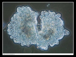
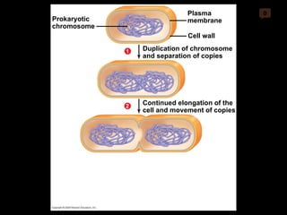
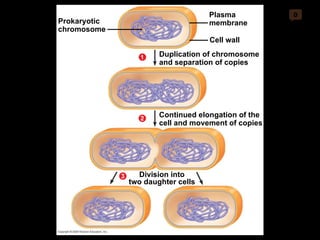
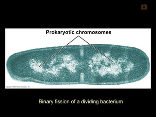
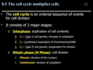
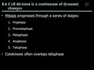
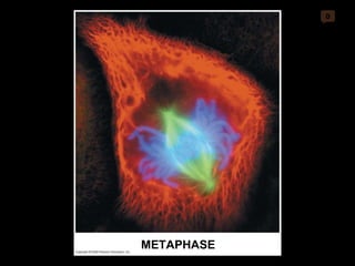
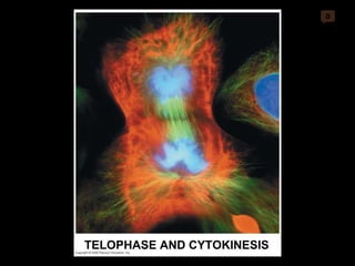
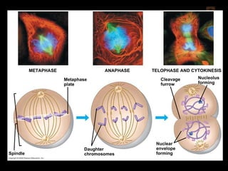
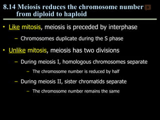
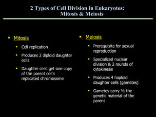
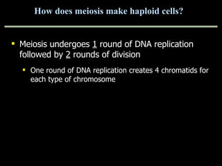
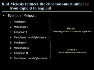
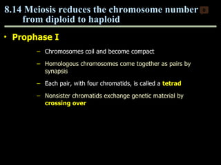
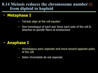
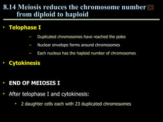
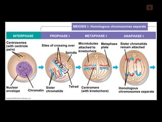
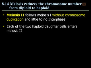
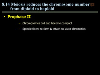
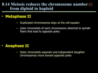
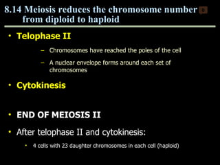
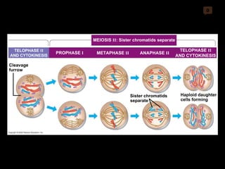
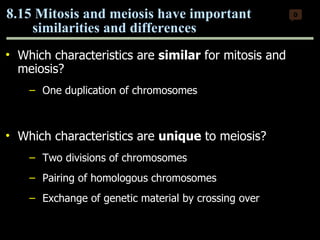
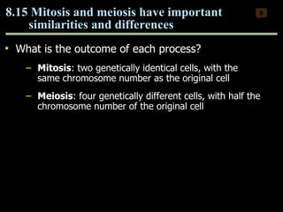
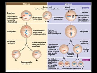
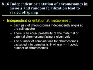
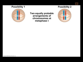
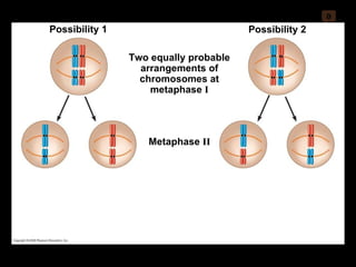
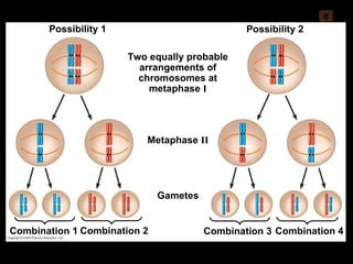
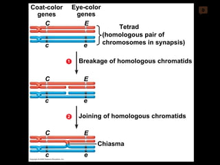
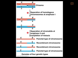
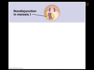
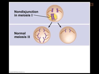
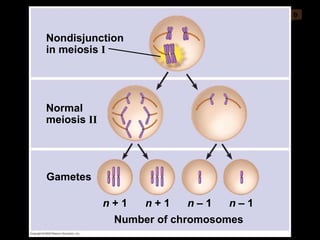
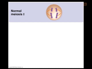
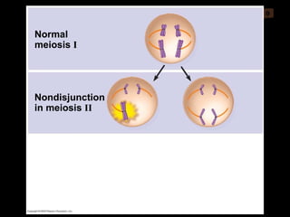
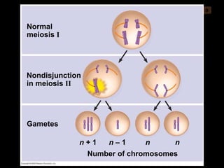
Mitosis and meiosis are both cell division processes in eukaryotes. Mitosis produces two identical daughter cells through chromosome duplication and separation, while meiosis reduces the chromosome number through two cell divisions. Meiosis results in four haploid daughter cells through homologous chromosome separation in meiosis I and sister chromatid separation in meiosis II. Both processes involve the duplication of chromosomes followed by their orderly separation through different stages including prophase, metaphase, anaphase and telophase.
Henrietta Lacks' immortal HeLa cells are used widely in medical research to study cancer, viruses, and other cell processes. Cells reproduce through mitosis, which duplicates DNA and divides the cell into two identical daughter cells. Mitosis maintains the chromosome number while meiosis halves it, producing gametes for sexual reproduction. Meiosis involves two nuclear divisions, mixing parental chromosomes and alleles in offspring.
Cells undergo mitosis or meiosis to divide. Mitosis produces two identical daughter cells from one parent cell during normal cell growth and repair. Meiosis produces four haploid gametes from one diploid cell for sexual reproduction. During meiosis, homologous chromosomes pair up and may exchange DNA segments through crossing over, introducing genetic variation into the gametes. The first meiotic division separates homologous chromosomes, while the second division separates sister chromatids to produce four unique haploid cells.
The document summarizes key aspects of the cell cycle and cell division. It discusses:
1) The cell cycle consists of interphase and the M phase where the cell divides. Interphase includes DNA replication in S phase to prepare for division.
2) Mitosis involves chromosome duplication and separation followed by cytokinesis to divide the cytoplasm. Meiosis produces gametes with half the normal chromosome number.
3) The mitotic spindle forms during cell division and uses microtubules to separate chromosomes between daughter cells.
Cells undergo mitosis and meiosis to divide. Mitosis produces two identical daughter cells through prophase, metaphase, anaphase and telophase and is used for growth and repair. Meiosis produces four non-identical haploid gametes through two divisions and crossing over, which contributes to genetic variation important for sexual reproduction. Regulators like cyclins and CDKs control the cell cycle.
1. The document discusses the process of cell division through mitosis and meiosis.
2. Mitosis is the process by which somatic body cells divide. It occurs in several phases: interphase, prophase, metaphase, anaphase, telophase, and cytokinesis.
3. Meiosis produces gametes like eggs and sperm, which have half the number of chromosomes as body cells. It involves two cell divisions and results in four daughter cells with unique combinations of chromosomes from each parent.
Mitosis is the process of cell division that results in two daughter cells with identical genetic material to the original parent cell. It occurs in eukaryotic cells and involves several phases - prophase, metaphase, anaphase and telophase. Cytokinesis then separates the daughter cells. Mitosis is important for tissue growth, repair and regeneration, asexual reproduction and embryonic development. Cyclins and cyclin-dependent kinases control the progression of cells through the cell cycle phases. Disruptions can lead to uncontrolled cell division and cancer. Smoking is strongly correlated with increased lung cancer rates due to carcinogens in tobacco smoke.
This document summarizes the cell cycle and cell division. It explains that the cell cycle includes interphase, where the cell grows and DNA is replicated, and cell division which includes mitosis of the nucleus and cytokinesis of the cytoplasm. Mitosis produces two identical daughter cells through the stages of prophase, metaphase, anaphase and telophase. Meiosis reduces the chromosome number by half and produces gametes like sperm and egg cells through two cell divisions. Fertilization restores the full chromosome number in the zygote.
The document discusses the cell cycle, which involves growth, functioning, and division of cells. It has two main types of cell division - mitosis and meiosis. Mitosis produces two identical cells and is involved in growth and repair. Meiosis produces gametes through two divisions and involves genetic mixing through crossing over. Precise control mechanisms regulate the cell cycle, and errors can lead to genetic conditions.
1) Bacterial cell division involves coordinating the segregation of replicated DNA and division of cytoplasmic material to generate progeny with identical genetic material. This requires spatial and temporal coordination of events.
2) The divisome, consisting of 10-15 proteins including FtsZ, assembles at midcell after DNA replication begins to assist in DNA separation and synthesize a new cell wall to divide the daughter cells.
3) There are multiple mechanisms that regulate divisome assembly to prevent it from occurring too early or in the same location as segregating chromosomes.
Cell division ensures the passage of genetic information from one generation of cells to the next. Mitosis and meiosis are the two main types of cell division. Mitosis produces somatic cells for growth and repair through binary fission in prokaryotes or nuclear division and cytokinesis in eukaryotes. Meiosis produces gametes through two rounds of nuclear division followed by cytokinesis, resulting in four haploid cells each with half the number of chromosomes of the original cell. Sexual reproduction requires the fusion of gametes during fertilization to form a diploid zygote and increase genetic variation in offspring.
Cells need to divide through the process of mitosis in order to repair and replace damaged or dead cells, to grow and develop, and for sexual reproduction. Mitosis involves the replication of DNA and the division of the cell into two identical daughter cells. The cell cycle is tightly regulated by internal and external factors to ensure proper cell division, growth and function. Deregulation of the cell cycle controls can lead to cancer if cells divide uncontrollably and form tumors that may metastasize and spread to other parts of the body. Treatments for cancer include surgery, chemotherapy, and radiation therapy.
The document discusses the cell cycle and its key stages and processes. It notes that the cell cycle consists of interphase, where the cell grows and DNA replicates, and the M phase where the cell divides. Interphase contains the G1, S, and G2 phases where the cell prepares for division. The M phase contains mitosis, where the nucleus and cytoplasm divide. Mitosis further consists of prophase, prometaphase, metaphase, anaphase and telophase stages. Meiosis is also discussed, which reduces the chromosome number in germ cells and involves two cell divisions.
Prokaryotic cells lack a nucleus and membrane-bound organelles. They have a cell wall, plasma membrane, cytoplasm containing ribosomes and a nucleoid region of naked DNA. Prokaryotic cells range from 1-10 micrometers and reproduce through binary fission rather than mitosis. They have varied metabolisms including fermentation, aerobic respiration, photosynthesis, and nitrogen fixation.
B.Sc. Biochemistry II Cellular Biochemistry Unit 3 Cell CycleRai University
The document discusses the cell cycle and cell division. It begins by explaining that all cells come from pre-existing cells and that cells divide through mitosis or binary fission to grow, repair damage, or replace old cells. The cell cycle consists of interphase, where the cell grows and DNA replicates, and mitosis, where the cell divides. Meiosis produces gametes through two cell divisions and results in four haploid cells rather than two identical diploid cells as in mitosis. The key stages and purposes of the cell cycle, mitosis, and meiosis are summarized.
Mitosis is a type of cell division that produces two daughter cells with identical chromosomes to the parent cell. It occurs in somatic cells for growth, repair, and replacement of damaged cells. The process involves nuclear division followed by cytokinesis. Uncontrolled mitosis can lead to tumors and cancer if not properly regulated by genes. Cloning uses techniques based on mitosis to produce genetically identical organisms through asexual reproduction and has applications in increasing crop yields, though it has disadvantages like reduced genetic variation and disease resistance.
This document provides an overview of genetics and the cell cycle. It discusses the stages of the cell cycle, including interphase (G1, S, G2 phases), mitosis, and cytokinesis. It describes cell division in eukaryotes and prokaryotes. Cancer is discussed, including characteristics of cancer cells, origins of cancer from mutations, and roles of oncogenes and tumor suppressor genes in regulating the cell cycle.
Cell division occurs through mitosis and meiosis and leads to growth, repair, asexual reproduction and sexual reproduction. Mitosis involves prophase, metaphase, anaphase and telophase and results in two identical daughter cells. Uncontrolled mitosis can lead to cancer due to genetic mutations. Cloning uses cell division to produce genetically identical copies of organisms and has applications in microbes, plants and animals.
The document provides an overview of meiosis cell division. It defines meiosis as a type of cell division that produces gametes with half the normal number of chromosomes. Meiosis occurs in two stages, Meiosis I and Meiosis II, and has four phases - prophase, metaphase, anaphase and telophase. In meiosis I, homologous chromosomes pair and may exchange genetic material through crossing over, resulting in genetic variation. This reduces the chromosome number from diploid to haploid. Meiosis II then divides the haploid cells into four haploid daughter cells.
Cell division occurs through mitosis and meiosis. Mitosis produces two identical daughter cells and is important for growth, repair, and asexual reproduction. It involves interphase, prophase, metaphase, anaphase, telophase, and cytokinesis. Meiosis produces gametes with half the number of chromosomes and involves two cell divisions. It ensures genetic variation through independent assortment and crossing over during prophase I. Meiosis results in four haploid cells from one diploid cell.
This document summarizes the key structures of prokaryotic and eukaryotic cells. Prokaryotic cells lack a nucleus and organelles, containing only a cell membrane, cell wall, cytoplasm, and sometimes structures like flagella or pili. Eukaryotic cells contain a nucleus enclosed within a nuclear envelope, as well as other membrane-bound organelles like the endoplasmic reticulum, Golgi apparatus, mitochondria, chloroplasts, lysosomes, and peroxisomes that carry out specialized functions. The document describes the roles of these various organelles in eukaryotic cells.
The document discusses cell division and inheritance, including how staining reveals nuclei, chromosomes are always present but only visible during cell division, and mitosis duplicates chromosomes to produce identical cells through nuclear envelope changes and chromatid separation.
The document discusses different types of cell division: binary fission, mitosis, and meiosis. Binary fission is how prokaryotic cells divide, splitting their single DNA strand to form two identical daughter cells. Mitosis and meiosis are forms of cell division in eukaryotes. Mitosis produces two identical daughter cells through the phases of interphase, prophase, metaphase, anaphase and telophase. Meiosis involves two cell divisions and results in four haploid cells with half the normal genetic material.
The document summarizes key points about the cell cycle and cell division. It discusses the different phases of the cell cycle including interphase and the M phase. Interphase consists of G1, S, and G2 phases. The M phase refers to mitosis which is divided into prophase, metaphase, anaphase and telophase. It also describes the process of cytokinesis in plant and animal cells. Meiosis is defined as a type of cell division that reduces chromosome number by half and involves two cell divisions - Meiosis I and Meiosis II. The stages of meiosis I including prophase I, metaphase I, anaphase I and telophase I are outlined. The significance of mitosis
Cell division occurs through mitosis and meiosis. Mitosis produces two identical daughter cells from one parent cell during growth and repair. Meiosis reduces the chromosome number by half and produces genetic variation through independent assortment and crossing over during gamete formation for sexual reproduction. The cell cycle is tightly regulated and consists of interphase, mitosis, and cytokinesis. Errors in meiosis can result in genetic disorders.
This document compares and contrasts mitosis and meiosis. Mitosis is a process of asexual reproduction that occurs in body cells and results in two identical diploid daughter cells. Meiosis is a type of sexual reproduction that occurs only in germ cells and results in four haploid daughter cells through two cell divisions. The key differences are that meiosis involves a reduction in chromosome number, genetic recombination through crossing over, and the production of haploid gametes rather than diploid somatic cells.
Multicellular organisms are made up of many different types of cells that cooperate and communicate to form organized systems. In humans there are over 10 trillion cells comprising 200 types of tissues. Each cell originates from a single fertilized egg cell and differentiates through activation of certain genes while others remain inactive, allowing cells to perform specialized functions. Bacteria also regulate genes in response to environmental signals, turning groups on and off. The lac operon in E. coli is a well-studied example of genetic control, where a repressor protein either allows or prevents transcription of genes involved in lactose metabolism depending on the presence of lactose.
This document provides an overview of cell membranes and their structure and functions. It begins by explaining that the plasma membrane forms the boundary of the cell and is selectively permeable. It then discusses how membranes are fluid mosaics composed of phospholipids and proteins. Various types of membrane proteins and their functions are also described, including transport proteins that allow passive and active movement of substances across the membrane. The document concludes by explaining how membrane structure results in selective permeability and the different mechanisms of passive and active transport.
This document provides a list of 21 architectural works and the architects or theorists associated with them. It includes seminal works that advanced modern and postmodern architecture in the 20th century, addressing topics like ornamentation, functionalism, complexity, diagramming, and relationships between buildings and landscapes. Each entry lists a short description or key concept related to the architectural work.
Development of cancer therapeutics is often carried out in 2D cultures prior to testing on animal model. In comparison to 2D cultures, discuss the potential of using 3D in vitro models for drug efficiency testing.
The document discusses different types of cell cultures, including primary cultures derived from animal tissue, continuous cultures comprised of cell lines, and normal diploid cells with a finite lifespan. It describes common cell lines used in research, including HeLa cells from cervical carcinoma and CHO cells from hamster ovary. The key components of maintaining cell cultures are discussed, such as temperature, atmosphere, sterile conditions, culture medium, and growth equipment. Tasks demonstrate observing cell morphology, quantifying cell density using a hemocytometer, and counting chromosomes in CHO cells, which are shown to have an aneuploid number due to long-term cultivation.
This document provides a review of key topics covered in Biology 163's 2nd semester, including:
1) Asexual and sexual reproduction - Asexual reproduction produces identical offspring from one parent faster, while sexual reproduction involves two parents and produces genetically diverse offspring over more time.
2) Genetics concepts - Terms like heterozygous, homozygous, dominant, recessive, codominant, and sex-linked traits are defined. Punnett squares, laws of segregation and independent assortment are also covered.
3) Evolution - Topics like natural selection, convergent evolution, and speciation are summarized, explaining how beneficial traits increase over generations through differential survival and reproduction.
4)
This document discusses techniques for in vitro clonal propagation of fruit crops. It covers the basics of micropropagation, which involves four stages: establishment, shoot multiplication, root formation, and acclimatization. Various tissue culture techniques are described that can be used for clonal propagation, including meristem culture, shoot tip micrografting, anther culture, embryo culture, ovary/ovule culture, callus culture, cell suspension culture, and protoplast culture. Requirements for facilities, media preparation, and procedures for each stage of micropropagation are provided. The document aims to inform the reader about the various in vitro techniques that can be used for commercial clonal propagation of fruit crops.
1) The chapter discusses the cellular basis of reproduction and inheritance. It covers topics like cell division, the cell cycle, meiosis, and alterations in chromosome structure.
2) The key stages of the cell cycle are described, including interphase and the phases of mitosis (prophase, metaphase, anaphase, telophase). Cytokinesis is the final step that divides the cytoplasm.
3) Meiosis is introduced as reducing the number of chromosomes by half to form gametes, while mitosis replicates chromosomes to form body cells. Homologous chromosomes pair and may exchange genetic material.
Commercial exploitation of micro its technologyPawan Nagar
Micropropagation is a tissue culture technique where whole plants are regenerated from small plant tissues or cells grown in a sterile nutrient culture medium. It allows for rapid multiplication of elite plant varieties in a short period of time compared to traditional propagation methods. The document outlines the various steps involved in micropropagation including selection of explant material, surface sterilization, initiation and establishment of aseptic culture, multiplication of shoots, rooting of plantlets, and acclimatization of plantlets in the greenhouse. Micropropagation has many advantages for commercial horticulture including production of disease-free plants, continuous propagation year-round, export of pathogen-free plants, and long-term germplasm storage.
This document summarizes an interview with Terry Orr-Weaver, a biologist who studies chromosome partitioning during cell division and DNA replication. Some key points:
- Orr-Weaver switched from yeast to fruit flies as her model organism to study how cell division and pattern formation are coordinated during development.
- Her research group uses genetics, biochemistry, and cell biology approaches like microscopy to study these processes directly. For example, they tagged a protein involved in DNA replication to see where it localizes in cells.
- Meiosis reduces the chromosome number from diploid to haploid by having an extra round of chromosome separation where maternal and paternal chromosomes are separated. This ensures sperm and eggs have one copy of
Plant tissue culture is the process of maintaining or growing plant cells, tissues or organs under sterile conditions on a nutrient culture medium of known composition. It involves techniques like cell culture, organ culture or meristem culture to produce clones of a plant through micropropagation. The key steps are selection of explant tissue from a donor plant, sterilization, establishment of the explant on a culture medium, multiplication through cell division and shoot formation, rooting of shoots, and acclimatization of plantlets in soil. Micropropagation allows for rapid mass multiplication of plant materials while maintaining genetic uniformity.
Mitosis and meiosis are both cell division processes but have key differences. Mitosis produces identical daughter cells through prophase, metaphase, anaphase and telophase and is used for growth and repair. Meiosis produces gametes through two rounds of division and results in non-identical haploid cells through prophase I, metaphase I, anaphase I, telophase I and then a second round of division. The stages of meiosis include homologous chromosome pairing and genetic recombination which do not occur in mitosis. Both processes rely on spindle fibers to separate chromosomes but achieve different end products - growth of somatic cells for mitosis versus genetic diversity in offspring for meiosis.
Application of plant tissue culture/ micro-propagationSushil Nyaupane
Tissue culture is the process of growing cells or tissues in sterile conditions. It allows for rapid cloning of plant materials. Plant tissue culture involves excising plant parts and growing them on nutrient media. This allows for mass multiplication of plant materials irrespective of season. Some key developments include Haberlandt's proposal of plant cell culture in 1902, and Murashige and Skoog's nutrient medium in 1962. Micropropagation is now used for conservation of rare species, producing disease-free plants, mutation breeding, and more. The future of this technique remains promising.
This document provides an overview of plant tissue culture. It defines tissue culture as the in vitro cultivation of plant cells or tissues under aseptic conditions on a nutrient medium. The history and key figures in the development of plant tissue culture are discussed. Details are provided on nutrient requirements, preparation and sterilization of culture media, basic laboratory requirements, establishment of cultures from explants, and types of growth and cultures. The advantages and applications of plant tissue culture are also summarized.
This document contains protocols for various plant tissue culture techniques. It discusses the introduction to plant tissue culture, sterilization techniques used, and then outlines 8 specific protocols: 1) tissue culture media preparation, 2) explant preparation and surface sterilization, 3) embryo culture, 4) culture of anther for haploid production, 5) meristem culture, 6) meristem tip culture for virus-free plants, 7) induction of somatic embryogenesis, and 8) protoplast isolation, culture, and regeneration. The goal of these protocols is to describe the principles and procedures of different plant tissue culture methods.
Tissue culture is a technique where cells, tissues or whole plants are grown in a sterile nutrient culture medium under controlled conditions. It allows for rapid vegetative propagation of plants. Key steps include sterilizing equipment and explants, preparing nutrient medium, subculturing to promote growth, and rooting and hardening plantlets. Tissue culture has many applications like mass multiplication of crops and plants, eliminating diseases, and genetic modification. It is used commercially for propagating crops but contamination and rooting difficulties can be issues.
KEY CONCEPTS
13.1 Offspring acquire genes from parents by inheriting
chromosomes
13.2 Fertilization and meiosis alternate in sexual life cycles
13.3 Meiosis reduces the number of chromosome sets from diploid to haploid
13.4 Genetic variation produced in sexual life cycles contributes to evolution
The document discusses the process of cell division through mitosis and cytokinesis. It explains that eukaryotic cells undergo a cell cycle that includes interphase, where the cell grows and duplicates its DNA, and the mitotic phase. During mitosis, the cell divides into two daughter cells through four main phases - prophase, metaphase, anaphase and telophase. Cytokinesis then separates the cytoplasmic components of the parent cell. Cancer cells are able to divide uncontrollably due to mutations in cell cycle regulation genes.
The document discusses mitosis and cell division. It describes the stages of mitosis including prophase, metaphase, anaphase and telophase. It also discusses the importance of mitosis for growth and repair of organisms. Uncontrolled mitosis can lead to cancer if cells divide rapidly and unregulated. Knowledge of mitosis is applied in cloning to produce genetically identical individuals.
1. Cellular reproduction can occur through binary fission in prokaryotes and mitosis in eukaryotes, which produces genetically identical daughter cells. 2. Meiosis produces gametes with half the normal number of chromosomes which can fuse during fertilization to form genetically unique offspring. 3. The cell cycle is regulated by cyclin-dependent kinases and checkpoints ensure DNA replication and chromosome separation occur properly.
This document provides information about the cell cycle and its regulation. It discusses the key phases of the cell cycle including interphase, mitosis, and meiosis. Interphase consists of G1, S, and G2 phases where the cell grows and DNA is replicated. Mitosis involves nuclear division into two daughter cells with identical chromosomes. Meiosis involves two cell divisions resulting in four daughter cells each with half the original chromosome number, allowing for genetic variation. The document defines important cell cycle concepts and compares the processes of mitosis and meiosis.
Cell division is a fundamental process that occurs in both prokaryotic and eukaryotic cells. In prokaryotes, cell division occurs through binary fission where the chromosome replicates and the cell membrane pinches inward to form two daughter cells. Eukaryotes undergo either mitosis or meiosis. Mitosis produces genetically identical daughter cells through replication of DNA followed by nuclear and cellular division. The process of mitosis consists of prophase, prometaphase, metaphase, anaphase and telophase where the chromosomes and cytoplasm are divided.
This document provides an overview of cell division through mitosis and meiosis. It defines key terms like interphase, prophase, metaphase, anaphase, telophase and cytokinesis. It explains the stages and importance of both mitosis and meiosis. Specifically, mitosis produces genetically identical daughter cells through the division of the nucleus, while meiosis reduces chromosome number by half to produce haploid gametes through two divisions. Uncontrolled mitosis can lead to cancer if chromosomes do not separate properly.
Mitosis allows for growth, repair, and asexual reproduction in organisms. It involves nuclear division followed by cytoplasmic division. The cell cycle consists of interphase, where the cell grows and prepares to divide, and mitosis, where the nucleus and cytoplasm divide. Mitosis ensures each new cell has an identical set of chromosomes through carefully regulated phases including prophase, metaphase, anaphase and telophase. Uncontrolled mitosis can lead to tumor growth and cancer.
Infer the significance of cell division.
Differentiate a DNA molecule, a chromosome, and a chromatid.
Characterize the phases of the cell cycle and their control points.
Describe the major events associated with stages of mitosis.
Explain the process of cytokinesis.
Learning Objectives
Describe the role of apoptosis in the life cycle of a cell.
Relate cancer as a result of the malfunction of the cell during the cell cycle.
The document summarizes key aspects of the cell cycle and cell division. It discusses how cell division results in genetically identical daughter cells through DNA replication and chromosome separation. The mitotic phase alternates with interphase in the cell cycle. Mitosis is divided into prophase, prometaphase, metaphase, anaphase and telophase. Cytokinesis then divides the cytoplasm. The cell cycle is regulated by a molecular control system involving cyclins and cyclin-dependent kinases. Cancer cells exhibit deregulated cell cycle control and proliferation.
Mitosis is cell division that produces two daughter cells identical to the parent cell. It occurs in somatic cells and involves the four phases of prophase, metaphase, anaphase and telophase. Meiosis is a type of cell division that produces gametes with half the number of chromosomes, and occurs in germ cells. Meiosis has two rounds of division, Meiosis I and Meiosis II, which separates homologous chromosomes and sister chromatids respectively to generate four haploid daughter cells from one diploid parent cell. Mitosis and meiosis are important for growth, tissue repair, sexual reproduction, and genetic variation.
Chapter-6Cell Cycle and DivisionCell Divisio.docxchristinemaritza
Chapter-6
Cell Cycle and Division
Cell Division
Cells reproduce by cell division, in which a parent cell normally gives rise to two daughter cells
Each daughter cell receives a complete set of hereditary information (DNA) from the parent cell and about half its cytoplasm
The hereditary information DNA is usually identical with that of the parent cell
The cell division of eukaryotic cells by which organisms grow or increase in number is called mitotic cell division
After cell division, the daughter cells may differentiate, becoming specialized for specific functions
The repeating pattern of divide, grow, and differentiate, then divide again is called the cell cycle
Most multicellular organisms have three categories of cells
1. stem cells
2. Other cells capable of dividing
3. Permanently differentiated cells
1.Stem cells :
- have two important characteristics: self-renewal, and the ability to differentiate into a variety of cell types
-Stem cells self-renew because they retain the ability to divide, perhaps for the entire life of the organism
-Some stem cells in early embryos can produce any of the specialized cell types of the entire body
2. Other cells capable of dividing
-Some cells other than stem cells are capable of continuing to divide, but typically differentiate into only one or two different cell types
-Dividing liver cells, for example, can only become more liver cells
3. Permanently differentiated cells
-Permanently differentiated cells differentiate and never divide again
-For example, most heart and brain cells cannot divide
CELL CYCLE
Both prokaryotic and eukaryotic cells have cell cycles that include growth, metabolic activity, DNA replication, and cell division
However, they have major structural and functional differences
Eukaryotic chromosome
Eukaryotic chromosomes are separated from the cytoplasm by a membrane-bound nucleus
Eukaryotic cells always have multiple chromosomes
Eukaryotic chromosomes are longer and have more DNA than prokaryotic chromosomes (human chromosomes are 10 to 80 times longer and have 10 to 50 times more DNA)
Genes
Genes are segments of the DNA of a chromosome
Genes are sequences of DNA from hundreds to thousands of nucleotides long
Each gene occupies a specific place, or locus (plural, loci) on the chromosome
Two important parts of chromosome
Two telomeres
One centromere
It temporarily holds two daughter DNA double helices together after DNA replication
It is the attachment site for microtubules that move the chromosomes during cell division
Homologous
11
Chromosomes that contain the same genes are called homologous chromosomes, or homologues
Cells with pairs of homologous chromosomes are called diploid, which means “double”
Cells with half the number of chromosomes are called haploid
Human Chromosomes
A typical human cell has ...
Cell division occurs through two main processes - mitosis and meiosis. Mitosis produces two identical daughter cells during regular cell growth and reproduction. It has four phases: prophase, metaphase, anaphase and telophase. Meiosis produces gamete cells like eggs and sperm, which have half the normal number of chromosomes to ensure fertility. During meiosis, one cell undergoes two cell divisions to produce four daughter cells each with half the original number of chromosomes. This allows for genetic variation in offspring. Cell division is essential for growth, repair and reproduction of living organisms.
Mitosis allows for growth, repair, and asexual reproduction in organisms. It is the process by which a cell divides its nucleus to form two daughter cells with identical genetic material. Mitosis consists of prophase, metaphase, anaphase, and telophase, where the chromosomes condense and separate. Cytokinesis then divides the cytoplasm to complete cell division. Uncontrolled mitosis can lead to tumor growth and cancer development.
The document discusses cellular reproduction and the cell cycle. It explains that cells require genetic instructions from DNA to survive and divide. There are two main types of cells - prokaryotic and eukaryotic. Eukaryotic cells undergo mitotic cell division, which involves interphase where DNA is replicated, followed by mitosis where the cell divides into two identical daughter cells through nuclear division and cytoplasmic division. Mitosis ensures each daughter cell receives a complete copy of genetic material and maintains chromosome number.
1) The cell cycle and cell division are essential for the growth, development, and repair of multicellular organisms. Cell division results in genetically identical daughter cells through mitosis or nonidentical gametes through meiosis.
2) The cell cycle consists of interphase, where the cell grows and DNA replicates, and mitosis, where the genetic material divides. Cyclins and cyclin-dependent kinases regulate progression through the cell cycle via checkpoints.
3) Cancer cells evade normal cell cycle controls, allowing uncontrolled growth and division that can lead to tumor formation and metastasis.
Multicellular organisms develop from a single cell known as zygote by the process of mitosis. Asexual reproduction in some organisms like amoeba and vegetative reproduction in plants takes place by mitosis. This type of cell division involves many steps and it does not alter the genetic material.
Cell division occurs through two main processes - mitosis and meiosis. Mitosis produces two identical daughter cells during normal growth and tissue repair. Meiosis produces four non-identical haploid daughter cells from a single diploid parent cell, which occurs during gamete formation. This introduces genetic diversity when gametes fuse during fertilization. The key events of meiosis include homologous chromosome pairing during prophase I and their subsequent separation in anaphase I, followed by two rounds of chromosome separation to form four unique haploid cells.
This document discusses the cell cycle and cell division. It begins by explaining the importance of cell division for reproduction and growth in both unicellular and multicellular organisms. It then describes the main stages and roles of the cell cycle, including interphase and the mitotic phase. Key details are provided on DNA replication in S phase, chromosome behavior, and the stages of mitosis. The document emphasizes the critical roles of the mitotic spindle and centrosomes in proper chromosome separation during cell division. Diagrams illustrate these various stages and structures discussed in the text.
THE SACRIFICE HOW PRO-PALESTINE PROTESTS STUDENTS ARE SACRIFICING TO CHANGE T...indexPub
The recent surge in pro-Palestine student activism has prompted significant responses from universities, ranging from negotiations and divestment commitments to increased transparency about investments in companies supporting the war on Gaza. This activism has led to the cessation of student encampments but also highlighted the substantial sacrifices made by students, including academic disruptions and personal risks. The primary drivers of these protests are poor university administration, lack of transparency, and inadequate communication between officials and students. This study examines the profound emotional, psychological, and professional impacts on students engaged in pro-Palestine protests, focusing on Generation Z's (Gen-Z) activism dynamics. This paper explores the significant sacrifices made by these students and even the professors supporting the pro-Palestine movement, with a focus on recent global movements. Through an in-depth analysis of printed and electronic media, the study examines the impacts of these sacrifices on the academic and personal lives of those involved. The paper highlights examples from various universities, demonstrating student activism's long-term and short-term effects, including disciplinary actions, social backlash, and career implications. The researchers also explore the broader implications of student sacrifices. The findings reveal that these sacrifices are driven by a profound commitment to justice and human rights, and are influenced by the increasing availability of information, peer interactions, and personal convictions. The study also discusses the broader implications of this activism, comparing it to historical precedents and assessing its potential to influence policy and public opinion. The emotional and psychological toll on student activists is significant, but their sense of purpose and community support mitigates some of these challenges. However, the researchers call for acknowledging the broader Impact of these sacrifices on the future global movement of FreePalestine.
Temple of Asclepius in Thrace. Excavation resultsKrassimira Luka
The temple and the sanctuary around were dedicated to Asklepios Zmidrenus. This name has been known since 1875 when an inscription dedicated to him was discovered in Rome. The inscription is dated in 227 AD and was left by soldiers originating from the city of Philippopolis (modern Plovdiv).
This document provides an overview of wound healing, its functions, stages, mechanisms, factors affecting it, and complications.
A wound is a break in the integrity of the skin or tissues, which may be associated with disruption of the structure and function.
Healing is the body’s response to injury in an attempt to restore normal structure and functions.
Healing can occur in two ways: Regeneration and Repair
There are 4 phases of wound healing: hemostasis, inflammation, proliferation, and remodeling. This document also describes the mechanism of wound healing. Factors that affect healing include infection, uncontrolled diabetes, poor nutrition, age, anemia, the presence of foreign bodies, etc.
Complications of wound healing like infection, hyperpigmentation of scar, contractures, and keloid formation.
Level 3 NCEA - NZ: A Nation In the Making 1872 - 1900 SML.pptHenry Hollis
The History of NZ 1870-1900.
Making of a Nation.
From the NZ Wars to Liberals,
Richard Seddon, George Grey,
Social Laboratory, New Zealand,
Confiscations, Kotahitanga, Kingitanga, Parliament, Suffrage, Repudiation, Economic Change, Agriculture, Gold Mining, Timber, Flax, Sheep, Dairying,
Gender and Mental Health - Counselling and Family Therapy Applications and In...PsychoTech Services
A proprietary approach developed by bringing together the best of learning theories from Psychology, design principles from the world of visualization, and pedagogical methods from over a decade of training experience, that enables you to: Learn better, faster!
A Free 200-Page eBook ~ Brain and Mind Exercise.pptxOH TEIK BIN
(A Free eBook comprising 3 Sets of Presentation of a selection of Puzzles, Brain Teasers and Thinking Problems to exercise both the mind and the Right and Left Brain. To help keep the mind and brain fit and healthy. Good for both the young and old alike.
Answers are given for all the puzzles and problems.)
With Metta,
Bro. Oh Teik Bin 🙏🤓🤔🥰
9. 0 Prokaryotic chromosome Duplication of chromosome and separation of copies Cell wall Plasma membrane 1
10. 0 Prokaryotic chromosome Duplication of chromosome and separation of copies Cell wall Plasma membrane 1 Continued elongation of the cell and movement of copies 2
11. 0 Prokaryotic chromosome Duplication of chromosome and separation of copies Cell wall Plasma membrane 1 Continued elongation of the cell and movement of copies 2 Division into two daughter cells 3
37. 0 Cleavage furrow Contracting ring of microfilaments Daughter cells Cleavage furrow
38. 0 Cell plate Daughter cells Cell wall Vesicles containing cell wall material Daughter nucleus Cell plate forming Wall of parent cell New cell wall
39.
40.
41. 0 G 1 checkpoint Control system M S G 2 G 1 M checkpoint G 2 checkpoint G 0
42.
43. 0 G 1 checkpoint Control system M S G 2 G 1 Receptor protein Signal transduction pathway Relay proteins Plasma membrane Growth factor
44.
45.
46. 0 A tumor grows from a single cancer cell. Cancer cells spread through lymph and blood vessels to other parts of the body. Cancer cells invade neighboring tissue. Tumor Glandular tissue Lymph vessels Blood vessel
47.
48.
49.
50.
51. 0 Sister chromatids One duplicated chromosome Centromere Homologous pair of chromosomes
52.
53. 0 Haploid gametes ( n = 23) n Egg cell Sperm cell Fertilization Meiosis Multicellular diploid adults (2 n = 46) Mitosis and development n 2 n Diploid zygote (2 n = 46)
54.
55.
56.
57.
58.
59.
60.
61. 0 Centrosomes (with centriole pairs) PROPHASE I Microtubules attached to kinetochore INTERPHASE Sites of crossing over Metaphase plate Spindle MEIOSIS I : Homologous chromosomes separate METAPHASE I Sister chromatids remain attached ANAPHASE I Nuclear envelope Sister chromatids Centromere (with kinetochore) Homologous chromosomes separate Chromatin Tetrad
67. 0 PROPHASE I MEIOSIS II : Sister chromatids separate METAPHASE II ANAPHASE II Cleavage furrow TELOPHASE II AND CYTOKINESIS Sister chromatids separate Haploid daughter cells forming TELOPHASE II AND CYTOKINESIS
68.
69.
70. 0 Prophase Metaphase I Metaphase 2 n = 4 Tetrads align at the metaphase plate Duplicated chromosome (two sister chromatids) Parent cell (before chromosome duplication) Chromosome duplication Chromosomes align at the metaphase plate Anaphase Telophase Sister chromatids separate during anaphase Daughter cells of mitosis 2 n 2 n n Chromosome duplication Site of crossing over Tetrad formed by synapsis of homologous chromosomes M EIOSIS Prophase I Anaphase I Telophase I M ITOSIS M EIOSIS I Haploid n = 2 Daughter cells of meiosis I M EIOSIS II n n n Daughter cells of meiosis II Homologous chromosomes separate ( anaphase I ); sister chroma- tids remain together No further chromosomal duplication; sister chromatids separate ( anaphase II )
71.
72. 0 Two equally probable arrangements of chromosomes at metaphase I Possibility 1 Possibility 2
73. 0 Two equally probable arrangements of chromosomes at metaphase I Possibility 1 Possibility 2 Metaphase II
74. 0 Two equally probable arrangements of chromosomes at metaphase I Possibility 1 Possibility 2 Metaphase II Combination 1 Gametes Combination 2 Combination 3 Combination 4
79. 0 Breakage of homologous chromatids Coat-color genes Eye-color genes C (homologous pair of chromosomes in synapsis) E c e Tetrad C E c e Joining of homologous chromatids 2 C E c e Chiasma 1
80. 0 Separation of homologous chromosomes at anaphase I C E c e Chiasma Separation of chromatids at anaphase II and completion of meiosis C E c e c E C e c e c E C E C e Parental type of chromosome Gametes of four genetic types Recombinant chromosome Parental type of chromosome Recombinant chromosome 4 3
81.
82.
83. 0 Packed red and white blood cells Centrifuge Blood culture Fluid 1 Blood culture is centrifuged to separate the blood cells From the culture fluid
84. 0 Packed red and white blood cells Centrifuge Blood culture Fluid 1 Hypotonic solution 2 Fluid is discarded, and a hypotonic solution is mixed with The cells. This makes the RBC burst, the WBC swell but Do not burst, and their chromosomes spread out
85. 0 Packed red and white blood cells Centrifuge Blood culture Fluid 1 Hypotonic solution 2 3 Fixative White blood cells Stain Another centrifugation separates the WBC. The fluid Containing the remnants of RBCs is discarded. A preservative Is mixed w/ the WBC and a drop of the cell suspension is Spread on a microscope slide, dried, and stained.
87. 0 Centromere Sister chromatids Pair of homologous chromosomes 5 Digital photograph of chromosomes is obtained and a computer Sorts them by size and shape. Resulting karyotype is below. 22 pairs of autosomes, 2 sex chromosomes
106. Need Chemistry Help? Contact Veronica Walker for FREE tutoring! [email_address] Need Biology Help?? Contact Veronica Walker for FREE tutoring! [email_address]
Editor's Notes
Most eukaryotic organisms are capable of both asexual and sexual reproduction. Students may be surprised to learn that asexual reproduction plays a major role in the life cycles of many organisms. For example, the unicellular algae Chlamydomonas generates an increased population by asexual reproduction when conditions are favorable for cell division. In unfavorable conditions, the organism undergoes sexual reproduction. This has the advantage of producing a new combination of genes and traits that could be advantageous for survival under new environmental conditions. Student Misconceptions and Concerns 1. As the authors note in Module 8.1, biologists use the term daughter to indicate offspring and not gender. Students with little experience in this terminology can easily become confused. 2. Some basic familiarity or faint memory of mitosis and meiosis might result in a lack of enthusiasm for mitosis and meiosis in some of your students. Consider beginning such lectures with important topics related to cellular reproduction. For example, cancer cells reproduce uncontrollably, stem cells have the capacity to regenerate lost or damaged tissues, and the study of embryonic stem cells is variously restricted and regulated. Teaching Tips 1. Sometimes the most basic questions can challenge students and get them focused on the subject at hand. Consider asking your students why we expect that dogs will produce dogs, cats will produce more cats, and chickens will only produce chickens. Why does like produce like?
Figure 8.1A A single-celled amoeba producing a genetically identical offspring cell through asexual reproduction. This figure shows an amoeba reproducing asexually. Ask students to suggest other examples of asexual reproduction. Possibilities include growth of a plant from a seed, replacement of skin cells, growth of an embryo or fetus from a fertilized egg.
Figure 8.1B Sexual reproduction produces offspring with unique combinations of genes. Members of this family show similar traits but have differences in appearance. Variations include skin color and hair curling. Many genes contribute to skin color, influencing the number, size, and shape of melanosomes, organelles that synthesize and store the pigment melanin, as well as the type of melanin produced. Hair curling is related to at least one gene with incomplete dominance. When alleles for curly and straight are combined in a heterozygous individual, wavy hair is observed.
Student Misconceptions and Concerns 1. Some basic familiarity or faint memory of mitosis and meiosis might result in a lack of enthusiasm for mitosis and meiosis in some of your students. Consider beginning such lectures with important topics related to cellular reproduction. For example, cancer cells reproduce uncontrollably, stem cells have the capacity to regenerate lost or damaged tissues, and the study of embryonic stem cells is variously restricted and regulated. Teaching Tips 1. Virchow’s principle of “every cell from a cell” is worth thinking through with your class. Students might expect that, like automobiles, computers, and cell phones, parts are constructed and cells are assembled. In our society, few nonliving products are generated only from existing products (try to think of such examples). For example, you do not need a painting to paint or a house to construct a house. Yet this is a common expectation in biology. 2. Students who think through Virchow’s principle might ask how the first cells formed. They might wonder further whether the same environments that produced these cells are still in existence. The conditions on Earth when life first formed were very different from those we know today. Chapter 15 addresses the origin and early evolution of life on Earth.
The process of binary fission is very rapid. E. coli cells divide every 20 minutes under optimal environmental conditions. The antibiotic penicillin inhibits the growth of the bacterial cell wall. Cells can duplicate their internal contents, including the chromosome, but will burst when they become too large for the existing cell wall. Student Misconceptions and Concerns 1. Some basic familiarity or faint memory of mitosis and meiosis might result in a lack of enthusiasm for mitosis and meiosis in some of your students. Consider beginning such lectures with important topics related to cellular reproduction. For example, cancer cells reproduce uncontrollably, stem cells have the capacity to regenerate lost or damaged tissues, and the study of embryonic stem cells is variously restricted and regulated. Teaching Tips Consider contrasting the timing of DNA replication and cytokinesis in prokaryotes and eukaryotes. In prokaryotes, addressed in Module 8.3, these processes are over-lapping. However, as revealed in the next few modules, these events are separate in eukaryotes.
Figure 8.3A Binary fission of a prokaryotic cell. This figure shows the steps in binary fission.
Figure 8.3B Electron micrograph of a dividing bacterium. The chromosomes of each of the two identical cells are visible in this electron micrograph.
Chromatin is compacted about 100,000 fold to produce the interphase/metaphase chromosome. If all the DNA in the human chromosomes were aligned, it would stretch for one meter. All of this DNA is condensed to fit into a nucleus that can only be seen with the aid of a microscope. The centromere has a unique DNA sequence involving repeated stretches of nucleotides. In biotechnological applications, artificial chromosomes can be produced that have a centromeric sequence. This chromosome will be properly distributed during cell division because the spindle fibers attach to the artificial centromeric sequence. Student Misconceptions and Concerns 1. Students often seem confused by the difference between a DNA molecule and a chromosome. This is especially problematic when discussing DNA replication. 2. Students are often confused by photographs of chromosomes. Such photographs, such as Figure 8.4B, typically show duplicated chromosomes during some aspect of cell division. It remains unclear to many why (a) chromosome structure is typically different between interphase G1 and the stages of division and (b) why chromosomes are not photographed during interphase (the stage in which chromosomes are typically first discussed) before the chromosomes duplicate. Teaching Tips 1. Figure 8.4C is an important point of reference for some basic terminology. Consider referring to it as you distinguish between a DNA molecule and a chromosome, unreplicated and replicated chromosomes, and the nature of sister chromatids.
Figure 8.4A A plant cell (from an African blood lily) just before division. This cell shows the coiling or compaction of chromatin, in prophase. Although the chromosomes have already duplicated, higher resolution is required to distinguish sister chromatids.
Figure 8.4B Electron micrograph of a duplicated chromosome. This electron micrograph provides a view of sister chromatids.
Differences in the length of the cell cycle can be instructive. Yeast cells have a 2-hour life cycle, while human cells in culture take about 24 hours to divide. Mitosis and cytokinesis represent a shorter section of the cycle, lasting one hour for cultured human cells. Teaching Tips 1.The authors note in Module 8.5 that each of your students consists of about 100 trillion cells. It is likely that this number is beyond comprehension for most of your students. Consider sharing several simple examples of the enormity of that number to try to make it more meaningful. For example, the U.S. population in 2008 is about 310 million people. To give every one of those people about $323,000, we will need a total of $100 trillion. Here is another example. If we give you $31,688 every second of your life, and you lived for 100 years, you would receive $100 trillion dollars. 2.The concepts of DNA replication and sister chromatids are often obstacles for many students. If you can find twist ties or other bendable wire, you can demonstrate or have students model the difference between (1) a chromosome before DNA replication and (2) sister chromatids after DNA replication. One piece of wire will represent a chromosome before replication. Two twist ties twisted about each other can represent sister chromatids. We have doubled the DNA, but the molecules remain attached (although not attached in the same way as the wire). You might also want to point out that when sister chromatids are separated, they are considered separate chromosomes. 3. In G1, the chromosomes have not duplicated. But by G2, chromosomes consist of sister chromatids. If you have created a demonstration of sister chromatids, relate DNA replication and sister chromatids to the cell cycle.
Figure 8.5 The eukaryotic cell cycle.
Teaching Tips 1. Students might keep better track of the sequence of events in a cell cycle by simply memorizing the letters IPPMAT: the first letters of interphase, prophase, prometaphase, metaphase, anaphase, and telophase are represented in this acronym. 2. The authors note that animals, but not plants, have a pair of centrioles in their centrosomes. They add that the role of centrioles in cell division is a mystery. Students might not appreciate all that remains to be explained in biology. Sharing the existence of such mysteries with them promotes critical thinking skills and helps them imagine a place for themselves in future research.
Centrioles give rise to basal bodies that are the foundations for cilia and flagella. They are found in animal cells but also in plants such as mosses and ferns that have swimming sperm. They are not found in flowering plants, showing that centrioles are not essential for spindle formation. There is other evidence, however, that suggests centrioles may influence progression through alternative stages in the cell cycle, including entry into the S phase and completion of cytokinesis. (Reviewed in A. W. Murray, 2001, “Centrioles at the Checkpoint,” Science , 291:1499–1501.) For the BioFlix Animation Mitosis, go to Animation and Video Files. Teaching Tips 1. Students might keep better track of the sequence of events in a cell cycle by simply memorizing the letters IPPMAT: the first letters of interphase, prophase, prometaphase, metaphase, anaphase, and telophase are represented in this acronym. 2. The authors note that animals, but not plants, have a pair of centrioles in their centrosomes. They add that the role of centrioles in cell division is a mystery. Students might not appreciate all that remains to be explained in biology. Sharing the existence of such mysteries with them promotes critical thinking skills and helps them imagine a place for themselves in future research.
Figure 8.6 The stages of cell division.
Applying Your Knowledge Human cells have 46 chromosomes. By the end of interphase, How many chromosomes are present in one cell? 46 How many chromatids are present in one cell? 92 (Each chromosome has been duplicated and consists of a pair of chromatids joined at the centromere.) Teaching Tips 1. Students might keep better track of the sequence of events in a cell cycle by simply memorizing the letters IPPMAT: the first letters of interphase, prophase, prometaphase, metaphase, anaphase, and telophase are represented in this acronym. 2. The authors note that animals, but not plants, have a pair of centrioles in their centrosomes. They add that the role of centrioles in cell division is a mystery. Students might not appreciate all that remains to be explained in biology. Sharing the existence of such mysteries with them promotes critical thinking skills and helps them imagine a place for themselves in future research.
Figure 8.6 The stages of cell division.
This material allows a review of cellular components. Students can be reminded that microtubules are composed of actin molecules and that actin and myosin work in concert for muscle cell contraction. They can also be reminded that vesicles have a lipid boundary that will contribute to the plasma membrane of the new plant cells. For the BLAST Animation Cytokinesis in Plants, go to Animation and Video Files. Teaching Tips 1. Many students think of mitosis and cytokinesis as one process. In some situations, mitosis occurs without subsequent cytokinesis. Challenge your students to predict the outcome of mitosis without cytokinesis (multinuclear cells called a syncytium). This occurs in human development during the formation of the placenta. 2. The authors make an analogy between a drawstring and the mechanism of cytokinesis in animal cells. Students seem to appreciate this association. Have your students think of a person tightening the drawstring of sweatpants so tight that they pinch themselves in two, or perhaps nearly so! The analogy is especially good because, like the drawstring just beneath the surface of the sweat pants, the microfilaments are just beneath the surface of the cell’s plasma membrane.
Figure 8.7A Cleavage of an animal cell.
Figure 8.7B Cell plate formation in a plant cell.
There is promising research on the use of growth factors to stimulate the regeneration of nerves by promoting axon growth. For example, the macrophage-derived protein oncomodulin has been shown to increase axon length in the optic nerve nearly twofold in vitro. (Laboratory of L. Benowitz, in Yin, et al., 2006, Nature Neuroscience 9, 843–852.) Teaching Tips 1. Students who closely examine a small abrasion on their skin might notice that the wound tends to heal from the outer edges inward. This space-filling mechanism is a natural example of density-dependant inhibition, which is also seen when cells in a cell culture dish stop dividing when they have formed a complete layer.
Student Misconceptions and Concerns 1. Students do not typically know that all cancers are genetically based. Consider making this clear early in your discussions. Challenge your students to explain how certain viruses can lead to cancer. Teaching Tips 1. The authors make an analogy between the cell cycle control system and the control device of an automatic washing machine. Each has a control system that triggers and coordinates key events in the cycle. However, as the authors note, unlike a washing machine, the components of the control system of a cell cycle are not all located in one place.
Figure 8.9A Mechanical model for the cell cycle control system.
Student Misconceptions and Concerns 1. Students do not typically know that all cancers are genetically based. Consider making this clear early in your discussions. Challenge your students to explain how certain viruses can lead to cancer. Teaching Tips 1. The authors make an analogy between the cell cycle control system and the control device of an automatic washing machine. Each has a control system that triggers and coordinates key events in the cycle. However, as the authors note, unlike a washing machine, the components of the control system of a cell cycle are not all located in one place.
Figure 8.9B How a growth factor signals the cell cycle control system.
Teaching Tips 1. Chemotherapy has some disastrous side effects. The drugs used to fight cancer attack rapidly dividing cells. Unfortunately for men, the cells that make sperm are also rapidly dividing. In some circumstances, chemotherapy can leave a man infertile (unable to produce viable sperm) but still able to produce an erection. Many other approaches are under consideration to attack cancers. You may wish to explore these as sidelights to your lecture. Good resources include cell biology and development textbooks.
Figure 8.10 Growth and metastasis of a malignant (cancerous) tumor of the breast.
Teaching Tips 1. Figure 8.11 visually summarizes key functions of mitosis. It is an important image to introduce mitosis or summarize mitosis after addressing its details.
Student Misconceptions and Concerns 1. Some students might conclude that sex chromosomes function only in determining the sex of the individual. As the authors note, sex chromosomes contain genes not involved in sex determination. Teaching Tips 1. Students might recall some basic genetics, remembering that for many traits a person receives a separate “signal” from mom and dad. These separate signals for the same trait are carried on the same portion of homologous chromosomes, such as the freckle trait discussed in Module 8.12. 2. Consider helping students through mitosis and meiosis by developing an analogy to pairs of shoes. In this case, any given species has a certain number of pairs of shoes, or homologous chromosomes. 3. In the shoe analogy, females have 23 pairs of matching shoes, while males have 22 matching pairs and 1 odd pair . . . Maybe a sandal and a sneaker!
Applying Your Knowledge Humans have 46 chromosomes; how many homologous pairs does that represent? 23 If there is one pair of sex chromosomes, how many pairs of autosomes are found in humans? 22 Student Misconceptions and Concerns 1. Some students might conclude that sex chromosomes function only in determining the sex of the individual. As the authors note, sex chromosomes contain genes not involved in sex determination. Teaching Tips 1. Students might recall some basic genetics, remembering that for many traits a person receives a separate “signal” from mom and dad. These separate signals for the same trait are carried on the same portion of homologous chromosomes, such as the freckle trait discussed in Module 8.12. 2. Consider helping students through mitosis and meiosis by developing an analogy to pairs of shoes. In this case, any given species has a certain number of pairs of shoes, or homologous chromosomes. 3. In the shoe analogy, females have 23 pairs of matching shoes, while males have 22 matching pairs and 1 odd pair . . . Maybe a sandal and a sneaker!
Figure 8.12 A homologous pair of chromosomes. This figure shows a pair of homologous chromosomes in their duplicated state. Each homologue is represented as a pair of chromatids. These two contexts for the word pair often confuse students. It would be helpful to introduce the term tetrad at this point, to emphasize both uses of the term pair .
Teaching Tips 1. You might want to get your students thinking by asking them why eggs and sperm are different. (This depends upon the species, but within vertebrates, eggs, and sperm are specialized for different tasks. Sperm are adapted to move to an egg and donate a nucleus. Eggs contain a nucleus and most of the cytoplasm of the future zygote. Thus eggs are typically larger, nonmotile, and full of cellular resources to sustain cell division and growth.)
Figure 8.13 The human life cycle.
Student Misconceptions and Concerns 1. Students might not immediately see the need for meiosis in sexual reproduction. Consider an example of what would happen over many generations if gametes were produced by mitosis. The resulting genetic doubling is prevented if each gamete has only half the genetic material of the adult cells. 2. How meiosis results in four haploid cells, yet mitosis yields two diploid cells, is often memorized but seldom understood. It can be explained like this. Consider a pair of chromosomes in a cell before any cell divisions. This pair of chromosomes duplicates such that two chromosomes become four (although each pair of sister chromatids are joined at their centromeres). Therefore, mitosis and meiosis each typically begin with four chromosomes. Mitosis divides once, producing two cells, each with two chromosomes. Meiosis divides twice, sorting the four chromosomes into four separate cells. Teaching Tips 1. In meiosis I and meiosis II, the processes begin with duplicated pairs of chromosomes. This pair becomes two pairs. The two pairs include four items. Sort this group into two subgroups, and you are back to two pairs. Divide again, and you have separated four items into four groups of one. This can work with the shoe analogy if you wish to continue the reference. A pair of shoes is “reproduced” and becomes two pairs. Mitosis sorts them back into two pairs of shoes. However, meiosis keeps sorting, eventually isolating each shoe. Each solitary shoe would then represent a gamete, which would then be matched with another similar shoe (gamete) to make a new pair of shoes (organism).
Sister chromatids are exact duplicates, but nonsister chromatids belong to different members of the homologous pair. Any one of the maternal chromatids is a nonsister to any of the paternal chromatids. Since maternal and paternal chromatids can have different versions of genes (alleles) at corresponding loci, crossing over potentially produces new genetic combinations, mixing maternal and paternal versions on the same chromatid. Student Misconceptions and Concerns 1. Students might not immediately see the need for meiosis in sexual reproduction. Consider an example of what would happen over many generations if gametes were produced by mitosis. The resulting genetic doubling is prevented if each gamete has only half the genetic material of the adult cells. 2. How meiosis results in four haploid cells, yet mitosis yields two diploid cells, is often memorized but seldom understood. It can be explained like this. Consider a pair of chromosomes in a cell before any cell divisions. This pair of chromosomes duplicates such that two chromosomes become four (although each pair of sister chromatids are joined at their centromeres). Therefore, mitosis and meiosis each typically begin with four chromosomes. Mitosis divides once, producing two cells, each with two chromosomes. Meiosis divides twice, sorting the four chromosomes into four separate cells. Teaching Tips 1. In meiosis I and meiosis II, the processes begin with duplicated pairs of chromosomes. This pair becomes two pairs. The two pairs include four items. Sort this group into two subgroups, and you are back to two pairs. Divide again, and you have separated four items into four groups of one. This can work with the shoe analogy if you wish to continue the reference. A pair of shoes is “reproduced” and becomes two pairs. Mitosis sorts them back into two pairs of shoes. However, meiosis keeps sorting, eventually isolating each shoe. Each solitary shoe would then represent a gamete, which would then be matched with another similar shoe (gamete) to make a new pair of shoes (organism).
Applying Your Knowledge Human cells have 46 chromosomes. At the end of Metaphase I, How many chromosomes are present in one cell? 46 How many chromatids are present in one cell? 92 Student Misconceptions and Concerns 1. Students might not immediately see the need for meiosis in sexual reproduction. Consider an example of what would happen over many generations if gametes were produced by mitosis. The resulting genetic doubling is prevented if each gamete has only half the genetic material of the adult cells. 2. How meiosis results in four haploid cells, yet mitosis yields two diploid cells, is often memorized but seldom understood. It can be explained like this. Consider a pair of chromosomes in a cell before any cell divisions. This pair of chromosomes duplicates such that two chromosomes become four (although each pair of sister chromatids are joined at their centromeres). Therefore, mitosis and meiosis each typically begin with four chromosomes. Mitosis divides once, producing two cells, each with two chromosomes. Meiosis divides twice, sorting the four chromosomes into four separate cells. Teaching Tips 1. In meiosis I and meiosis II, the processes begin with duplicated pairs of chromosomes. This pair becomes two pairs. The two pairs include four items. Sort this group into two subgroups, and you are back to two pairs. Divide again, and you have separated four items into four groups of one. This can work with the shoe analogy if you wish to continue the reference. A pair of shoes is “reproduced” and becomes two pairs. Mitosis sorts them back into two pairs of shoes. However, meiosis keeps sorting, eventually isolating each shoe. Each solitary shoe would then represent a gamete, which would then be matched with another similar shoe (gamete) to make a new pair of shoes (organism).
Applying Your Knowledge After telophase I and cytokinesis, How many chromosomes are present in one human cell? 23 How many chromatids are present in one human cell? 46 For the BioFlix Animation Meiosis, go to Animation and Video Files. Student Misconceptions and Concerns 1. Students might not immediately see the need for meiosis in sexual reproduction. Consider an example of what would happen over many generations if gametes were produced by mitosis. The resulting genetic doubling is prevented if each gamete has only half the genetic material of the adult cells. 2. How meiosis results in four haploid cells, yet mitosis yields two diploid cells, is often memorized but seldom understood. It can be explained like this. Consider a pair of chromosomes in a cell before any cell divisions. This pair of chromosomes duplicates such that two chromosomes become four (although each pair of sister chromatids are joined at their centromeres). Therefore, mitosis and meiosis each typically begin with four chromosomes. Mitosis divides once, producing two cells, each with two chromosomes. Meiosis divides twice, sorting the four chromosomes into four separate cells. Teaching Tips 1. In meiosis I and meiosis II, the processes begin with duplicated pairs of chromosomes. This pair becomes two pairs. The two pairs include four items. Sort this group into two subgroups, and you are back to two pairs. Divide again, and you have separated four items into four groups of one. This can work with the shoe analogy if you wish to continue the reference. A pair of shoes is “reproduced” and becomes two pairs. Mitosis sorts them back into two pairs of shoes. However, meiosis keeps sorting, eventually isolating each shoe. Each solitary shoe would then represent a gamete, which would then be matched with another similar shoe (gamete) to make a new pair of shoes (organism).
Figure 8.14 The stages of meiosis.
Meiosis II is similar to mitosis, starting with a haploid cell. In evolutionary terms, mitosis is the earlier process, being observed for haploid cells in the protist kingdom, for example. Student Misconceptions and Concerns 1. Students might not immediately see the need for meiosis in sexual reproduction. Consider an example of what would happen over many generations if gametes were produced by mitosis. The resulting genetic doubling is prevented if each gamete has only half the genetic material of the adult cells. 2. How meiosis results in four haploid cells, yet mitosis yields two diploid cells, is often memorized but seldom understood. It can be explained like this. Consider a pair of chromosomes in a cell before any cell divisions. This pair of chromosomes duplicates such that two chromosomes become four (although each pair of sister chromatids are joined at their centromeres). Therefore, mitosis and meiosis each typically begin with four chromosomes. Mitosis divides once, producing two cells, each with two chromosomes. Meiosis divides twice, sorting the four chromosomes into four separate cells. Teaching Tips 1. In meiosis I and meiosis II, the processes begin with duplicated pairs of chromosomes. This pair becomes two pairs. The two pairs include four items. Sort this group into two subgroups, and you are back to two pairs. Divide again, and you have separated four items into four groups of one. This can work with the shoe analogy if you wish to continue the reference. A pair of shoes is “reproduced” and becomes two pairs. Mitosis sorts them back into two pairs of shoes. However, meiosis keeps sorting, eventually isolating each shoe. Each solitary shoe would then represent a gamete, which would then be matched with another similar shoe (gamete) to make a new pair of shoes (organism).
Student Misconceptions and Concerns 1. Students might not immediately see the need for meiosis in sexual reproduction. Consider an example of what would happen over many generations if gametes were produced by mitosis. The resulting genetic doubling is prevented if each gamete has only half the genetic material of the adult cells. 2. How meiosis results in four haploid cells, yet mitosis yields two diploid cells, is often memorized but seldom understood. It can be explained like this. Consider a pair of chromosomes in a cell before any cell divisions. This pair of chromosomes duplicates such that two chromosomes become four (although each pair of sister chromatids are joined at their centromeres). Therefore, mitosis and meiosis each typically begin with four chromosomes. Mitosis divides once, producing two cells, each with two chromosomes. Meiosis divides twice, sorting the four chromosomes into four separate cells. Teaching Tips 1. In meiosis I and meiosis II, the processes begin with duplicated pairs of chromosomes. This pair becomes two pairs. The two pairs include four items. Sort this group into two subgroups, and you are back to two pairs. Divide again, and you have separated four items into four groups of one. This can work with the shoe analogy if you wish to continue the reference. A pair of shoes is “reproduced” and becomes two pairs. Mitosis sorts them back into two pairs of shoes. However, meiosis keeps sorting, eventually isolating each shoe. Each solitary shoe would then represent a gamete, which would then be matched with another similar shoe (gamete) to make a new pair of shoes (organism).
Applying Your Knowledge After telophase II and cytokinesis, How many chromosomes are present in one human cell? 23 How many chromatids are present in one human cell? 0 Student Misconceptions and Concerns 1. Students might not immediately see the need for meiosis in sexual reproduction. Consider an example of what would happen over many generations if gametes were produced by mitosis. The resulting genetic doubling is prevented if each gamete has only half the genetic material of the adult cells. 2. How meiosis results in four haploid cells, yet mitosis yields two diploid cells, is often memorized but seldom understood. It can be explained like this. Consider a pair of chromosomes in a cell before any cell divisions. This pair of chromosomes duplicates such that two chromosomes become four (although each pair of sister chromatids are joined at their centromeres). Therefore, mitosis and meiosis each typically begin with four chromosomes. Mitosis divides once, producing two cells, each with two chromosomes. Meiosis divides twice, sorting the four chromosomes into four separate cells. Teaching Tips 1. In meiosis I and meiosis II, the processes begin with duplicated pairs of chromosomes. This pair becomes two pairs. The two pairs include four items. Sort this group into two subgroups, and you are back to two pairs. Divide again, and you have separated four items into four groups of one. This can work with the shoe analogy if you wish to continue the reference. A pair of shoes is “reproduced” and becomes two pairs. Mitosis sorts them back into two pairs of shoes. However, meiosis keeps sorting, eventually isolating each shoe. Each solitary shoe would then represent a gamete, which would then be matched with another similar shoe (gamete) to make a new pair of shoes (organism).
Figure 8.14 The stages of meiosis.
This slide can be used to generate class discussion in a variety of ways. The answers can be brought in by animation after each question is considered by the class. Or the answers on the slide can be deleted and responses generated by the class can be added to the slide during the presentation. Which characteristics are similar for mitosis and meiosis? One duplication of chromosomes Which characteristics are unique to meiosis? Two divisions of chromosomes Pairing of homologous chromosomes Exchange of genetic material by crossing over Student Misconceptions and Concerns 1. How meiosis results in four haploid cells, yet mitosis yields two diploid cells, is often memorized but seldom understood. It can be explained like this. Consider a pair of chromosomes in a cell before any cell divisions. This pair of chromosomes duplicates such that two chromosomes become four (although each pair of sister chromatids are joined at their centromeres). Therefore, mitosis and meiosis each typically begin with four chromosomes. Mitosis divides once, producing two cells, each with two chromosomes. Meiosis divides twice, sorting the four chromosomes into four separate cells. Teaching Tips 1. Consider emphasizing a crucial difference between the processes of mitosis and meiosis. In mitosis, sister chromatids separate at metaphase. In meiosis I metaphase, sister chromatids stay together, and homologous pairs of chromosomes separate. Consider sketching a comparison of the alignment of the chromosomes at mitosis metaphase and meiosis metaphase I. Figure 8.15 helps to make this important distinction. You might create a test question in which you ask students to draw several pairs of homologous chromosomes lined up at metaphase in mitosis versus meiosis I.
This slide can be used to generate class discussion in a variety of ways. The answers can be brought in by animation after each question is considered by the class. Or the answers on the slide can be deleted and responses generated by the class can be added to the slide during the presentation. What is the outcome of each process? Mitosis: two genetically identical cells, with the same chromosome number as the original cell Meiosis: four genetically different cells, with half the chromosome number of the original cell The reference to genetic differences between meiotic products assumes both crossing over and independent orientation of multiple pairs of chromosomes. These processes are detailed on slides that follow. Student Misconceptions and Concerns 1. How meiosis results in four haploid cells, yet mitosis yields two diploid cells, is often memorized but seldom understood. It can be explained like this. Consider a pair of chromosomes in a cell before any cell divisions. This pair of chromosomes duplicates such that two chromosomes become four (although each pair of sister chromatids are joined at their centromeres). Therefore, mitosis and meiosis each typically begin with four chromosomes. Mitosis divides once, producing two cells, each with two chromosomes. Meiosis divides twice, sorting the four chromosomes into four separate cells. Teaching Tips 1. Consider emphasizing a crucial difference between the processes of mitosis and meiosis. In mitosis, sister chromatids separate at metaphase. In meiosis I metaphase, sister chromatids stay together, and homologous pairs of chromosomes separate. Consider sketching a comparison of the alignment of the chromosomes at mitosis metaphase and meiosis metaphase I. Figure 8.15 helps to make this important distinction. You might create a test question in which you ask students to draw several pairs of homologous chromosomes lined up at metaphase in mitosis versus meiosis I.
Figure 8.15 Comparison of mitosis and meiosis.
The amount of genetic diversity possible for humans can be emphasized by the calculation described in the text. Based on independent orientation at metaphase I, the number of different eggs (and sperm) for humans is 2 23 = 8 million. So with the union of unique gametes, the probability of two siblings having exactly the same genetic profile is 1 out of 64 trillion possibilities. (Identical twins are not considered, as both individuals result from the same fertilization event.) This is an underestimate because it does not include the additional variability generated by crossing over. For the BLAST Animation Genetic Variation: Fusion of Gametes, go to Animation and Video Files. Teaching Tips 1. The possible number of combinations produced by independent orientation of human chromosomes at meiosis metaphase is 2 23 or 8,388,698. This number squared is more than 70 trillion. The authors rounded down to 8 million for 2 23 and squared this to estimate 64 trillion possible combinations. But more precisely, the number of possible zygotes produced by a single pair of reproducing humans, based solely on independent assortment and random fertilization is over 70 trillion! 2. Another way to represent the various combinations produced by independent orientation of chromosomes at meiosis metaphase I continues the shoe analogy. Imagine that you have two pairs of shoes. One pair is black, the other is white. You want to make a new pair drawing one shoe from each original pair. There are four possible pairs that can be made. You can have (1) the left black and left white, (2) the right black and right white, (3) the left black and right white, or (4) the right black and left white. Actually using two pairs of shoes from your students can inject humor and create a concrete example that reduces confusion. For an additional bit of humor, ask the class if 46 students want to contribute their shoes as you try to demonstrate all 8,388,608 combinations!
Figure 8.16 Results of the independent orientation of chromosomes at metaphase I.
The amount of genetic diversity possible for humans can be emphasized by the calculation described in the text. Based on independent orientation at metaphase I, the number of different eggs (and sperm) for humans is 2 23 = 8 million. So with the union of unique gametes, the probability of two siblings having exactly the same genetic profile is 1 out of 64 trillion possibilities. (Identical twins are not considered, as both individuals result from the same fertilization event.) This is an underestimate because it does not include the additional variability generated by crossing over. For the BLAST Animation Genetic Variation: Fusion of Gametes, go to Animation and Video Files. Teaching Tips 1. The possible number of combinations produced by independent orientation of human chromosomes at meiosis metaphase is 2 23 or 8,388,698. This number squared is more than 70 trillion. The authors rounded down to 8 million for 2 23 and squared this to estimate 64 trillion possible combinations. But more precisely, the number of possible zygotes produced by a single pair of reproducing humans, based solely on independent assortment and random fertilization is over 70 trillion! 2. Another way to represent the various combinations produced by independent orientation of chromosomes at meiosis metaphase I continues the shoe analogy. Imagine that you have two pairs of shoes. One pair is black, the other is white. You want to make a new pair drawing one shoe from each original pair. There are four possible pairs that can be made. You can have (1) the left black and left white, (2) the right black and right white, (3) the left black and right white, or (4) the right black and left white. Actually using two pairs of shoes from your students can inject humor and create a concrete example that reduces confusion. For an additional bit of humor, ask the class if 46 students want to contribute their shoes as you try to demonstrate all 8,388,608 combinations!
Teaching Tips 1. The possible number of combinations produced by independent orientation of human chromosomes at meiosis metaphase is 2 23 or 8,388,698. This number squared is more than 70 trillion. The authors rounded down to 8 million for 2 23 and squared this to estimate 64 trillion possible combinations. But more precisely, the number of possible zygotes produced by a single pair of reproducing humans, based solely on independent assortment and random fertilization is over 70 trillion! 2. Another way to represent the various combinations produced by independent orientation of chromosomes at meiosis metaphase I continues the shoe analogy. Imagine that you have two pairs of shoes. One pair is black, the other is white. You want to make a new pair drawing one shoe from each original pair. There are four possible pairs that can be made. You can have (1) the left black and left white, (2) the right black and right white, (3) the left black and right white, or (4) the right black and left white. Actually using two pairs of shoes from your students can inject humor and create a concrete example that reduces confusion. For an additional bit of humor, ask the class if 46 students want to contribute their shoes as you try to demonstrate all 8,388,608 combinations!
Students are often surprised at the frequency of recombination, so emphasizing the average of one to three crossover events per chromosome is instructive. This shows the likelihood that chromosomes with combinations of traits from both parents can be passed to offspring. Teaching Tips 1. If you wish to continue the shoe analogy, crossing over is somewhat like exchanging the shoelaces in a pair of shoes (although this analogy is quite limited). A point to make is that the shoes (chromosomes) before crossing over are what you inherited . . . either from the sperm or the egg; but, as a result of crossing over, you no longer pass along exactly what you inherited. Instead, you pass along a combination of homologous chromosomes (think of shoes with switched shoelaces). Critiquing this limited analogy may also help students to think through the process of crossing over. 2. In the shoe analogy, after exchanging shoelaces, we have “recombinant shoes”! 3. Challenge students to consider the number of unique humans that can be formed by the processes of the independent orientation of chromosomes, random fertilization, and crossing over. Without crossing over, we already calculated over 70 trillion possibilities. But as the text notes in Module 8.18, there are typically one to three crossover events for each human chromosome, and these can occur at many different places along the length of the chromosome. The potential number of combinations far exceeds any number that humans can comprehend, representing the truly unique nature of each human being (an important point that delights many students!)
Figure 8.18A Chiasmata.
Figure 8.18B How crossing over leads to genetic recombination. This figure shows the outcome of crossing over between the coat and eye color genes for the mice introduced in module 8.17. One of the parents contributes the genes for brown coat with black eyes, while the other parent donates the genes for white coat with pink eyes. Four genetically different chromosomes are produced, two parental versions carrying genes for brown coat with black eyes and white coat with pink eyes, and two recombinant versions carrying genes for brown coat with pink eyes and white coat with black eyes.
Student Misconceptions and Concerns 1. Before addressing karyotyping and nondisjunction events, consider reviewing the general structure and terminology associated with replicated chromosomes and the arrangement of chromosomes during metaphase of mitosis, meiosis I, and meiosis II. Figures 8.4B and 8.15 will be particularly helpful. A firm foundation in chromosome basics is necessary to understand the irregularities discussed in Modules 8.20–8.23. Teaching Tips 1. The Genetic Interest Group has a website devoted to human genetic disorders at www.gig.org.uk / . Its long list of Internet links is both extensive and comprehensive.
Figure 8.19 Preparation of a karyotype from a blood sample.
Student Misconceptions and Concerns 1. Before addressing karyotyping and nondisjunction events, consider reviewing the general structure and terminology associated with replicated chromosomes and the arrangement of chromosomes during metaphase of mitosis, meiosis I, and meiosis II. Figures 8.4B and 8.15 will be particularly helpful. A firm foundation in chromosome basics is necessary to understand the irregularities discussed in Modules 8.20–8.23. Teaching Tips 1. The Genetic Interest Group has a website devoted to human genetic disorders at www.gig.org.uk / . Its long list of Internet links is both extensive and comprehensive.
Figure 8.20A A karyotype for trisomy 21 (Down syndrome). This figure shows the karyotype of a female with Down syndrome. Chromosomes 1–20, 22, and 23 are shown in pairs while chromosome 21 is present in three copies.
Figure 8.20B A child with Down syndrome. This child shows characteristic facial features associated with Down syndrome, including a round face and flattened nose bridge.
Figure 8.20C Maternal age and incidence of Down syndrome. This figure shows the rise in incidence of Down syndrome with increasing maternal age. Studies demonstrate that a high frequency of cases is related to nondisjunction during meiosis I, but the mechanism for this increased occurrence in aging eggs has not yet been elucidated. As described in the text, there may be an age-related error in one of the checkpoints that coordinate the meiotic process. There is also some evidence for nondisjunction during sperm production since the incidence of Down syndrome is further increased when both the mother and father are over age 40.
Student Misconceptions and Concerns 1. Before addressing karyotyping and nondisjunction events, consider reviewing the general structure and terminology associated with replicated chromosomes and the arrangement of chromosomes during metaphase of mitosis, meiosis I, and meiosis II. Figures 8.4B and 8.15 will be particularly helpful. A firm foundation in chromosome basics is necessary to understand the irregularities discussed in Modules 8.20–8.23. Teaching Tips 1. The Genetic Interest Group has a website devoted to human genetic disorders at www.gig.org.uk / . Its long list of Internet links is both extensive and comprehensive. 2. 4. Students might be confused by the term nondisjunction. But simply put, it is an error in the sorting of chromosomes during mitosis or meiosis. Figure 8.21 illustrates two types of nondisjunction errors in meiosis.
Figure 8.21A Nondisjunction in meiosis I. If nondisjunction occurs during meiosis I, all gametes will have an abnormal number of chromosomes. Half will have two copies of the chromosome pair that failed to separate, and the other half will be missing copies of that chromosome. For Down syndrome, an egg with two copies of chromosome 21 can be fertilized by a sperm carrying one copy of chromosome 21. For the sex-chromosome abnormality Turner syndrome, an egg lacking a copy of the X or Y chromosome can be fertilized by a sperm carrying a copy of the X chromosome.
Figure 8.21B Nondisjunction in meiosis II. If nondisjunction occurs during meiosis II, two of the four products will have a balanced number of chromosomes. Of the other two products, one will have two copies of the same chromosome, and the other will be missing any copy of that chromosome.
Table 8.22 Abnormalities of Sex Chromosome Number in Humans. At fertilization, humans are chromosomally male or female but have presumptive gonads that can be influenced to become testes or ovaries. If the Y chromosome has been inherited, a series of genetic changes influences testis development. In the absence of a Y chromosome, the gonads become ovaries. Klinefelter syndrome results from two or more X chromosomes with one Y chromosome. Individuals with Turner syndrome (XO) are sterile, showing the importance of two X chromosomes during early development for the formation of functional sex organs. This influence must be exerted before X-chromosome inactivation, otherwise XX females would also be sterile.
Student Misconceptions and Concerns 1. Before addressing karyotyping and nondisjunction events, consider reviewing the general structure and terminology associated with replicated chromosomes and the arrangement of chromosomes during metaphase of mitosis, meiosis I, and meiosis II. Figures 8.4B and 8.15 will be particularly helpful. A firm foundation in chromosome basics is necessary to understand the irregularities discussed in Modules 8.20–8.23. Teaching Tips 1. The Genetic Interest Group has a website devoted to human genetic disorders at www.gig.org.uk / . Its long list of Internet links is both extensive and comprehensive. 2. 4. Students might be confused by the term nondisjunction. But simply put, it is an error in the sorting of chromosomes during mitosis or meiosis. Figure 8.21 illustrates two types of nondisjunction errors in meiosis.
Animal examples include fish, amphibians, and one species of rat. Student Misconceptions and Concerns 1. Before addressing karyotyping and nondisjunction events, consider reviewing the general structure and terminology associated with replicated chromosomes and the arrangement of chromosomes during metaphase of mitosis, meiosis I, and meiosis II. Figures 8.4B and 8.15 will be particularly helpful. A firm foundation in chromosome basics is necessary to understand the irregularities discussed in Modules 8.20–8.23. Teaching Tips 1. The Genetic Interest Group has a website devoted to human genetic disorders at www.gig.org.uk / . Its long list of Internet links is both extensive and comprehensive. 2. 4. Students might be confused by the term nondisjunction. But simply put, it is an error in the sorting of chromosomes during mitosis or meiosis. Figure 8.21 illustrates two types of nondisjunction errors in meiosis.
Reciprocal translocations involve exchange of segments between nonhomologous chromosomes, but the sizes of the segments do not need to be the same. Teaching Tips 1. Challenge students to create a sentence and then modify that sentence to represent (a) a deletion, (b) a duplication, and (c) an inversion as an analogy to these changes to a chromosome.
Figure 8.24A Alterations of chromosome structure involving one chromosome or a homologous pair.
Figure 8.24B Chromosomal translocation between nonhomologous chromosomes.
Figure 8.24C The translocation associated with chronic myelogenous leukemia. Familial Down syndrome is the result of a Robertsonian translocation. This is when the long arms of two nonhomologous chromosomes are joined to the same centromere. A translocation carrier for Down syndrome would have a translocated chromosome that has the long arm of chromosome 21 attached to another chromosome, such as chromosome 15. That individual would also have one complete copy of chromosome 21 and one complete copy of chromosome 15. Due to the translocated chromosome, this individual would have 45 chromosomes. If this parent produces a gamete containing the translocated chromosome along with the complete copy of 21, and the other parent provides single copies of 15 and 21, the offspring will have Down syndrome. The translocated chromosome provides a nearly complete third copy of chromosome 21.