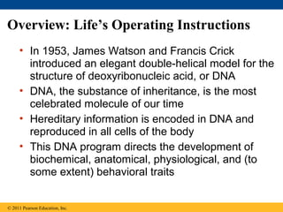
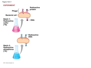
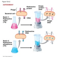
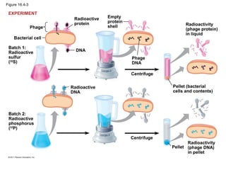
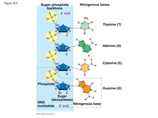
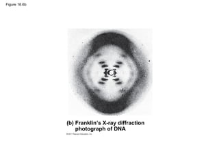
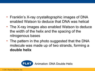
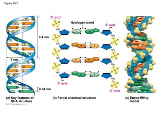
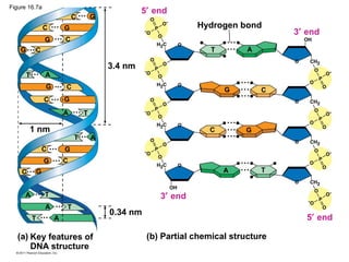
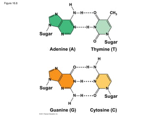
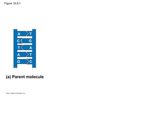
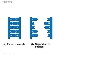
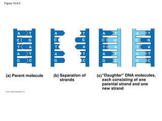
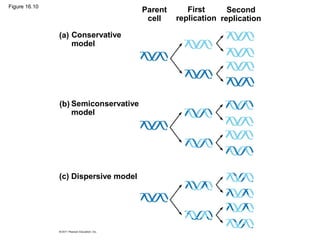
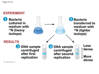
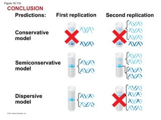
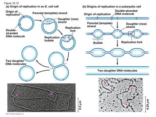
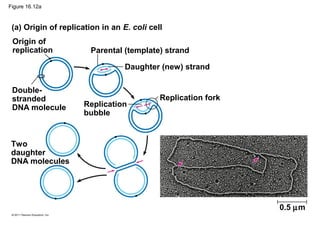
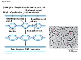
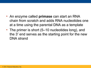
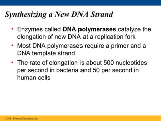
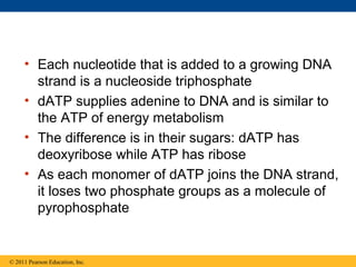
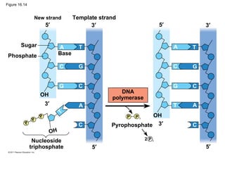
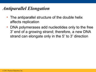
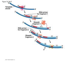
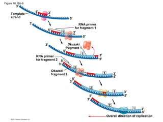
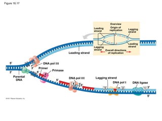
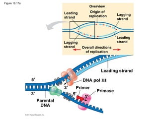
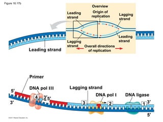
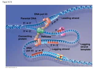
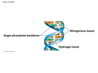
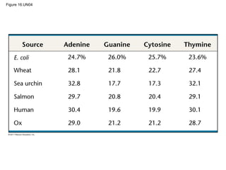
SlideShare a Scribd company logo
LECTURE PRESENTATIONS
For CAMPBELL BIOLOGY, NINTH EDITION
Jane B. Reece, Lisa A. Urry, Michael L. Cain, Steven A. Wasserman, Peter V. Minorsky, Robert B. Jackson
© 2011 Pearson Education, Inc.
Lectures by
Erin Barley
Kathleen Fitzpatrick
The Molecular Basis of
Inheritance
Chapter 16
Overview: Life’s Operating Instructions
• In 1953, James Watson and Francis Crick
introduced an elegant double-helical model for the
structure of deoxyribonucleic acid, or DNA
• DNA, the substance of inheritance, is the most
celebrated molecule of our time
• Hereditary information is encoded in DNA and
reproduced in all cells of the body
• This DNA program directs the development of
biochemical, anatomical, physiological, and (to
some extent) behavioral traits
© 2011 Pearson Education, Inc.
Figure 16.1
Concept 16.1: DNA is the genetic material
• Early in the 20th century, the identification of the
molecules of inheritance loomed as a major
challenge to biologists
© 2011 Pearson Education, Inc.
The Search for the Genetic Material:
Scientific Inquiry
• When T. H. Morgan’s group showed that genes
are located on chromosomes, the two components
of chromosomes—DNA and protein—became
candidates for the genetic material
• The key factor in determining the genetic material
was choosing appropriate experimental organisms
• The role of DNA in heredity was first discovered by
studying bacteria and the viruses that infect them
© 2011 Pearson Education, Inc.
Evidence That DNA Can Transform Bacteria
• The discovery of the genetic role of DNA began
with research by Frederick Griffith in 1928
• Griffith worked with two strains of a bacterium, one
pathogenic and one harmless
© 2011 Pearson Education, Inc.
• When he mixed heat-killed remains of the
pathogenic strain with living cells of the harmless
strain, some living cells became pathogenic
• He called this phenomenon transformation, now
defined as a change in genotype and phenotype
due to assimilation of foreign DNA
© 2011 Pearson Education, Inc.
Living S cells
(control)
Living R cells
(control)
Heat-killed
S cells
(control)
Mixture of
heat-killed
S cells and
living R cells
Mouse dies Mouse diesMouse healthy Mouse healthy
Living S cells
EXPERIMENT
RESULTS
Figure 16.2
• In 1944, Oswald Avery, Maclyn McCarty, and
Colin MacLeod announced that the transforming
substance was DNA
• Their conclusion was based on experimental
evidence that only DNA worked in transforming
harmless bacteria into pathogenic bacteria
• Many biologists remained skeptical, mainly
because little was known about DNA
© 2011 Pearson Education, Inc.
Evidence That Viral DNA Can Program Cells
• More evidence for DNA as the genetic material
came from studies of viruses that infect bacteria
• Such viruses, called bacteriophages (or phages),
are widely used in molecular genetics research
© 2011 Pearson Education, Inc.
Animation: Phage T2 Reproductive Cycle
Figure 16.3
Phage
head
Tail
sheath
Tail fiber
DNA
Bacterial
cell
100nm
• In 1952, Alfred Hershey and Martha Chase
performed experiments showing that DNA is the
genetic material of a phage known as T2
• To determine this, they designed an experiment
showing that only one of the two components of
T2 (DNA or protein) enters an E. coli cell during
infection
• They concluded that the injected DNA of the
phage provides the genetic information
© 2011 Pearson Education, Inc.
Animation: Hershey-Chase Experiment
Figure 16.4-1
Bacterial cell
Phage
Batch 1:
Radioactive
sulfur
(35
S)
DNA
Batch 2:
Radioactive
phosphorus
(32
P)
Radioactive
DNA
EXPERIMENT
Radioactive
protein
Figure 16.4-2
Bacterial cell
Phage
Batch 1:
Radioactive
sulfur
(35
S)
Radioactive
protein
DNA
Batch 2:
Radioactive
phosphorus
(32
P)
Radioactive
DNA
Empty
protein
shell
Phage
DNA
EXPERIMENT
Figure 16.4-3
Bacterial cell
Phage
Batch 1:
Radioactive
sulfur
(35
S)
Radioactive
protein
DNA
Batch 2:
Radioactive
phosphorus
(32
P)
Radioactive
DNA
Empty
protein
shell
Phage
DNA
Centrifuge
Centrifuge
Radioactivity
(phage protein)
in liquid
Pellet (bacterial
cells and contents)
Pellet
Radioactivity
(phage DNA)
in pellet
EXPERIMENT
Additional Evidence That DNA Is the
Genetic Material
• It was known that DNA is a polymer of
nucleotides, each consisting of a nitrogenous
base, a sugar, and a phosphate group
• In 1950, Erwin Chargaff reported that DNA
composition varies from one species to the next
• This evidence of diversity made DNA a more
credible candidate for the genetic material
© 2011 Pearson Education, Inc.
Animation: DNA and RNA Structure
• Two findings became known as Chargaff’s rules
– The base composition of DNA varies between
species
– In any species the number of A and T bases are
equal and the number of G and C bases are equal
• The basis for these rules was not understood until
the discovery of the double helix
© 2011 Pearson Education, Inc.
Figure 16.5
Sugar–phosphate
backbone
Nitrogenous bases
Thymine (T)
Adenine (A)
Cytosine (C)
Guanine (G)
Nitrogenous base
Phosphate
DNA
nucleotide
Sugar
(deoxyribose)
3′ end
5′ end
Building a Structural Model of DNA:
Scientific Inquiry
• After DNA was accepted as the genetic material,
the challenge was to determine how its structure
accounts for its role in heredity
• Maurice Wilkins and Rosalind Franklin were using
a technique called X-ray crystallography to study
molecular structure
• Franklin produced a picture of the DNA molecule
using this technique
© 2011 Pearson Education, Inc.
Figure 16.6
(a) Rosalind Franklin (b) Franklin’s X-ray diffraction
photograph of DNA
Figure 16.6a
(a) Rosalind Franklin
Figure 16.6b
(b) Franklin’s X-ray diffraction
photograph of DNA
• Franklin’s X-ray crystallographic images of DNA
enabled Watson to deduce that DNA was helical
• The X-ray images also enabled Watson to deduce
the width of the helix and the spacing of the
nitrogenous bases
• The pattern in the photo suggested that the DNA
molecule was made up of two strands, forming a
double helix
© 2011 Pearson Education, Inc.
Animation: DNA Double Helix
Figure 16.7
3.4 nm
1 nm
0.34 nm
Hydrogen bond
(a) Key features of
DNA structure
Space-filling
model
(c)(b) Partial chemical structure
3′ end
5′ end
3′ end
5′ end
T
T
A
A
G
G
C
C
C
C
C
C
C
C
C
C
C
G
G
G
G
G
G
G
G
G
T
T
T
T
T
T
A
A
A
A
A
A
3.4 nm
1 nm
0.34 nm
Hydrogen bond
(a) Key features of
DNA structure
(b) Partial chemical structure
3′ end
5′ end
3′ end
5′ end
T
T
A
A
G
G
C
C
C
C
C
C
C
C
C
C
C
G
G
G
G
G
G
G
G
G
T
T
T
T
T
T
A
A
A
A
A
A
Figure 16.7a
Figure 16.7b
(c) Space-filling model
• Watson and Crick built models of a double helix to
conform to the X-rays and chemistry of DNA
• Franklin had concluded that there were two outer
sugar-phosphate backbones, with the nitrogenous
bases paired in the molecule’s interior
• Watson built a model in which the backbones were
antiparallel (their subunits run in opposite
directions)
© 2011 Pearson Education, Inc.
• At first, Watson and Crick thought the bases paired
like with like (A with A, and so on), but such
pairings did not result in a uniform width
• Instead, pairing a purine with a pyrimidine resulted
in a uniform width consistent with the X-ray data
© 2011 Pearson Education, Inc.
Figure 16.UN01
Purine + purine: too wide
Pyrimidine + pyrimidine: too narrow
Purine + pyrimidine: width
consistent with X-ray data
• Watson and Crick reasoned that the pairing was
more specific, dictated by the base structures
• They determined that adenine (A) paired only with
thymine (T), and guanine (G) paired only with
cytosine (C)
• The Watson-Crick model explains Chargaff’s
rules: in any organism the amount of A = T, and
the amount of G = C
© 2011 Pearson Education, Inc.
Figure 16.8
Sugar
Sugar
Sugar
Sugar
Adenine (A) Thymine (T)
Guanine (G) Cytosine (C)
Concept 16.2: Many proteins work
together in DNA replication and repair
• The relationship between structure and function is
manifest in the double helix
• Watson and Crick noted that the specific base
pairing suggested a possible copying mechanism
for genetic material
© 2011 Pearson Education, Inc.
The Basic Principle: Base Pairing to a
Template Strand
• Since the two strands of DNA are complementary,
each strand acts as a template for building a new
strand in replication
• In DNA replication, the parent molecule unwinds,
and two new daughter strands are built based on
base-pairing rules
© 2011 Pearson Education, Inc.
Animation: DNA Replication Overview
Figure 16.9-1
(a) Parent molecule
A
A
A
T
T
T
C
C
G
G
Figure 16.9-2
(a) Parent molecule (b) Separation of
strands
A
A
A
A
A
A
T
T
T
T
T
T
C
C
C
C
G
G
G
G
Figure 16.9-3
(a) Parent molecule (b) Separation of
strands
(c)“Daughter” DNA molecules,
each consisting of one
parental strand and one
new strand
A
A
A
A
A
A
A
A
A
A
A
A
T
T
T
T
T
T
T
T
T
T
T
T
C
C
C
C
C
C
C
C
G
G
G
G
G
G
G
G
• Watson and Crick’s semiconservative model of
replication predicts that when a double helix
replicates, each daughter molecule will have one
old strand (derived or “conserved” from the parent
molecule) and one newly made strand
• Competing models were the conservative model
(the two parent strands rejoin) and the dispersive
model (each strand is a mix of old and new)
© 2011 Pearson Education, Inc.
Figure 16.10
(a) Conservative
model
(b) Semiconservative
model
(c) Dispersive model
Parent
cell
First
replication
Second
replication
• Experiments by Matthew Meselson and Franklin
Stahl supported the semiconservative model
• They labeled the nucleotides of the old strands
with a heavy isotope of nitrogen, while any new
nucleotides were labeled with a lighter isotope
© 2011 Pearson Education, Inc.
• The first replication produced a band of hybrid
DNA, eliminating the conservative model
• A second replication produced both light and
hybrid DNA, eliminating the dispersive model and
supporting the semiconservative model
© 2011 Pearson Education, Inc.
Figure 16.11
Bacteria
cultured in
medium with
15
N (heavy
isotope)
Bacteria
transferred to
medium with
14
N (lighter
isotope)
DNA sample
centrifuged
after first
replication
DNA sample
centrifuged
after second
replication
Less
dense
More
dense
Predictions: First replication Second replication
Conservative
model
Semiconservative
model
Dispersive
model
21
3 4
EXPERIMENT
RESULTS
CONCLUSION
Figure 16.11a
Bacteria
cultured in
medium with
15
N (heavy
isotope)
Bacteria
transferred to
medium with
14
N (lighter
isotope)
DNA sample
centrifuged
after first
replication
DNA sample
centrifuged
after second
replication
Less
dense
More
dense
21
3 4
EXPERIMENT
RESULTS
Figure 16.11b
Predictions: First replication Second replication
Conservative
model
Semiconservative
model
Dispersive
model
CONCLUSION
DNA Replication: A Closer Look
• The copying of DNA is remarkable in its speed
and accuracy
• More than a dozen enzymes and other proteins
participate in DNA replication
© 2011 Pearson Education, Inc.
Getting Started
• Replication begins at particular sites called
origins of replication, where the two DNA
strands are separated, opening up a replication
“bubble”
• A eukaryotic chromosome may have hundreds or
even thousands of origins of replication
• Replication proceeds in both directions from each
origin, until the entire molecule is copied
© 2011 Pearson Education, Inc.
Animation: Origins of Replication
Figure 16.12
(a) Origin of replication in an E. coli cell (b) Origins of replication in a eukaryotic cell
Origin of
replication
Parental (template) strand
Double-
stranded
DNA molecule
Daughter (new)
strand
Replication
fork
Replication
bubble
Two daughter
DNA molecules
Origin of replication
Double-stranded
DNA molecule
Parental (template)
strand
Daughter (new)
strand
Bubble Replication fork
Two daughter DNA molecules
0.5µm
0.25µm
Figure 16.12a
(a) Origin of replication in an E. coli cell
Origin of
replication Parental (template) strand
Double-
stranded
DNA molecule
Daughter (new) strand
Replication fork
Replication
bubble
Two
daughter
DNA molecules
0.5 µm
Figure 16.12b
(b) Origins of replication in a eukaryotic cell
Origin of replication
Double-stranded
DNA molecule
Parental (template)
strand
Daughter (new)
strand
Bubble Replication fork
Two daughter DNA molecules
0.25 µm
Figure 16.12c
0.5µm
Figure 16.12d
0.25µm
• At the end of each replication bubble is a
replication fork, a Y-shaped region where new
DNA strands are elongating
• Helicases are enzymes that untwist the double
helix at the replication forks
• Single-strand binding proteins bind to and
stabilize single-stranded DNA
• Topoisomerase corrects “overwinding” ahead of
replication forks by breaking, swiveling, and
rejoining DNA strands
© 2011 Pearson Education, Inc.
Figure 16.13
Topoisomerase
Primase
RNA
primer
Helicase
Single-strand binding
proteins
5′
3′
5′
5′3′
3′
• DNA polymerases cannot initiate synthesis of a
polynucleotide; they can only add nucleotides to
the 3′ end
• The initial nucleotide strand is a short RNA
primer
© 2011 Pearson Education, Inc.
• An enzyme called primase can start an RNA
chain from scratch and adds RNA nucleotides one
at a time using the parental DNA as a template
• The primer is short (5–10 nucleotides long), and
the 3′ end serves as the starting point for the new
DNA strand
© 2011 Pearson Education, Inc.
Synthesizing a New DNA Strand
• Enzymes called DNA polymerases catalyze the
elongation of new DNA at a replication fork
• Most DNA polymerases require a primer and a
DNA template strand
• The rate of elongation is about 500 nucleotides
per second in bacteria and 50 per second in
human cells
© 2011 Pearson Education, Inc.
• Each nucleotide that is added to a growing DNA
strand is a nucleoside triphosphate
• dATP supplies adenine to DNA and is similar to
the ATP of energy metabolism
• The difference is in their sugars: dATP has
deoxyribose while ATP has ribose
• As each monomer of dATP joins the DNA strand,
it loses two phosphate groups as a molecule of
pyrophosphate
© 2011 Pearson Education, Inc.
Figure 16.14
New strand Template strand
Sugar
Phosphate Base
Nucleoside
triphosphate
DNA
polymerase
Pyrophosphate
5′
5′
5′
5′
3′
3′
3′
3′
OH
OH
OH
P P i
2 P i
P
P
P
A
A
A
A
T T
T
T
C
C
C
C
C
C
G
G
G
G
Antiparallel Elongation
• The antiparallel structure of the double helix
affects replication
• DNA polymerases add nucleotides only to the free
3′ end of a growing strand; therefore, a new DNA
strand can elongate only in the 5′ to 3′ direction
© 2011 Pearson Education, Inc.
• Along one template strand of DNA, the DNA
polymerase synthesizes a leading strand
continuously, moving toward the replication fork
© 2011 Pearson Education, Inc.
Animation: Leading Strand
Figure 16.15
Leading
strand
Lagging
strand
Overview
Origin of replication Lagging
strand
Leading
strand
Primer
Overall directions
of replication
Origin of
replication
RNA primer
Sliding clamp
DNA pol III
Parental DNA
3′
5′
5′
3′
3′
5′
3′
5′
3′
5′
3′
5′
Figure 16.15a
Leading
strand
Lagging
strand
Overview
Origin of replication Lagging
strand
Leading
strand
Primer
Overall directions
of replication
Origin of
replication
RNA primer
Sliding clamp
DNA pol III
Parental DNA
3′
5′
5′
3′
3′
5′
3′
5′
3′
5′
3′
5′
Figure 16.15b
• To elongate the other new strand, called the
lagging strand, DNA polymerase must work in the
direction away from the replication fork
• The lagging strand is synthesized as a series of
segments called Okazaki fragments, which are
joined together by DNA ligase
© 2011 Pearson Education, Inc.
Animation: Lagging Strand
Origin of replication
Overview
Leading
strand
Leading
strand
Lagging
strand
Lagging strand
Overall directions
of replication
Template
strand
RNA primer
for fragment 1
Okazaki
fragment 1
RNA primer
for fragment 2
Okazaki
fragment 2
Overall direction of replication
3′
3′
3′
3′
3′
3′
3′
3′
3′
3′
3′
3′
5′
5′
5′
5′
5′
5′
5′
5′5′
5′
5′
5′
2
2
2
1
1
1
1
1
2
1
Figure 16.16
Figure 16.16a
Origin of replication
Overview
Leading
strand
Leading
strand
Lagging
strand
Lagging strand
Overall directions
of replication
1
2
Figure 16.16b-1
Template
strand
3′
3′5′
5′
Figure 16.16b-2
Template
strand
RNA primer
for fragment 1
3′
3′
3′
3′
5′
5′
5′
5′
1
Figure 16.16b-3
Template
strand
RNA primer
for fragment 1
Okazaki
fragment 1
3′
3′
3′
3′
3′
3′
5′
5′
5′
5′
5′
5′
1
1
Figure 16.16b-4
Template
strand
RNA primer
for fragment 1
Okazaki
fragment 1
RNA primer
for fragment 2
Okazaki
fragment 2
3′
3′
3′
3′
3′
3′
3′
3′
5′
5′
5′
5′
5′
5′
5′
5′
2
1
1
1
Figure 16.16b-5
Template
strand
RNA primer
for fragment 1
Okazaki
fragment 1
RNA primer
for fragment 2
Okazaki
fragment 2
3′
3′
3′
3′
3′
3′
3′
3′
3′
3′
3′
5′
5′
5′
5′
5′
5′
5′
5′
5′
5′5′
2
2
1
1
1
1
Figure 16.16b-6
Template
strand
RNA primer
for fragment 1
Okazaki
fragment 1
RNA primer
for fragment 2
Okazaki
fragment 2
Overall direction of replication
3′
3′
3′
3′
3′
3′
3′
3′
3′
3′
3′
3′
5′
5′
5′
5′
5′
5′
5′
5′
5′
5′5′
5′
2
2
2
1
1
1
1
1
Figure 16.17
Overview
Leading
strand
Origin of
replication Lagging
strand
Leading
strandLagging
strand Overall directions
of replicationLeading strand
DNA pol III
DNA pol III Lagging strand
DNA pol I DNA ligase
Primer
Primase
Parental
DNA
5′
5′
5′
5′
5′
3′
3′
3′
3′
3′3 2 1
4
Figure 16.17a
Overview
Leading
strand
Origin of
replication Lagging
strand
Leading
strandLagging
strand Overall directions
of replication
Leading strand
DNA pol III
Primer
Primase
Parental
DNA
5′
5′3′
3′
3′
Overview
Leading
strand
Origin of
replication Lagging
strand
Leading
strandLagging
strand Overall directions
of replicationLeading strand
Primer
DNA pol III
DNA pol I
Lagging strand
DNA ligase
5′
5′
5′
3′
3′
3′ 3
4
2 1
Figure 16.17b
The DNA Replication Complex
• The proteins that participate in DNA replication
form a large complex, a “DNA replication machine”
• The DNA replication machine may be stationary
during the replication process
• Recent studies support a model in which DNA
polymerase molecules “reel in” parental DNA and
“extrude” newly made daughter DNA molecules
© 2011 Pearson Education, Inc.
Animation: DNA Replication Review
Figure 16.18
Parental DNA
DNA pol III
Leading strand
Connecting
protein
Helicase
Lagging strandDNA
pol III
Lagging
strand
template
5′
5′
5′
5′
5′
5′
3′ 3′
3′
3′
3′
3′
Proofreading and Repairing DNA
• DNA polymerases proofread newly made DNA,
replacing any incorrect nucleotides
• In mismatch repair of DNA, repair enzymes
correct errors in base pairing
• DNA can be damaged by exposure to harmful
chemical or physical agents such as cigarette
smoke and X-rays; it can also undergo
spontaneous changes
• In nucleotide excision repair, a nuclease cuts
out and replaces damaged stretches of DNA
© 2011 Pearson Education, Inc.
Figure 16.19
Nuclease
DNA
polymerase
DNA
ligase
5′
5′
5′
5′
5′
5′
5′
5′
3′
3′
3′
3′
3′
3′
3′
3′
Evolutionary Significance of Altered DNA
Nucleotides
• Error rate after proofreading repair is low but not
zero
• Sequence changes may become permanent and
can be passed on to the next generation
• These changes (mutations) are the source of the
genetic variation upon which natural selection
operates
© 2011 Pearson Education, Inc.
Replicating the Ends of DNA Molecules
• Limitations of DNA polymerase create problems
for the linear DNA of eukaryotic chromosomes
• The usual replication machinery provides no way
to complete the 5′ ends, so repeated rounds of
replication produce shorter DNA molecules with
uneven ends
• This is not a problem for prokaryotes, most of
which have circular chromosomes
© 2011 Pearson Education, Inc.
Figure 16.20
Ends of parental
DNA strands
Leading strand
Lagging strand
Last fragment Next-to-last fragment
Lagging strand RNA primer
Parental strand
Removal of primers and
replacement with DNA
where a 3′ end is available
Second round
of replication
Further rounds
of replication
New leading strand
New lagging strand
Shorter and shorter daughter molecules
3′
3′
3′
3′
3′
5′
5′
5′
5′
5′
Figure 16.20a
Ends of parental
DNA strands
Leading strand
Lagging strand
Last fragment Next-to-last fragment
Lagging strand RNA primer
Parental strand
Removal of primers and
replacement with DNA
where a 3′ end is available
3′
3′
3′
5′
5′
5′
Figure 16.20b
Second round
of replication
Further rounds
of replication
New leading strand
New lagging strand
Shorter and shorter daughter molecules
3′
3′
3′
5′
5′
5′
• Eukaryotic chromosomal DNA molecules have
special nucleotide sequences at their ends called
telomeres
• Telomeres do not prevent the shortening of DNA
molecules, but they do postpone the erosion of
genes near the ends of DNA molecules
• It has been proposed that the shortening of
telomeres is connected to aging
© 2011 Pearson Education, Inc.
Figure 16.21
1 µm
• If chromosomes of germ cells became shorter in
every cell cycle, essential genes would eventually
be missing from the gametes they produce
• An enzyme called telomerase catalyzes the
lengthening of telomeres in germ cells
© 2011 Pearson Education, Inc.
• The shortening of telomeres might protect cells
from cancerous growth by limiting the number of
cell divisions
• There is evidence of telomerase activity in cancer
cells, which may allow cancer cells to persist
© 2011 Pearson Education, Inc.
Concept 16.3 A chromosome consists of a
DNA molecule packed together with proteins
• The bacterial chromosome is a double-stranded,
circular DNA molecule associated with a small
amount of protein
• Eukaryotic chromosomes have linear DNA
molecules associated with a large amount of
protein
• In a bacterium, the DNA is “supercoiled” and found
in a region of the cell called the nucleoid
© 2011 Pearson Education, Inc.
• Chromatin, a complex of DNA and protein,
is found in the nucleus of eukaryotic cells
• Chromosomes fit into the nucleus through
an elaborate, multilevel system of packing
© 2011 Pearson Education, Inc.
Animation: DNA Packing
Figure 16.22a
DNA double helix
(2 nm in diameter)
DNA, the double helix
Nucleosome
(10 nm in diameter)
Histones
Histones
Histone
tail
H1
Nucleosomes, or “beads on
a string” (10-nm fiber)
Figure 16.22b
30-nm fiber
30-nm fiber
Loops Scaffold
300-nm fiber
Chromatid
(700 nm)
Replicated
chromosome
(1,400 nm)
Looped domains
(300-nm fiber) Metaphase
chromosome
Figure 16.22c
DNA double helix (2 nm in diameter)
Figure 16.22d
Nucleosome (10 nm in diameter)
Figure 16.22e
30-nm fiber
Figure 16.22f
Loops Scaffold
Figure 16.22g
Chromatid
(700 nm)
• Chromatin undergoes changes in packing during
the cell cycle
• At interphase, some chromatin is organized into a
10-nm fiber, but much is compacted into a 30-nm
fiber, through folding and looping
• Though interphase chromosomes are not highly
condensed, they still occupy specific restricted
regions in the nucleus
© 2011 Pearson Education, Inc.
Figure 16.23
5µm
Figure 16.23a
Figure 16.23b
Figure 16.23c
5µm
• Most chromatin is loosely packed in the nucleus
during interphase and condenses prior to mitosis
• Loosely packed chromatin is called euchromatin
• During interphase a few regions of chromatin
(centromeres and telomeres) are highly
condensed into heterochromatin
• Dense packing of the heterochromatin makes it
difficult for the cell to express genetic information
coded in these regions
© 2011 Pearson Education, Inc.
• Histones can undergo chemical modifications that
result in changes in chromatin organization
© 2011 Pearson Education, Inc.
Figure 16.UN02
Sugar-phosphate backbone
Nitrogenous bases
Hydrogen bond
G
G
G
G
C
C
C
C
A
A
A
A
T
T
T
T
Figure 16.UN03
DNA pol III synthesizes
leading strand continuously
Parental
DNA DNA pol III starts DNA
synthesis at 3′ end of primer,
continues in 5′ → 3′ direction
Origin of
replication
Helicase
Primase synthesizes
a short RNA primer
DNA pol I replaces the RNA
primer with DNA nucleotides
3′
3′
3′
5′
5′
5′
5′
Lagging strand synthesized
in short Okazaki fragments,
later joined by DNA ligase
Figure 16.UN04
Figure 16.UN05
Figure 16.UN06
Figure 16.UN07

More Related Content

What's hot

13 meiosis and sexual life cycles
13 meiosis and sexual life cycles13 meiosis and sexual life cycles
13 meiosis and sexual life cycleskindarspirit
 
21 genomes and their evolution
21 genomes and their evolution21 genomes and their evolution
21 genomes and their evolutionkindarspirit
 
14 mendel and the gene idea
14 mendel and the gene idea14 mendel and the gene idea
14 mendel and the gene ideakindarspirit
 
Ch 9: Cell Respiration and Fermentation
Ch 9: Cell Respiration and FermentationCh 9: Cell Respiration and Fermentation
Ch 9: Cell Respiration and Fermentation
veneethmathew
 
Ch 13: Meiosis and Sexual Life Cycles
Ch 13: Meiosis and Sexual Life CyclesCh 13: Meiosis and Sexual Life Cycles
Ch 13: Meiosis and Sexual Life Cycles
veneethmathew
 
Ch 5: The Structure and Function of Large Biological Molecules
Ch 5: The Structure and Function of Large Biological MoleculesCh 5: The Structure and Function of Large Biological Molecules
Ch 5: The Structure and Function of Large Biological Molecules
veneethmathew
 
Ch 6: A Tour of the Cell
Ch 6: A Tour of the CellCh 6: A Tour of the Cell
Ch 6: A Tour of the Cell
veneethmathew
 
Biology in Focus - Chapter 14
Biology in Focus - Chapter 14Biology in Focus - Chapter 14
Biology in Focus - Chapter 14
mpattani
 
Ch 10: Photosynthesis
Ch 10: PhotosynthesisCh 10: Photosynthesis
Ch 10: Photosynthesis
veneethmathew
 
24 lecture origin_of_species
24 lecture origin_of_species24 lecture origin_of_species
24 lecture origin_of_speciesveneethmathew
 
Ch 4: Carbon and Diversity
Ch 4: Carbon and DiversityCh 4: Carbon and Diversity
Ch 4: Carbon and Diversity
veneethmathew
 
22 lecture descent_with_modification
22 lecture descent_with_modification22 lecture descent_with_modification
22 lecture descent_with_modificationveneethmathew
 
Ch 7: Membrane Structure and Function
Ch 7: Membrane Structure and Function Ch 7: Membrane Structure and Function
Ch 7: Membrane Structure and Function
veneethmathew
 
17 - From Gene to Protein
17 - From Gene to Protein17 - From Gene to Protein
17 - From Gene to Proteinkindarspirit
 
23 lecture evolution_populations
23 lecture evolution_populations23 lecture evolution_populations
23 lecture evolution_populationsveneethmathew
 
Ch. 51: Animal Behavior
Ch. 51: Animal BehaviorCh. 51: Animal Behavior
Ch. 51: Animal Behavior
AndreDominic1
 
23 the evolution of populations
23 the evolution of populations23 the evolution of populations
23 the evolution of populationskindarspirit
 

What's hot (20)

13 meiosis and sexual life cycles
13 meiosis and sexual life cycles13 meiosis and sexual life cycles
13 meiosis and sexual life cycles
 
21 genomes and their evolution
21 genomes and their evolution21 genomes and their evolution
21 genomes and their evolution
 
19 - Viruses
19 - Viruses19 - Viruses
19 - Viruses
 
14 mendel and the gene idea
14 mendel and the gene idea14 mendel and the gene idea
14 mendel and the gene idea
 
20 lecture biotech
20 lecture biotech20 lecture biotech
20 lecture biotech
 
Ch 9: Cell Respiration and Fermentation
Ch 9: Cell Respiration and FermentationCh 9: Cell Respiration and Fermentation
Ch 9: Cell Respiration and Fermentation
 
Ch 13: Meiosis and Sexual Life Cycles
Ch 13: Meiosis and Sexual Life CyclesCh 13: Meiosis and Sexual Life Cycles
Ch 13: Meiosis and Sexual Life Cycles
 
Ch 5: The Structure and Function of Large Biological Molecules
Ch 5: The Structure and Function of Large Biological MoleculesCh 5: The Structure and Function of Large Biological Molecules
Ch 5: The Structure and Function of Large Biological Molecules
 
Ch 6: A Tour of the Cell
Ch 6: A Tour of the CellCh 6: A Tour of the Cell
Ch 6: A Tour of the Cell
 
Biology in Focus - Chapter 14
Biology in Focus - Chapter 14Biology in Focus - Chapter 14
Biology in Focus - Chapter 14
 
Ch 10: Photosynthesis
Ch 10: PhotosynthesisCh 10: Photosynthesis
Ch 10: Photosynthesis
 
24 lecture origin_of_species
24 lecture origin_of_species24 lecture origin_of_species
24 lecture origin_of_species
 
Ch 4: Carbon and Diversity
Ch 4: Carbon and DiversityCh 4: Carbon and Diversity
Ch 4: Carbon and Diversity
 
22 lecture descent_with_modification
22 lecture descent_with_modification22 lecture descent_with_modification
22 lecture descent_with_modification
 
Ch 7: Membrane Structure and Function
Ch 7: Membrane Structure and Function Ch 7: Membrane Structure and Function
Ch 7: Membrane Structure and Function
 
17 - From Gene to Protein
17 - From Gene to Protein17 - From Gene to Protein
17 - From Gene to Protein
 
23 lecture evolution_populations
23 lecture evolution_populations23 lecture evolution_populations
23 lecture evolution_populations
 
Ch. 51: Animal Behavior
Ch. 51: Animal BehaviorCh. 51: Animal Behavior
Ch. 51: Animal Behavior
 
23 the evolution of populations
23 the evolution of populations23 the evolution of populations
23 the evolution of populations
 
12 the cell cycle
12 the cell cycle12 the cell cycle
12 the cell cycle
 

Similar to Ch 16: The Molecular Basis of Inheritance

Molecular biology for post graduates.ppt
Molecular biology for post graduates.pptMolecular biology for post graduates.ppt
Molecular biology for post graduates.ppt
AhmedKasem39
 
16themolecularbasisofinheritance 130311053514-phpapp02
16themolecularbasisofinheritance 130311053514-phpapp0216themolecularbasisofinheritance 130311053514-phpapp02
16themolecularbasisofinheritance 130311053514-phpapp02
Cleophas Rwemera
 
L5 DNA structure and replication.pptx
L5 DNA structure and replication.pptxL5 DNA structure and replication.pptx
L5 DNA structure and replication.pptx
Abdulkarim803288
 
The molecular basis of inheritance
The molecular basis of inheritanceThe molecular basis of inheritance
The molecular basis of inheritance
tanzeem khan
 
Chapter 16 molecular inheritance
Chapter 16 molecular inheritanceChapter 16 molecular inheritance
Chapter 16 molecular inheritanceHeather Fogell
 
Ap Chapter 16
Ap Chapter 16Ap Chapter 16
Ap Chapter 16
smithbio
 
16 molecularinheritance text
16 molecularinheritance text16 molecularinheritance text
16 molecularinheritance text
Rv26121972
 
Chapter16 inheritance-151125150159-lva1-app6891 (1)
Chapter16 inheritance-151125150159-lva1-app6891 (1)Chapter16 inheritance-151125150159-lva1-app6891 (1)
Chapter16 inheritance-151125150159-lva1-app6891 (1)
Cleophas Rwemera
 
Chapter 16: Molecular Basis of Inheritance
Chapter 16: Molecular Basis of InheritanceChapter 16: Molecular Basis of Inheritance
Chapter 16: Molecular Basis of Inheritance
Angel Vega
 
Dna and replication
Dna and replication Dna and replication
Dna and replication
university of karachi
 
Principles of microbial genetics
Principles of microbial geneticsPrinciples of microbial genetics
Principles of microbial genetics
Rinaldo John
 
A complete PPT on DNA
A complete PPT on DNA A complete PPT on DNA
A complete PPT on DNA
University of Mumbai
 
Dna History And Replication Review
Dna History And Replication ReviewDna History And Replication Review
Dna History And Replication ReviewCrystal Wood
 
Chapter 16
Chapter 16Chapter 16
Chapter 16ktanaka2
 
Chapter 16
Chapter 16Chapter 16
Chapter 16ktanaka2
 
DNA Structure.ppt
DNA Structure.pptDNA Structure.ppt
DNA Structure.ppt
EsterDad
 
13lecturepresentation 151216140648
13lecturepresentation 15121614064813lecturepresentation 151216140648
13lecturepresentation 151216140648
Cleophas Rwemera
 

Similar to Ch 16: The Molecular Basis of Inheritance (20)

Molecular biology for post graduates.ppt
Molecular biology for post graduates.pptMolecular biology for post graduates.ppt
Molecular biology for post graduates.ppt
 
16themolecularbasisofinheritance 130311053514-phpapp02
16themolecularbasisofinheritance 130311053514-phpapp0216themolecularbasisofinheritance 130311053514-phpapp02
16themolecularbasisofinheritance 130311053514-phpapp02
 
L5 DNA structure and replication.pptx
L5 DNA structure and replication.pptxL5 DNA structure and replication.pptx
L5 DNA structure and replication.pptx
 
The molecular basis of inheritance
The molecular basis of inheritanceThe molecular basis of inheritance
The molecular basis of inheritance
 
Chapter 16 molecular inheritance
Chapter 16 molecular inheritanceChapter 16 molecular inheritance
Chapter 16 molecular inheritance
 
Ap Chapter 16
Ap Chapter 16Ap Chapter 16
Ap Chapter 16
 
16 molecularinheritance text
16 molecularinheritance text16 molecularinheritance text
16 molecularinheritance text
 
Dn ato protein
Dn ato proteinDn ato protein
Dn ato protein
 
Chapter16 inheritance-151125150159-lva1-app6891 (1)
Chapter16 inheritance-151125150159-lva1-app6891 (1)Chapter16 inheritance-151125150159-lva1-app6891 (1)
Chapter16 inheritance-151125150159-lva1-app6891 (1)
 
Chapter 16: Molecular Basis of Inheritance
Chapter 16: Molecular Basis of InheritanceChapter 16: Molecular Basis of Inheritance
Chapter 16: Molecular Basis of Inheritance
 
Dna and replication
Dna and replication Dna and replication
Dna and replication
 
Principles of microbial genetics
Principles of microbial geneticsPrinciples of microbial genetics
Principles of microbial genetics
 
A complete PPT on DNA
A complete PPT on DNA A complete PPT on DNA
A complete PPT on DNA
 
Dna History And Replication Review
Dna History And Replication ReviewDna History And Replication Review
Dna History And Replication Review
 
4.1 DNA structure
4.1 DNA structure4.1 DNA structure
4.1 DNA structure
 
4.1 DNA Structure
4.1 DNA Structure4.1 DNA Structure
4.1 DNA Structure
 
Chapter 16
Chapter 16Chapter 16
Chapter 16
 
Chapter 16
Chapter 16Chapter 16
Chapter 16
 
DNA Structure.ppt
DNA Structure.pptDNA Structure.ppt
DNA Structure.ppt
 
13lecturepresentation 151216140648
13lecturepresentation 15121614064813lecturepresentation 151216140648
13lecturepresentation 151216140648
 

More from veneethmathew

26 lecture phylogeny
26 lecture phylogeny26 lecture phylogeny
26 lecture phylogenyveneethmathew
 
25 lecture history_of_life
25 lecture history_of_life25 lecture history_of_life
25 lecture history_of_lifeveneethmathew
 
21 lecture genome_and_evolution
21 lecture genome_and_evolution21 lecture genome_and_evolution
21 lecture genome_and_evolutionveneethmathew
 
Ch 3: Water and Life
Ch 3: Water and LifeCh 3: Water and Life
Ch 3: Water and Life
veneethmathew
 
Ch 2: The Chemical Context of Life
Ch 2: The Chemical Context of LifeCh 2: The Chemical Context of Life
Ch 2: The Chemical Context of Life
veneethmathew
 
Ch 1: Themes in the Study of Life
Ch 1: Themes in the Study of LifeCh 1: Themes in the Study of Life
Ch 1: Themes in the Study of Life
veneethmathew
 

More from veneethmathew (8)

27 lecture bacteria
27 lecture bacteria27 lecture bacteria
27 lecture bacteria
 
26 lecture phylogeny
26 lecture phylogeny26 lecture phylogeny
26 lecture phylogeny
 
25 lecture history_of_life
25 lecture history_of_life25 lecture history_of_life
25 lecture history_of_life
 
21 lecture genome_and_evolution
21 lecture genome_and_evolution21 lecture genome_and_evolution
21 lecture genome_and_evolution
 
19 lecture viruses
19 lecture viruses19 lecture viruses
19 lecture viruses
 
Ch 3: Water and Life
Ch 3: Water and LifeCh 3: Water and Life
Ch 3: Water and Life
 
Ch 2: The Chemical Context of Life
Ch 2: The Chemical Context of LifeCh 2: The Chemical Context of Life
Ch 2: The Chemical Context of Life
 
Ch 1: Themes in the Study of Life
Ch 1: Themes in the Study of LifeCh 1: Themes in the Study of Life
Ch 1: Themes in the Study of Life
 

Ch 16: The Molecular Basis of Inheritance

  • 1. LECTURE PRESENTATIONS For CAMPBELL BIOLOGY, NINTH EDITION Jane B. Reece, Lisa A. Urry, Michael L. Cain, Steven A. Wasserman, Peter V. Minorsky, Robert B. Jackson © 2011 Pearson Education, Inc. Lectures by Erin Barley Kathleen Fitzpatrick The Molecular Basis of Inheritance Chapter 16
  • 2. Overview: Life’s Operating Instructions • In 1953, James Watson and Francis Crick introduced an elegant double-helical model for the structure of deoxyribonucleic acid, or DNA • DNA, the substance of inheritance, is the most celebrated molecule of our time • Hereditary information is encoded in DNA and reproduced in all cells of the body • This DNA program directs the development of biochemical, anatomical, physiological, and (to some extent) behavioral traits © 2011 Pearson Education, Inc.
  • 4. Concept 16.1: DNA is the genetic material • Early in the 20th century, the identification of the molecules of inheritance loomed as a major challenge to biologists © 2011 Pearson Education, Inc.
  • 5. The Search for the Genetic Material: Scientific Inquiry • When T. H. Morgan’s group showed that genes are located on chromosomes, the two components of chromosomes—DNA and protein—became candidates for the genetic material • The key factor in determining the genetic material was choosing appropriate experimental organisms • The role of DNA in heredity was first discovered by studying bacteria and the viruses that infect them © 2011 Pearson Education, Inc.
  • 6. Evidence That DNA Can Transform Bacteria • The discovery of the genetic role of DNA began with research by Frederick Griffith in 1928 • Griffith worked with two strains of a bacterium, one pathogenic and one harmless © 2011 Pearson Education, Inc.
  • 7. • When he mixed heat-killed remains of the pathogenic strain with living cells of the harmless strain, some living cells became pathogenic • He called this phenomenon transformation, now defined as a change in genotype and phenotype due to assimilation of foreign DNA © 2011 Pearson Education, Inc.
  • 8. Living S cells (control) Living R cells (control) Heat-killed S cells (control) Mixture of heat-killed S cells and living R cells Mouse dies Mouse diesMouse healthy Mouse healthy Living S cells EXPERIMENT RESULTS Figure 16.2
  • 9. • In 1944, Oswald Avery, Maclyn McCarty, and Colin MacLeod announced that the transforming substance was DNA • Their conclusion was based on experimental evidence that only DNA worked in transforming harmless bacteria into pathogenic bacteria • Many biologists remained skeptical, mainly because little was known about DNA © 2011 Pearson Education, Inc.
  • 10. Evidence That Viral DNA Can Program Cells • More evidence for DNA as the genetic material came from studies of viruses that infect bacteria • Such viruses, called bacteriophages (or phages), are widely used in molecular genetics research © 2011 Pearson Education, Inc. Animation: Phage T2 Reproductive Cycle
  • 12. • In 1952, Alfred Hershey and Martha Chase performed experiments showing that DNA is the genetic material of a phage known as T2 • To determine this, they designed an experiment showing that only one of the two components of T2 (DNA or protein) enters an E. coli cell during infection • They concluded that the injected DNA of the phage provides the genetic information © 2011 Pearson Education, Inc. Animation: Hershey-Chase Experiment
  • 13. Figure 16.4-1 Bacterial cell Phage Batch 1: Radioactive sulfur (35 S) DNA Batch 2: Radioactive phosphorus (32 P) Radioactive DNA EXPERIMENT Radioactive protein
  • 14. Figure 16.4-2 Bacterial cell Phage Batch 1: Radioactive sulfur (35 S) Radioactive protein DNA Batch 2: Radioactive phosphorus (32 P) Radioactive DNA Empty protein shell Phage DNA EXPERIMENT
  • 15. Figure 16.4-3 Bacterial cell Phage Batch 1: Radioactive sulfur (35 S) Radioactive protein DNA Batch 2: Radioactive phosphorus (32 P) Radioactive DNA Empty protein shell Phage DNA Centrifuge Centrifuge Radioactivity (phage protein) in liquid Pellet (bacterial cells and contents) Pellet Radioactivity (phage DNA) in pellet EXPERIMENT
  • 16. Additional Evidence That DNA Is the Genetic Material • It was known that DNA is a polymer of nucleotides, each consisting of a nitrogenous base, a sugar, and a phosphate group • In 1950, Erwin Chargaff reported that DNA composition varies from one species to the next • This evidence of diversity made DNA a more credible candidate for the genetic material © 2011 Pearson Education, Inc. Animation: DNA and RNA Structure
  • 17. • Two findings became known as Chargaff’s rules – The base composition of DNA varies between species – In any species the number of A and T bases are equal and the number of G and C bases are equal • The basis for these rules was not understood until the discovery of the double helix © 2011 Pearson Education, Inc.
  • 18. Figure 16.5 Sugar–phosphate backbone Nitrogenous bases Thymine (T) Adenine (A) Cytosine (C) Guanine (G) Nitrogenous base Phosphate DNA nucleotide Sugar (deoxyribose) 3′ end 5′ end
  • 19. Building a Structural Model of DNA: Scientific Inquiry • After DNA was accepted as the genetic material, the challenge was to determine how its structure accounts for its role in heredity • Maurice Wilkins and Rosalind Franklin were using a technique called X-ray crystallography to study molecular structure • Franklin produced a picture of the DNA molecule using this technique © 2011 Pearson Education, Inc.
  • 20. Figure 16.6 (a) Rosalind Franklin (b) Franklin’s X-ray diffraction photograph of DNA
  • 22. Figure 16.6b (b) Franklin’s X-ray diffraction photograph of DNA
  • 23. • Franklin’s X-ray crystallographic images of DNA enabled Watson to deduce that DNA was helical • The X-ray images also enabled Watson to deduce the width of the helix and the spacing of the nitrogenous bases • The pattern in the photo suggested that the DNA molecule was made up of two strands, forming a double helix © 2011 Pearson Education, Inc. Animation: DNA Double Helix
  • 24. Figure 16.7 3.4 nm 1 nm 0.34 nm Hydrogen bond (a) Key features of DNA structure Space-filling model (c)(b) Partial chemical structure 3′ end 5′ end 3′ end 5′ end T T A A G G C C C C C C C C C C C G G G G G G G G G T T T T T T A A A A A A
  • 25. 3.4 nm 1 nm 0.34 nm Hydrogen bond (a) Key features of DNA structure (b) Partial chemical structure 3′ end 5′ end 3′ end 5′ end T T A A G G C C C C C C C C C C C G G G G G G G G G T T T T T T A A A A A A Figure 16.7a
  • 27. • Watson and Crick built models of a double helix to conform to the X-rays and chemistry of DNA • Franklin had concluded that there were two outer sugar-phosphate backbones, with the nitrogenous bases paired in the molecule’s interior • Watson built a model in which the backbones were antiparallel (their subunits run in opposite directions) © 2011 Pearson Education, Inc.
  • 28. • At first, Watson and Crick thought the bases paired like with like (A with A, and so on), but such pairings did not result in a uniform width • Instead, pairing a purine with a pyrimidine resulted in a uniform width consistent with the X-ray data © 2011 Pearson Education, Inc.
  • 29. Figure 16.UN01 Purine + purine: too wide Pyrimidine + pyrimidine: too narrow Purine + pyrimidine: width consistent with X-ray data
  • 30. • Watson and Crick reasoned that the pairing was more specific, dictated by the base structures • They determined that adenine (A) paired only with thymine (T), and guanine (G) paired only with cytosine (C) • The Watson-Crick model explains Chargaff’s rules: in any organism the amount of A = T, and the amount of G = C © 2011 Pearson Education, Inc.
  • 31. Figure 16.8 Sugar Sugar Sugar Sugar Adenine (A) Thymine (T) Guanine (G) Cytosine (C)
  • 32. Concept 16.2: Many proteins work together in DNA replication and repair • The relationship between structure and function is manifest in the double helix • Watson and Crick noted that the specific base pairing suggested a possible copying mechanism for genetic material © 2011 Pearson Education, Inc.
  • 33. The Basic Principle: Base Pairing to a Template Strand • Since the two strands of DNA are complementary, each strand acts as a template for building a new strand in replication • In DNA replication, the parent molecule unwinds, and two new daughter strands are built based on base-pairing rules © 2011 Pearson Education, Inc. Animation: DNA Replication Overview
  • 34. Figure 16.9-1 (a) Parent molecule A A A T T T C C G G
  • 35. Figure 16.9-2 (a) Parent molecule (b) Separation of strands A A A A A A T T T T T T C C C C G G G G
  • 36. Figure 16.9-3 (a) Parent molecule (b) Separation of strands (c)“Daughter” DNA molecules, each consisting of one parental strand and one new strand A A A A A A A A A A A A T T T T T T T T T T T T C C C C C C C C G G G G G G G G
  • 37. • Watson and Crick’s semiconservative model of replication predicts that when a double helix replicates, each daughter molecule will have one old strand (derived or “conserved” from the parent molecule) and one newly made strand • Competing models were the conservative model (the two parent strands rejoin) and the dispersive model (each strand is a mix of old and new) © 2011 Pearson Education, Inc.
  • 38. Figure 16.10 (a) Conservative model (b) Semiconservative model (c) Dispersive model Parent cell First replication Second replication
  • 39. • Experiments by Matthew Meselson and Franklin Stahl supported the semiconservative model • They labeled the nucleotides of the old strands with a heavy isotope of nitrogen, while any new nucleotides were labeled with a lighter isotope © 2011 Pearson Education, Inc.
  • 40. • The first replication produced a band of hybrid DNA, eliminating the conservative model • A second replication produced both light and hybrid DNA, eliminating the dispersive model and supporting the semiconservative model © 2011 Pearson Education, Inc.
  • 41. Figure 16.11 Bacteria cultured in medium with 15 N (heavy isotope) Bacteria transferred to medium with 14 N (lighter isotope) DNA sample centrifuged after first replication DNA sample centrifuged after second replication Less dense More dense Predictions: First replication Second replication Conservative model Semiconservative model Dispersive model 21 3 4 EXPERIMENT RESULTS CONCLUSION
  • 42. Figure 16.11a Bacteria cultured in medium with 15 N (heavy isotope) Bacteria transferred to medium with 14 N (lighter isotope) DNA sample centrifuged after first replication DNA sample centrifuged after second replication Less dense More dense 21 3 4 EXPERIMENT RESULTS
  • 43. Figure 16.11b Predictions: First replication Second replication Conservative model Semiconservative model Dispersive model CONCLUSION
  • 44. DNA Replication: A Closer Look • The copying of DNA is remarkable in its speed and accuracy • More than a dozen enzymes and other proteins participate in DNA replication © 2011 Pearson Education, Inc.
  • 45. Getting Started • Replication begins at particular sites called origins of replication, where the two DNA strands are separated, opening up a replication “bubble” • A eukaryotic chromosome may have hundreds or even thousands of origins of replication • Replication proceeds in both directions from each origin, until the entire molecule is copied © 2011 Pearson Education, Inc. Animation: Origins of Replication
  • 46. Figure 16.12 (a) Origin of replication in an E. coli cell (b) Origins of replication in a eukaryotic cell Origin of replication Parental (template) strand Double- stranded DNA molecule Daughter (new) strand Replication fork Replication bubble Two daughter DNA molecules Origin of replication Double-stranded DNA molecule Parental (template) strand Daughter (new) strand Bubble Replication fork Two daughter DNA molecules 0.5µm 0.25µm
  • 47. Figure 16.12a (a) Origin of replication in an E. coli cell Origin of replication Parental (template) strand Double- stranded DNA molecule Daughter (new) strand Replication fork Replication bubble Two daughter DNA molecules 0.5 µm
  • 48. Figure 16.12b (b) Origins of replication in a eukaryotic cell Origin of replication Double-stranded DNA molecule Parental (template) strand Daughter (new) strand Bubble Replication fork Two daughter DNA molecules 0.25 µm
  • 51. • At the end of each replication bubble is a replication fork, a Y-shaped region where new DNA strands are elongating • Helicases are enzymes that untwist the double helix at the replication forks • Single-strand binding proteins bind to and stabilize single-stranded DNA • Topoisomerase corrects “overwinding” ahead of replication forks by breaking, swiveling, and rejoining DNA strands © 2011 Pearson Education, Inc.
  • 53. • DNA polymerases cannot initiate synthesis of a polynucleotide; they can only add nucleotides to the 3′ end • The initial nucleotide strand is a short RNA primer © 2011 Pearson Education, Inc.
  • 54. • An enzyme called primase can start an RNA chain from scratch and adds RNA nucleotides one at a time using the parental DNA as a template • The primer is short (5–10 nucleotides long), and the 3′ end serves as the starting point for the new DNA strand © 2011 Pearson Education, Inc.
  • 55. Synthesizing a New DNA Strand • Enzymes called DNA polymerases catalyze the elongation of new DNA at a replication fork • Most DNA polymerases require a primer and a DNA template strand • The rate of elongation is about 500 nucleotides per second in bacteria and 50 per second in human cells © 2011 Pearson Education, Inc.
  • 56. • Each nucleotide that is added to a growing DNA strand is a nucleoside triphosphate • dATP supplies adenine to DNA and is similar to the ATP of energy metabolism • The difference is in their sugars: dATP has deoxyribose while ATP has ribose • As each monomer of dATP joins the DNA strand, it loses two phosphate groups as a molecule of pyrophosphate © 2011 Pearson Education, Inc.
  • 57. Figure 16.14 New strand Template strand Sugar Phosphate Base Nucleoside triphosphate DNA polymerase Pyrophosphate 5′ 5′ 5′ 5′ 3′ 3′ 3′ 3′ OH OH OH P P i 2 P i P P P A A A A T T T T C C C C C C G G G G
  • 58. Antiparallel Elongation • The antiparallel structure of the double helix affects replication • DNA polymerases add nucleotides only to the free 3′ end of a growing strand; therefore, a new DNA strand can elongate only in the 5′ to 3′ direction © 2011 Pearson Education, Inc.
  • 59. • Along one template strand of DNA, the DNA polymerase synthesizes a leading strand continuously, moving toward the replication fork © 2011 Pearson Education, Inc. Animation: Leading Strand
  • 60. Figure 16.15 Leading strand Lagging strand Overview Origin of replication Lagging strand Leading strand Primer Overall directions of replication Origin of replication RNA primer Sliding clamp DNA pol III Parental DNA 3′ 5′ 5′ 3′ 3′ 5′ 3′ 5′ 3′ 5′ 3′ 5′
  • 61. Figure 16.15a Leading strand Lagging strand Overview Origin of replication Lagging strand Leading strand Primer Overall directions of replication
  • 62. Origin of replication RNA primer Sliding clamp DNA pol III Parental DNA 3′ 5′ 5′ 3′ 3′ 5′ 3′ 5′ 3′ 5′ 3′ 5′ Figure 16.15b
  • 63. • To elongate the other new strand, called the lagging strand, DNA polymerase must work in the direction away from the replication fork • The lagging strand is synthesized as a series of segments called Okazaki fragments, which are joined together by DNA ligase © 2011 Pearson Education, Inc. Animation: Lagging Strand
  • 64. Origin of replication Overview Leading strand Leading strand Lagging strand Lagging strand Overall directions of replication Template strand RNA primer for fragment 1 Okazaki fragment 1 RNA primer for fragment 2 Okazaki fragment 2 Overall direction of replication 3′ 3′ 3′ 3′ 3′ 3′ 3′ 3′ 3′ 3′ 3′ 3′ 5′ 5′ 5′ 5′ 5′ 5′ 5′ 5′5′ 5′ 5′ 5′ 2 2 2 1 1 1 1 1 2 1 Figure 16.16
  • 65. Figure 16.16a Origin of replication Overview Leading strand Leading strand Lagging strand Lagging strand Overall directions of replication 1 2
  • 67. Figure 16.16b-2 Template strand RNA primer for fragment 1 3′ 3′ 3′ 3′ 5′ 5′ 5′ 5′ 1
  • 68. Figure 16.16b-3 Template strand RNA primer for fragment 1 Okazaki fragment 1 3′ 3′ 3′ 3′ 3′ 3′ 5′ 5′ 5′ 5′ 5′ 5′ 1 1
  • 69. Figure 16.16b-4 Template strand RNA primer for fragment 1 Okazaki fragment 1 RNA primer for fragment 2 Okazaki fragment 2 3′ 3′ 3′ 3′ 3′ 3′ 3′ 3′ 5′ 5′ 5′ 5′ 5′ 5′ 5′ 5′ 2 1 1 1
  • 70. Figure 16.16b-5 Template strand RNA primer for fragment 1 Okazaki fragment 1 RNA primer for fragment 2 Okazaki fragment 2 3′ 3′ 3′ 3′ 3′ 3′ 3′ 3′ 3′ 3′ 3′ 5′ 5′ 5′ 5′ 5′ 5′ 5′ 5′ 5′ 5′5′ 2 2 1 1 1 1
  • 71. Figure 16.16b-6 Template strand RNA primer for fragment 1 Okazaki fragment 1 RNA primer for fragment 2 Okazaki fragment 2 Overall direction of replication 3′ 3′ 3′ 3′ 3′ 3′ 3′ 3′ 3′ 3′ 3′ 3′ 5′ 5′ 5′ 5′ 5′ 5′ 5′ 5′ 5′ 5′5′ 5′ 2 2 2 1 1 1 1 1
  • 72. Figure 16.17 Overview Leading strand Origin of replication Lagging strand Leading strandLagging strand Overall directions of replicationLeading strand DNA pol III DNA pol III Lagging strand DNA pol I DNA ligase Primer Primase Parental DNA 5′ 5′ 5′ 5′ 5′ 3′ 3′ 3′ 3′ 3′3 2 1 4
  • 73. Figure 16.17a Overview Leading strand Origin of replication Lagging strand Leading strandLagging strand Overall directions of replication Leading strand DNA pol III Primer Primase Parental DNA 5′ 5′3′ 3′ 3′
  • 74. Overview Leading strand Origin of replication Lagging strand Leading strandLagging strand Overall directions of replicationLeading strand Primer DNA pol III DNA pol I Lagging strand DNA ligase 5′ 5′ 5′ 3′ 3′ 3′ 3 4 2 1 Figure 16.17b
  • 75. The DNA Replication Complex • The proteins that participate in DNA replication form a large complex, a “DNA replication machine” • The DNA replication machine may be stationary during the replication process • Recent studies support a model in which DNA polymerase molecules “reel in” parental DNA and “extrude” newly made daughter DNA molecules © 2011 Pearson Education, Inc. Animation: DNA Replication Review
  • 76. Figure 16.18 Parental DNA DNA pol III Leading strand Connecting protein Helicase Lagging strandDNA pol III Lagging strand template 5′ 5′ 5′ 5′ 5′ 5′ 3′ 3′ 3′ 3′ 3′ 3′
  • 77. Proofreading and Repairing DNA • DNA polymerases proofread newly made DNA, replacing any incorrect nucleotides • In mismatch repair of DNA, repair enzymes correct errors in base pairing • DNA can be damaged by exposure to harmful chemical or physical agents such as cigarette smoke and X-rays; it can also undergo spontaneous changes • In nucleotide excision repair, a nuclease cuts out and replaces damaged stretches of DNA © 2011 Pearson Education, Inc.
  • 79. Evolutionary Significance of Altered DNA Nucleotides • Error rate after proofreading repair is low but not zero • Sequence changes may become permanent and can be passed on to the next generation • These changes (mutations) are the source of the genetic variation upon which natural selection operates © 2011 Pearson Education, Inc.
  • 80. Replicating the Ends of DNA Molecules • Limitations of DNA polymerase create problems for the linear DNA of eukaryotic chromosomes • The usual replication machinery provides no way to complete the 5′ ends, so repeated rounds of replication produce shorter DNA molecules with uneven ends • This is not a problem for prokaryotes, most of which have circular chromosomes © 2011 Pearson Education, Inc.
  • 81. Figure 16.20 Ends of parental DNA strands Leading strand Lagging strand Last fragment Next-to-last fragment Lagging strand RNA primer Parental strand Removal of primers and replacement with DNA where a 3′ end is available Second round of replication Further rounds of replication New leading strand New lagging strand Shorter and shorter daughter molecules 3′ 3′ 3′ 3′ 3′ 5′ 5′ 5′ 5′ 5′
  • 82. Figure 16.20a Ends of parental DNA strands Leading strand Lagging strand Last fragment Next-to-last fragment Lagging strand RNA primer Parental strand Removal of primers and replacement with DNA where a 3′ end is available 3′ 3′ 3′ 5′ 5′ 5′
  • 83. Figure 16.20b Second round of replication Further rounds of replication New leading strand New lagging strand Shorter and shorter daughter molecules 3′ 3′ 3′ 5′ 5′ 5′
  • 84. • Eukaryotic chromosomal DNA molecules have special nucleotide sequences at their ends called telomeres • Telomeres do not prevent the shortening of DNA molecules, but they do postpone the erosion of genes near the ends of DNA molecules • It has been proposed that the shortening of telomeres is connected to aging © 2011 Pearson Education, Inc.
  • 86. • If chromosomes of germ cells became shorter in every cell cycle, essential genes would eventually be missing from the gametes they produce • An enzyme called telomerase catalyzes the lengthening of telomeres in germ cells © 2011 Pearson Education, Inc.
  • 87. • The shortening of telomeres might protect cells from cancerous growth by limiting the number of cell divisions • There is evidence of telomerase activity in cancer cells, which may allow cancer cells to persist © 2011 Pearson Education, Inc.
  • 88. Concept 16.3 A chromosome consists of a DNA molecule packed together with proteins • The bacterial chromosome is a double-stranded, circular DNA molecule associated with a small amount of protein • Eukaryotic chromosomes have linear DNA molecules associated with a large amount of protein • In a bacterium, the DNA is “supercoiled” and found in a region of the cell called the nucleoid © 2011 Pearson Education, Inc.
  • 89. • Chromatin, a complex of DNA and protein, is found in the nucleus of eukaryotic cells • Chromosomes fit into the nucleus through an elaborate, multilevel system of packing © 2011 Pearson Education, Inc. Animation: DNA Packing
  • 90. Figure 16.22a DNA double helix (2 nm in diameter) DNA, the double helix Nucleosome (10 nm in diameter) Histones Histones Histone tail H1 Nucleosomes, or “beads on a string” (10-nm fiber)
  • 91. Figure 16.22b 30-nm fiber 30-nm fiber Loops Scaffold 300-nm fiber Chromatid (700 nm) Replicated chromosome (1,400 nm) Looped domains (300-nm fiber) Metaphase chromosome
  • 92. Figure 16.22c DNA double helix (2 nm in diameter)
  • 93. Figure 16.22d Nucleosome (10 nm in diameter)
  • 97. • Chromatin undergoes changes in packing during the cell cycle • At interphase, some chromatin is organized into a 10-nm fiber, but much is compacted into a 30-nm fiber, through folding and looping • Though interphase chromosomes are not highly condensed, they still occupy specific restricted regions in the nucleus © 2011 Pearson Education, Inc.
  • 102. • Most chromatin is loosely packed in the nucleus during interphase and condenses prior to mitosis • Loosely packed chromatin is called euchromatin • During interphase a few regions of chromatin (centromeres and telomeres) are highly condensed into heterochromatin • Dense packing of the heterochromatin makes it difficult for the cell to express genetic information coded in these regions © 2011 Pearson Education, Inc.
  • 103. • Histones can undergo chemical modifications that result in changes in chromatin organization © 2011 Pearson Education, Inc.
  • 104. Figure 16.UN02 Sugar-phosphate backbone Nitrogenous bases Hydrogen bond G G G G C C C C A A A A T T T T
  • 105. Figure 16.UN03 DNA pol III synthesizes leading strand continuously Parental DNA DNA pol III starts DNA synthesis at 3′ end of primer, continues in 5′ → 3′ direction Origin of replication Helicase Primase synthesizes a short RNA primer DNA pol I replaces the RNA primer with DNA nucleotides 3′ 3′ 3′ 5′ 5′ 5′ 5′ Lagging strand synthesized in short Okazaki fragments, later joined by DNA ligase

Editor's Notes

  1. Figure 16.1 How was the structure of DNA determined?
  2. Figure 16.2 Inquiry: Can a genetic trait be transferred between different bacterial strains?
  3. Figure 16.3 Viruses infecting a bacterial cell.
  4. Figure 16.4 Inquiry: Is protein or DNA the genetic material of phage T2?
  5. Figure 16.4 Inquiry: Is protein or DNA the genetic material of phage T2?
  6. Figure 16.4 Inquiry: Is protein or DNA the genetic material of phage T2?
  7. Figure 16.5 The structure of a DNA strand.
  8. Figure 16.6 Rosalind Franklin and her X-ray diffraction photo of DNA.
  9. Figure 16.6 Rosalind Franklin and her X-ray diffraction photo of DNA.
  10. Figure 16.6 Rosalind Franklin and her X-ray diffraction photo of DNA.
  11. Figure 16.7 The double helix.
  12. Figure 16.7 The double helix.
  13. Figure 16.7 The double helix.
  14. Figure 16.UN01 In-text figure, p. 310
  15. Figure 16.8 Base pairing in DNA.
  16. Figure 16.9 A model for DNA replication: the basic concept.
  17. Figure 16.9 A model for DNA replication: the basic concept.
  18. Figure 16.9 A model for DNA replication: the basic concept.
  19. Figure 16.10 Three alternative models of DNA replication.
  20. Figure 16.11 Inquiry: Does DNA replication follow the conservative, semiconservative, or dispersive model?
  21. Figure 16.11 Inquiry: Does DNA replication follow the conservative, semiconservative, or dispersive model?
  22. Figure 16.11 Inquiry: Does DNA replication follow the conservative, semiconservative, or dispersive model?
  23. Figure 16.12 Origins of replication in E. coli and eukaryotes.
  24. Figure 16.12 Origins of replication in E. coli and eukaryotes.
  25. Figure 16.12 Origins of replication in E. coli and eukaryotes.
  26. Figure 16.12 Origins of replication in E. coli and eukaryotes.
  27. Figure 16.12 Origins of replication in E. coli and eukaryotes.
  28. Figure 16.13 Some of the proteins involved in the initiation of DNA replication.
  29. Figure 16.14 Incorporation of a nucleotide into a DNA strand.
  30. Figure 16.15 Synthesis of the leading strand during DNA replication.
  31. Figure 16.15 Synthesis of the leading strand during DNA replication.
  32. Figure 16.15 Synthesis of the leading strand during DNA replication.
  33. Figure 16.16 Synthesis of the lagging strand.
  34. Figure 16.16 Synthesis of the lagging strand.
  35. Figure 16.16 Synthesis of the lagging strand.
  36. Figure 16.16 Synthesis of the lagging strand.
  37. Figure 16.16 Synthesis of the lagging strand.
  38. Figure 16.16 Synthesis of the lagging strand.
  39. Figure 16.16 Synthesis of the lagging strand.
  40. Figure 16.16 Synthesis of the lagging strand.
  41. Figure 16.17 A summary of bacterial DNA replication.
  42. Figure 16.17 A summary of bacterial DNA replication.
  43. Figure 16.17 A summary of bacterial DNA replication.
  44. Figure 16.18 A current model of the DNA replication complex.
  45. Figure 16.19 Nucleotide excision repair of DNA damage.
  46. Figure 16.20 Shortening of the ends of linear DNA molecules.
  47. Figure 16.20 Shortening of the ends of linear DNA molecules.
  48. Figure 16.20 Shortening of the ends of linear DNA molecules.
  49. Figure 16.21 Telomeres.
  50. For the Cell Biology Video Cartoon and Stick Model of a Nucleosomal Particle, go to Animation and Video Files.
  51. Figure 16.22 Exploring: Chromatin Packing in a Eukaryotic Chromosome
  52. Figure 16.22 Exploring: Chromatin Packing in a Eukaryotic Chromosome
  53. Figure 16.22 Exploring: Chromatin Packing in a Eukaryotic Chromosome
  54. Figure 16.22 Exploring: Chromatin Packing in a Eukaryotic Chromosome
  55. Figure 16.22 Exploring: Chromatin Packing in a Eukaryotic Chromosome
  56. Figure 16.22 Exploring: Chromatin Packing in a Eukaryotic Chromosome
  57. Figure 16.22 Exploring: Chromatin Packing in a Eukaryotic Chromosome
  58. Figure 16.23 Impact: Painting Chromosomes
  59. Figure 16.23 Impact: Painting Chromosomes
  60. Figure 16.23 Impact: Painting Chromosomes
  61. Figure 16.23 Impact: Painting Chromosomes
  62. Figure 16.UN02 Summary figure, Concept 16.1
  63. Figure 16.UN03 Summary figure, Concept 16.1 2
  64. Figure 16.UN04 Test Your Understanding, question 10
  65. Figure 16.UN05 Test Your Understanding, question 12
  66. Figure 16.UN06 Appendix A: answer to Figure 16.17 legend question
  67. Figure 16.UN07 Appendix A: answer to Test Your Understanding, question 12