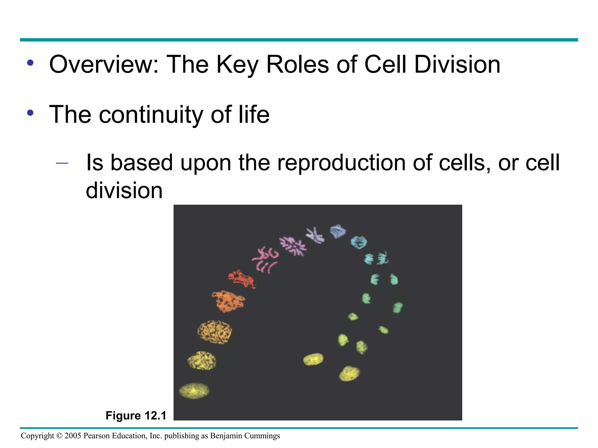
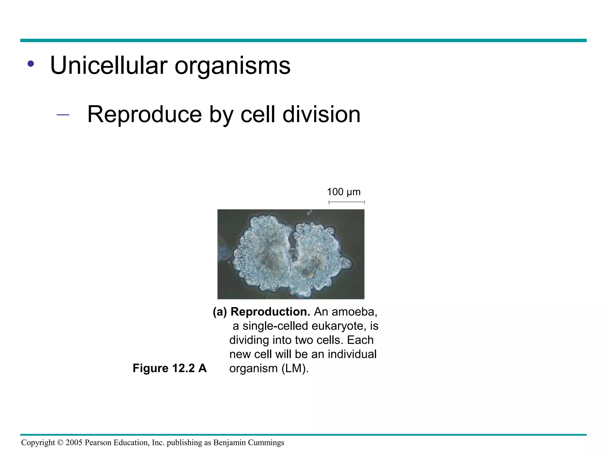
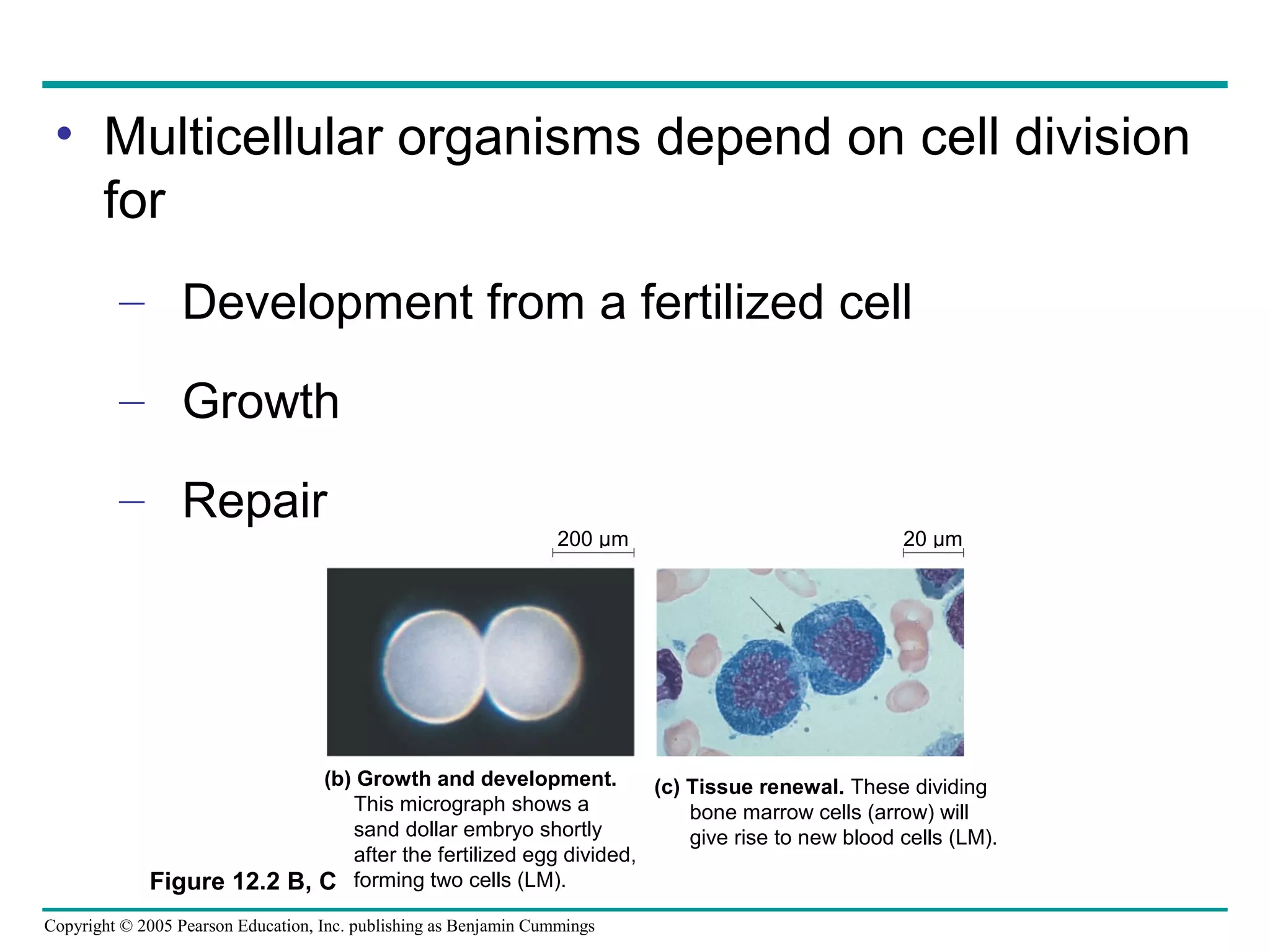
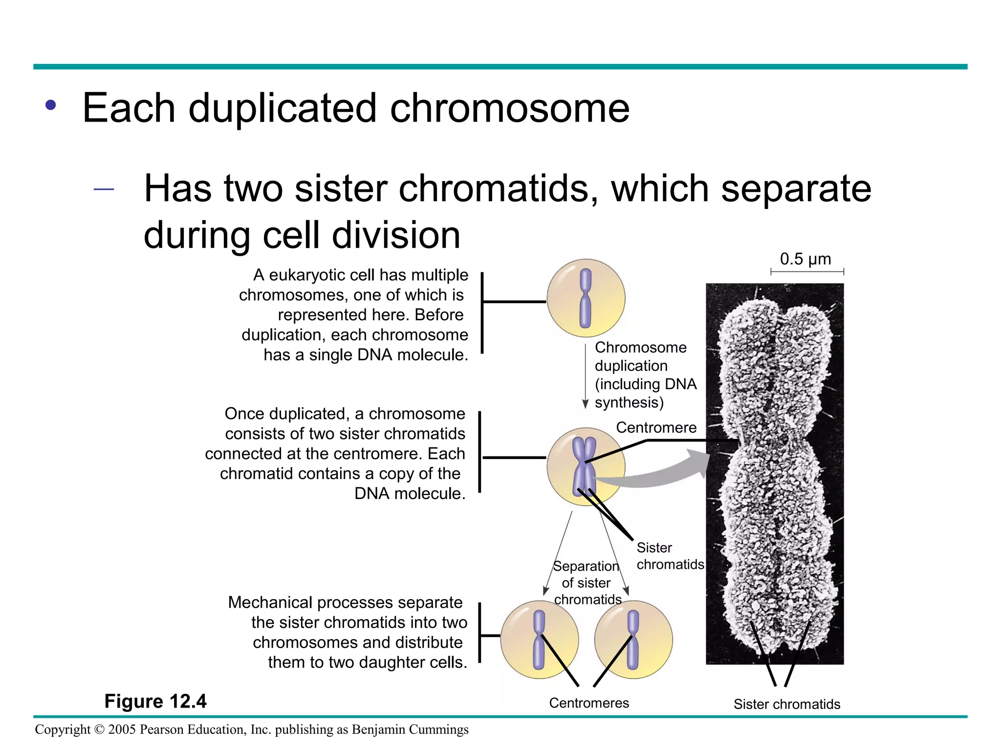
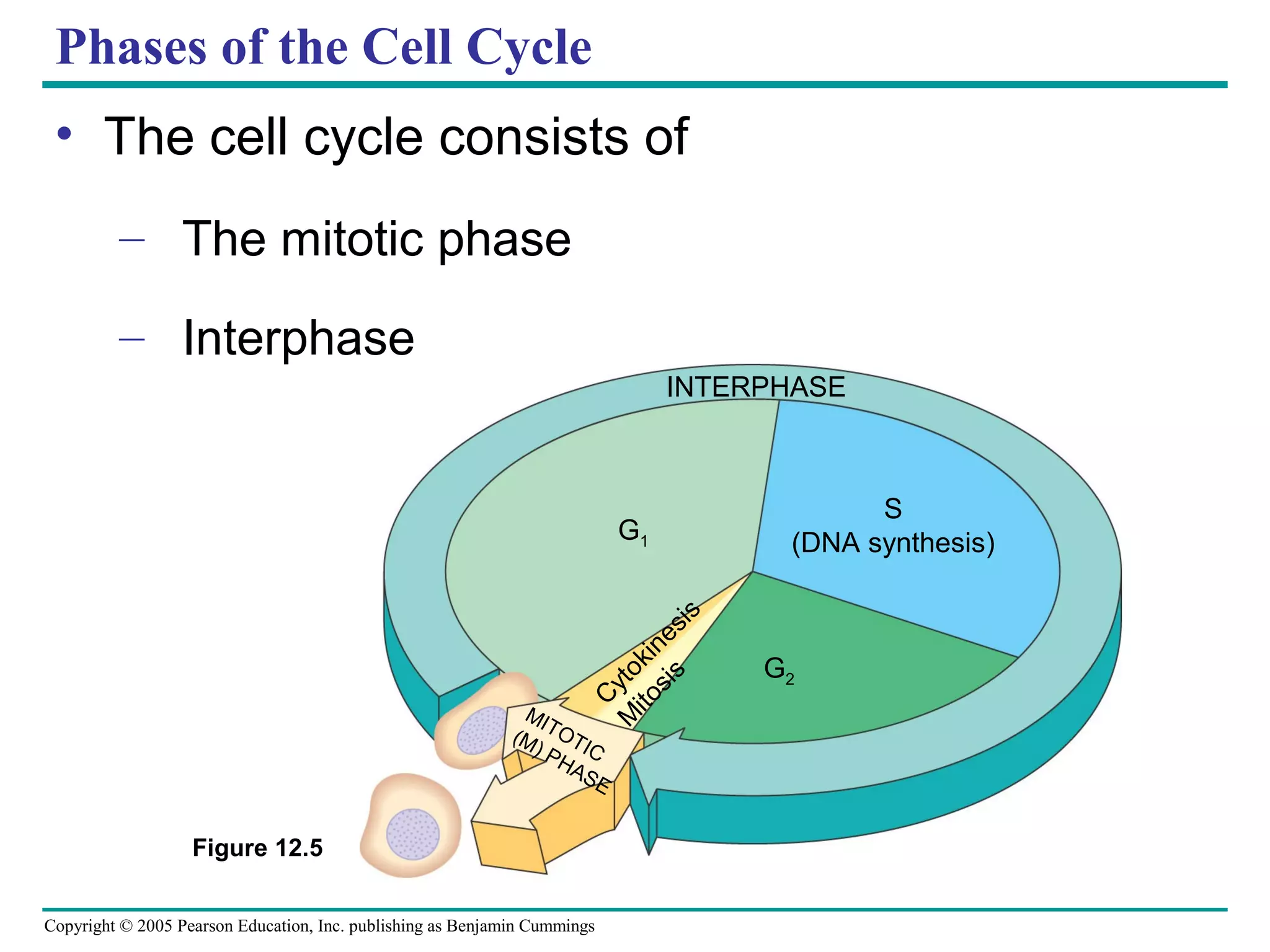
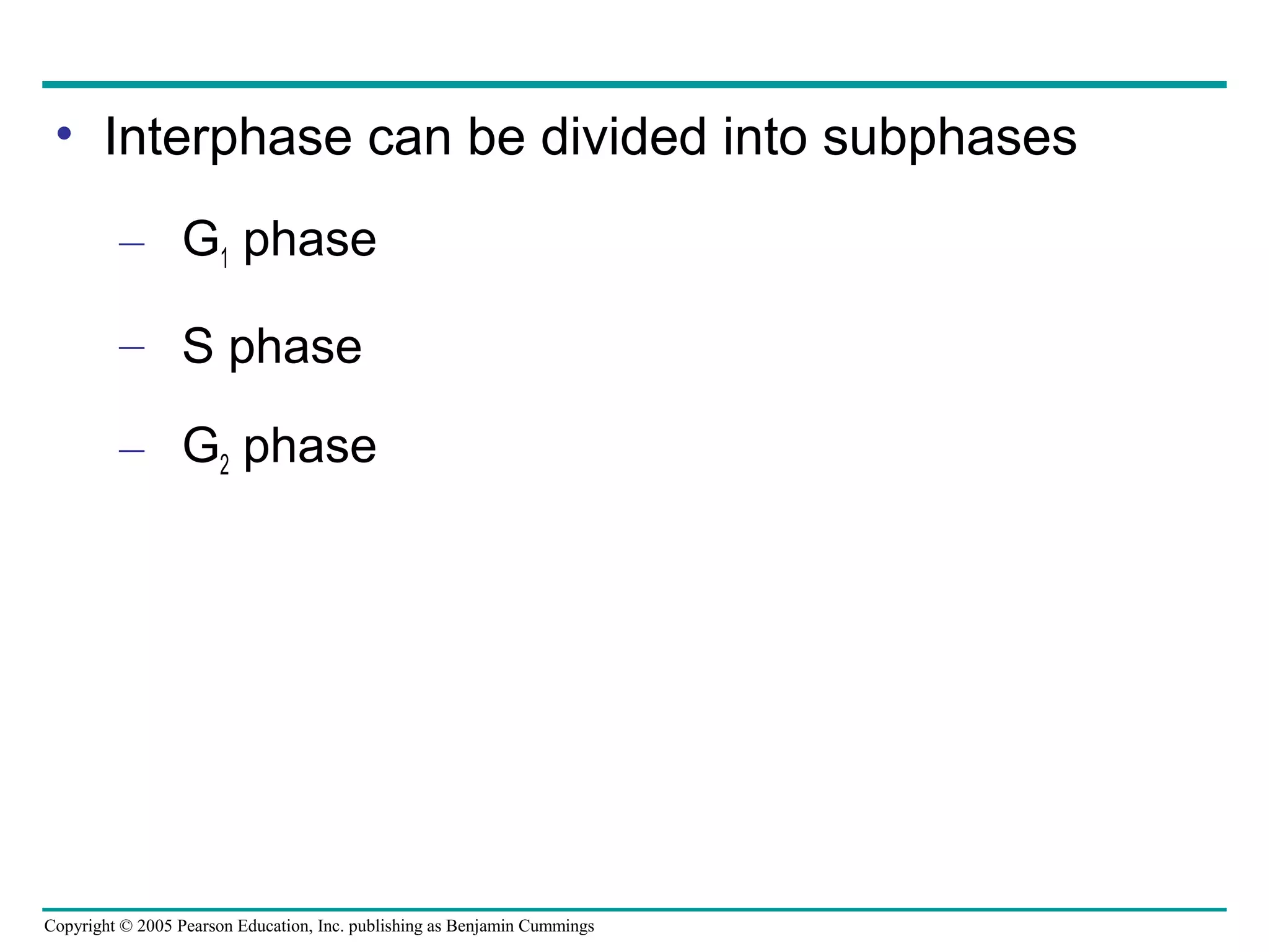
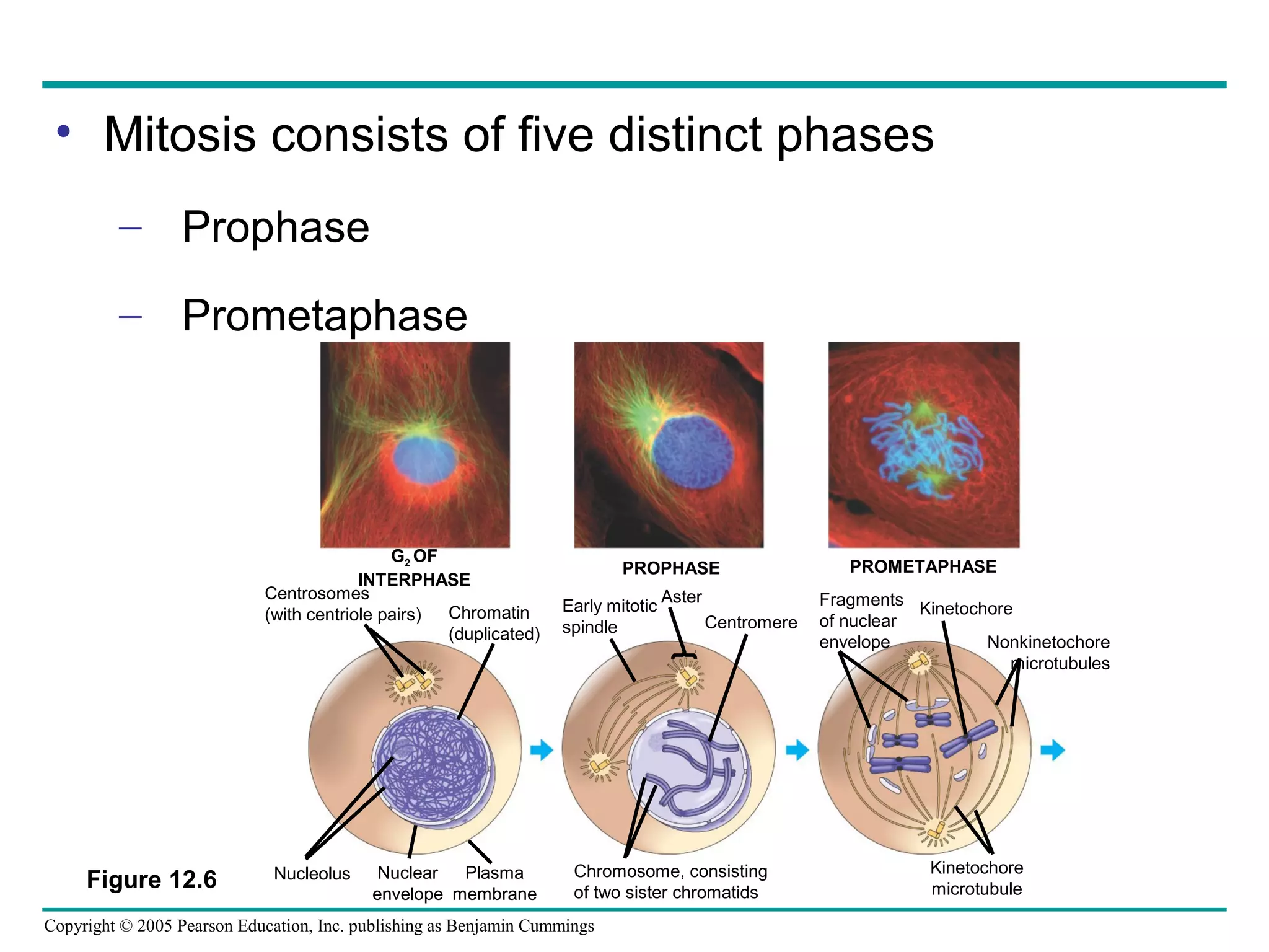
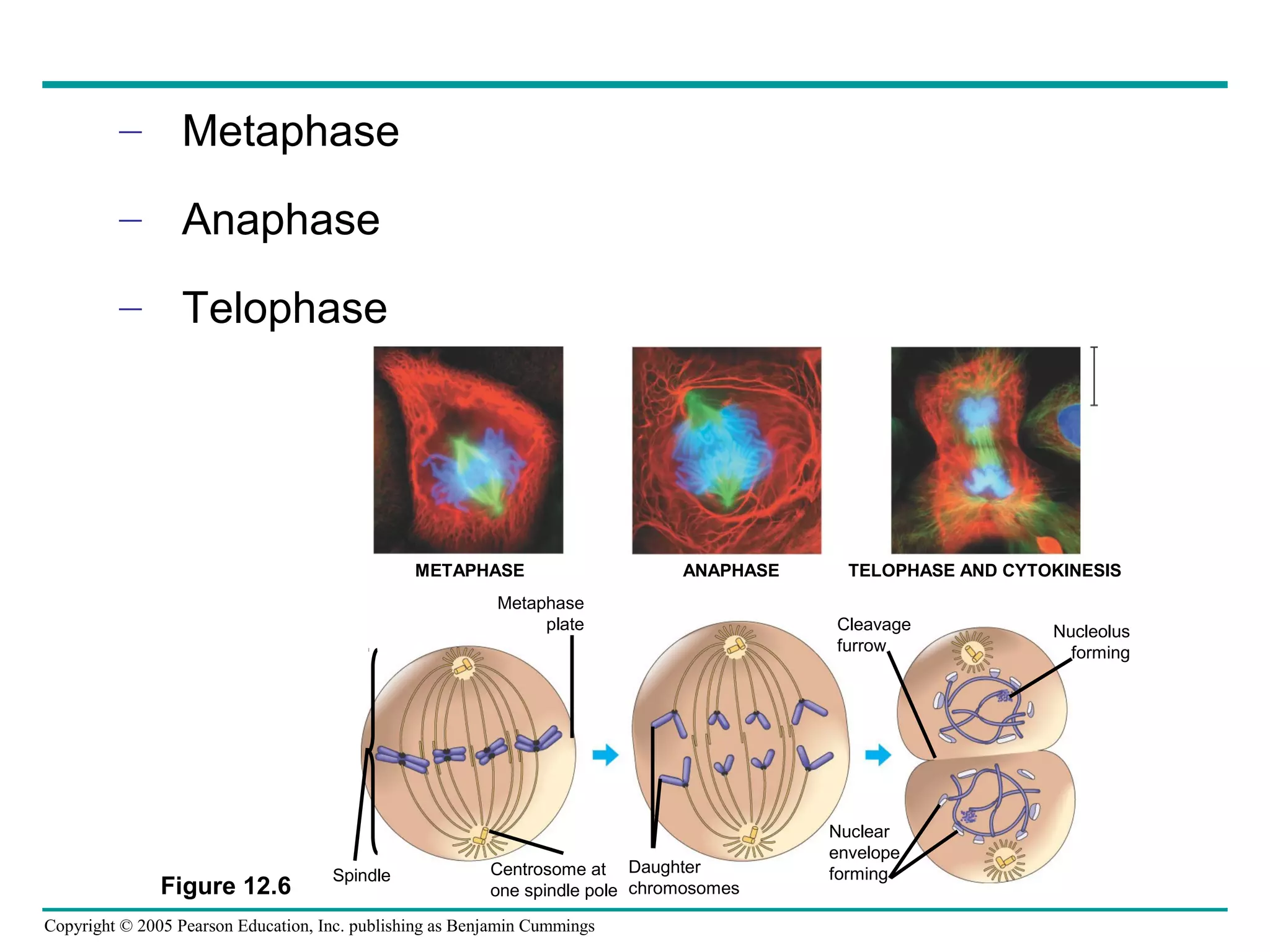
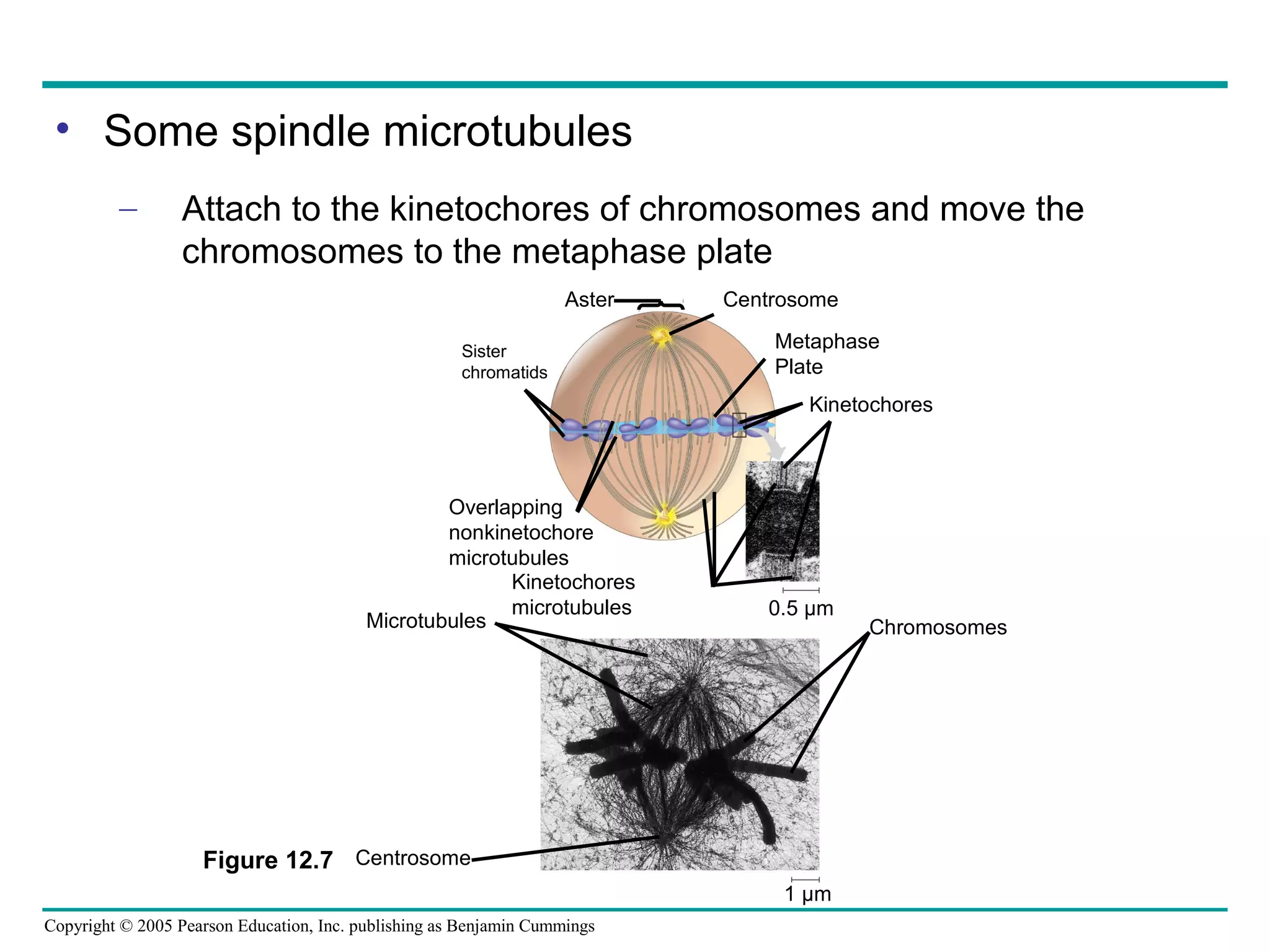
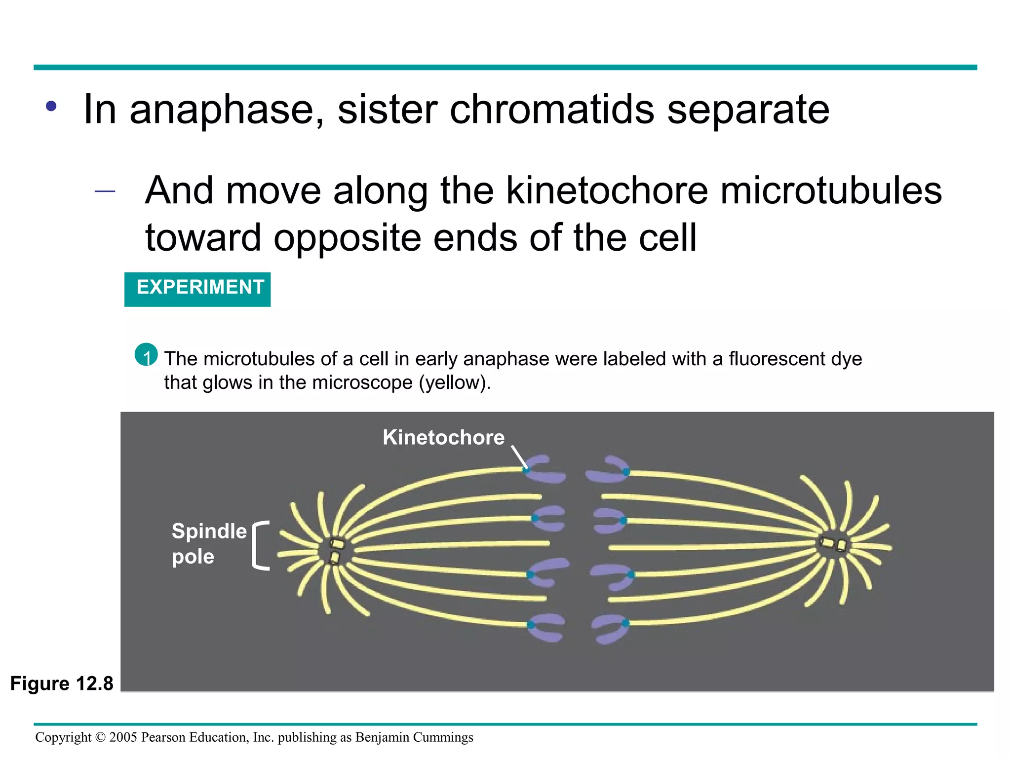
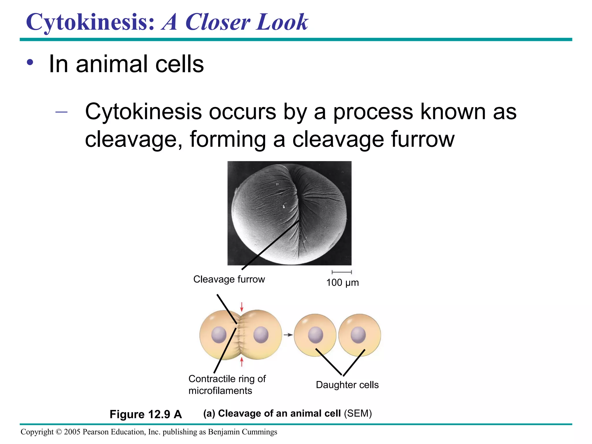
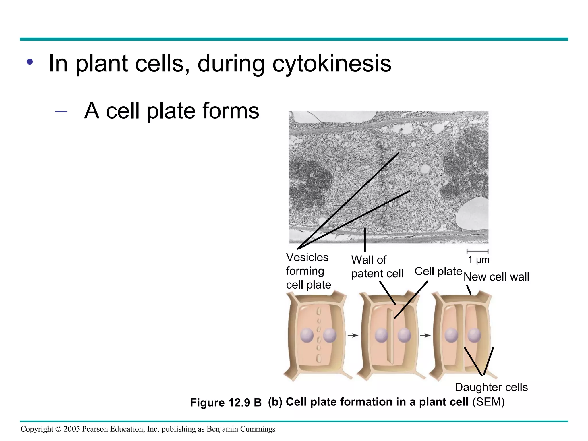
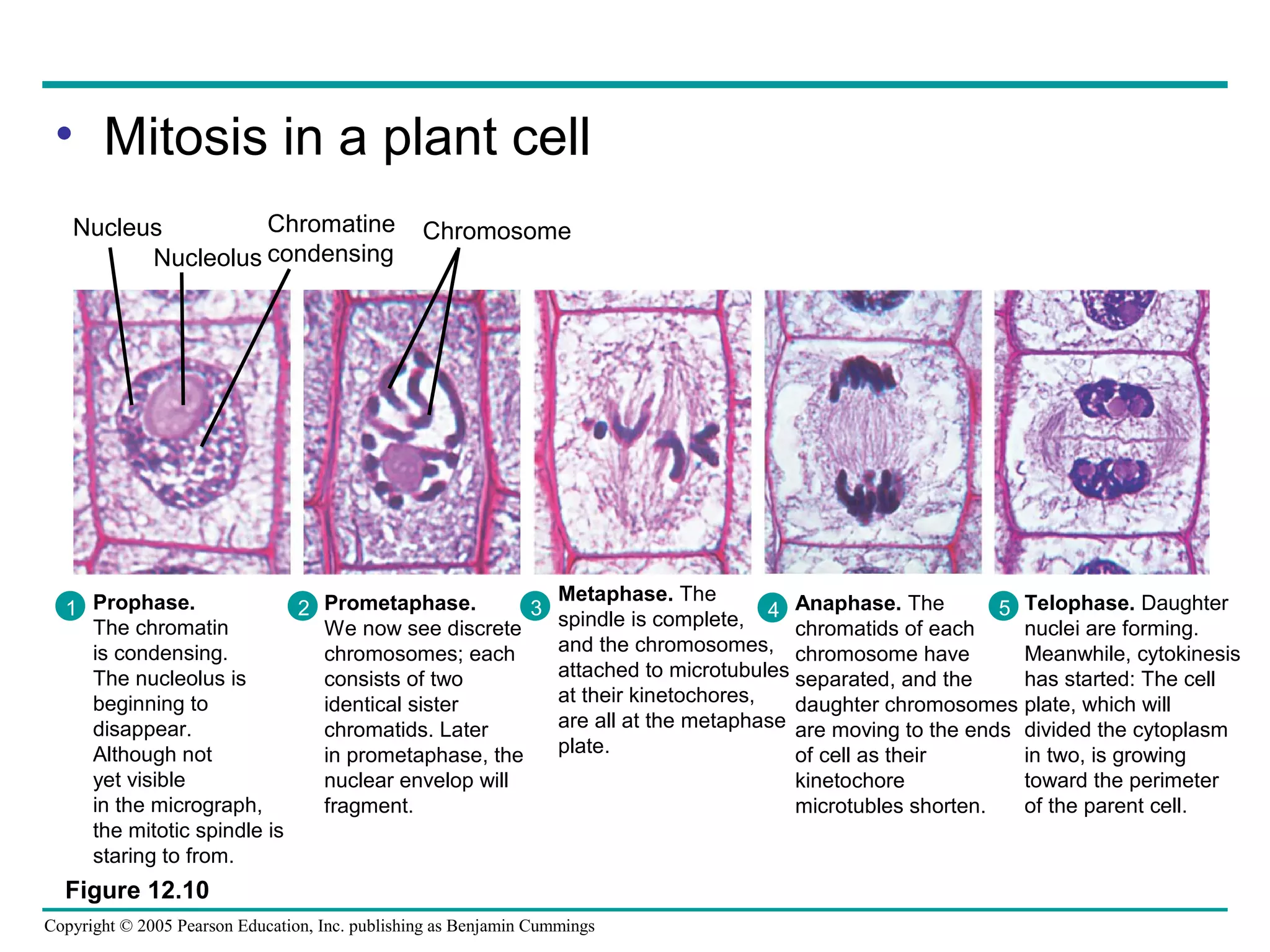
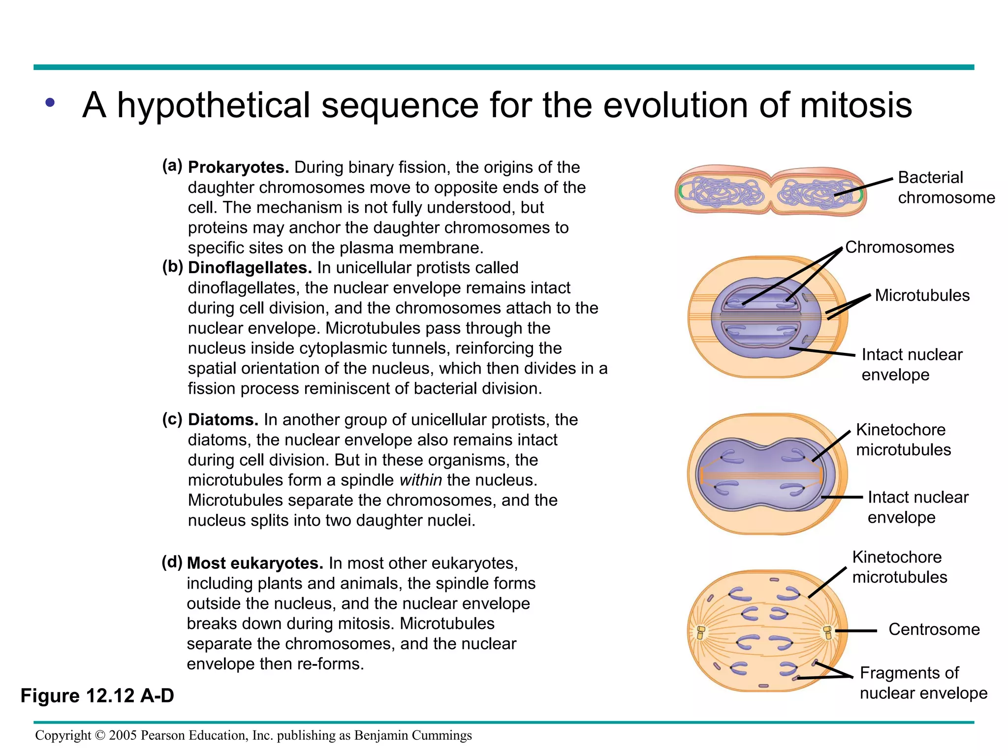
1) The document discusses the cell cycle and cell division in eukaryotic cells. It describes the key phases of the cell cycle - interphase and the mitotic phase - and explains how cells duplicate their DNA and separate their chromosomes during cell division.
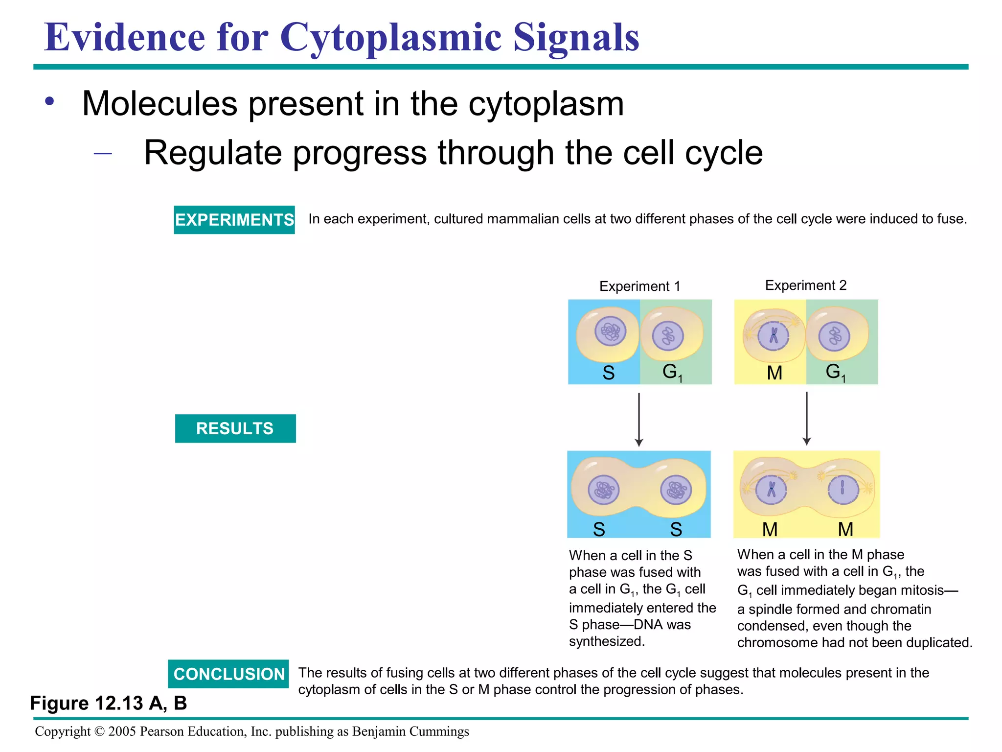
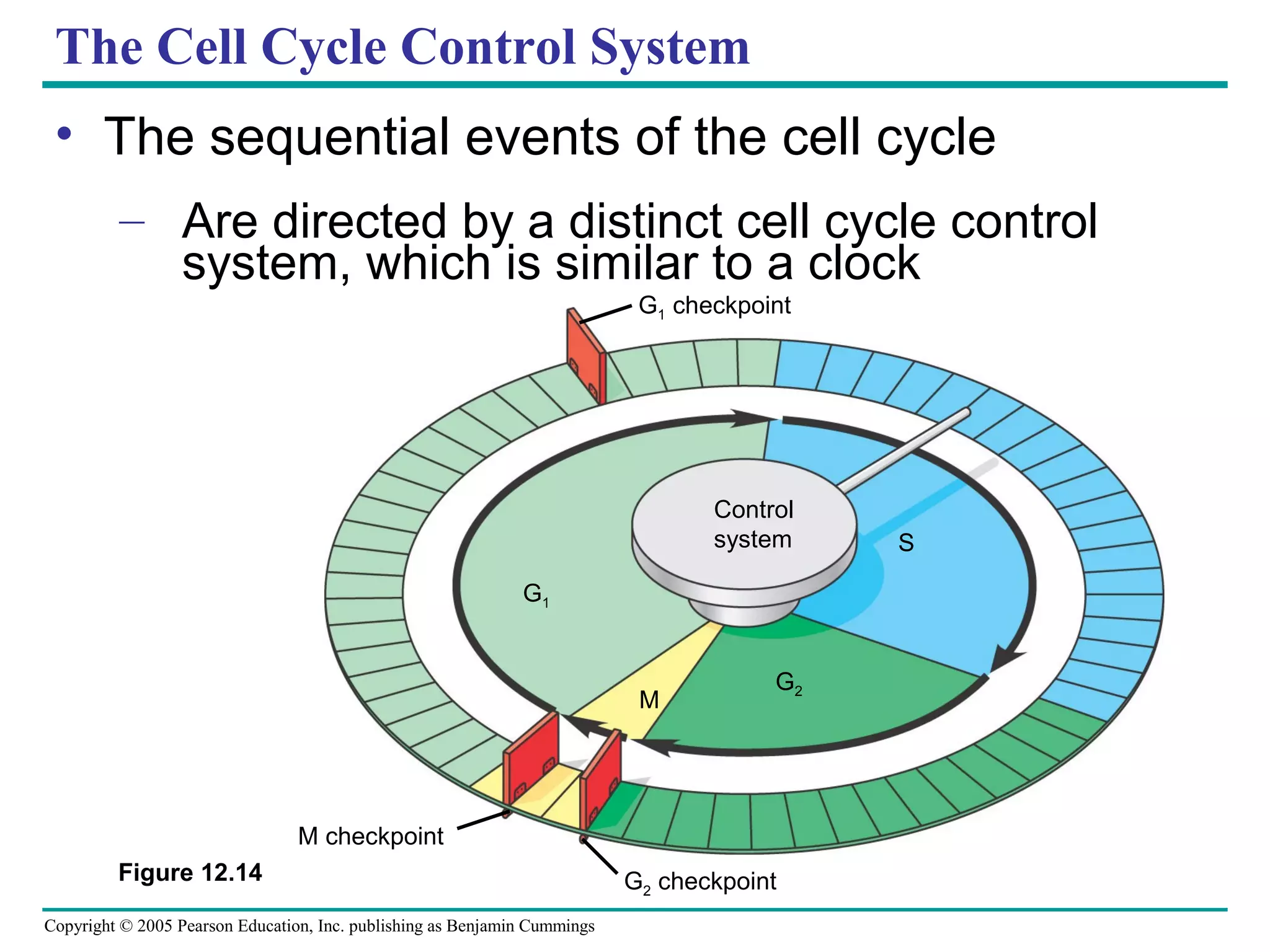
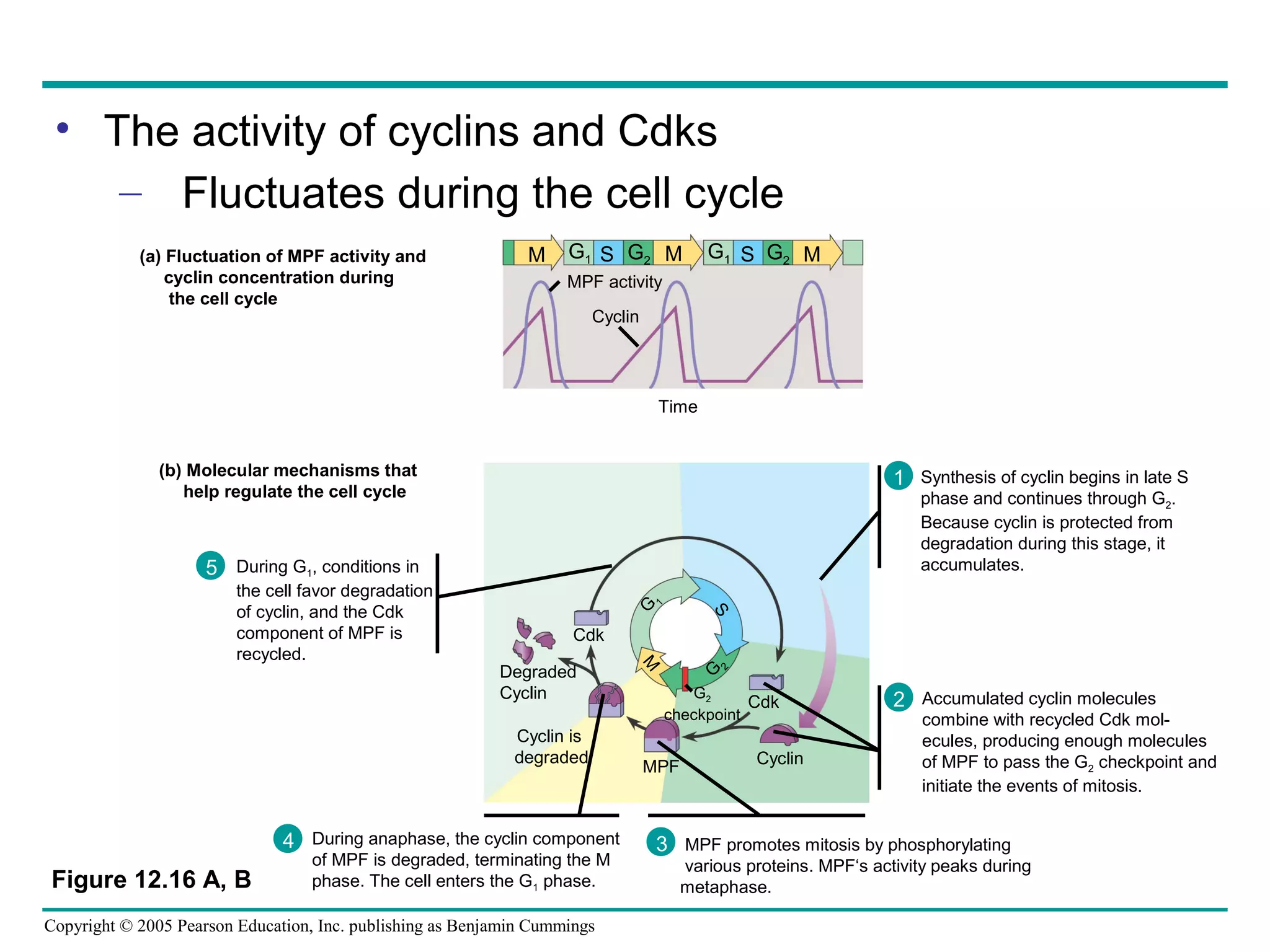
2) Cyclins and cyclin-dependent kinases play important roles in regulating progression through the cell cycle by controlling checkpoints between each phase. Their activity fluctuates throughout the cell cycle.
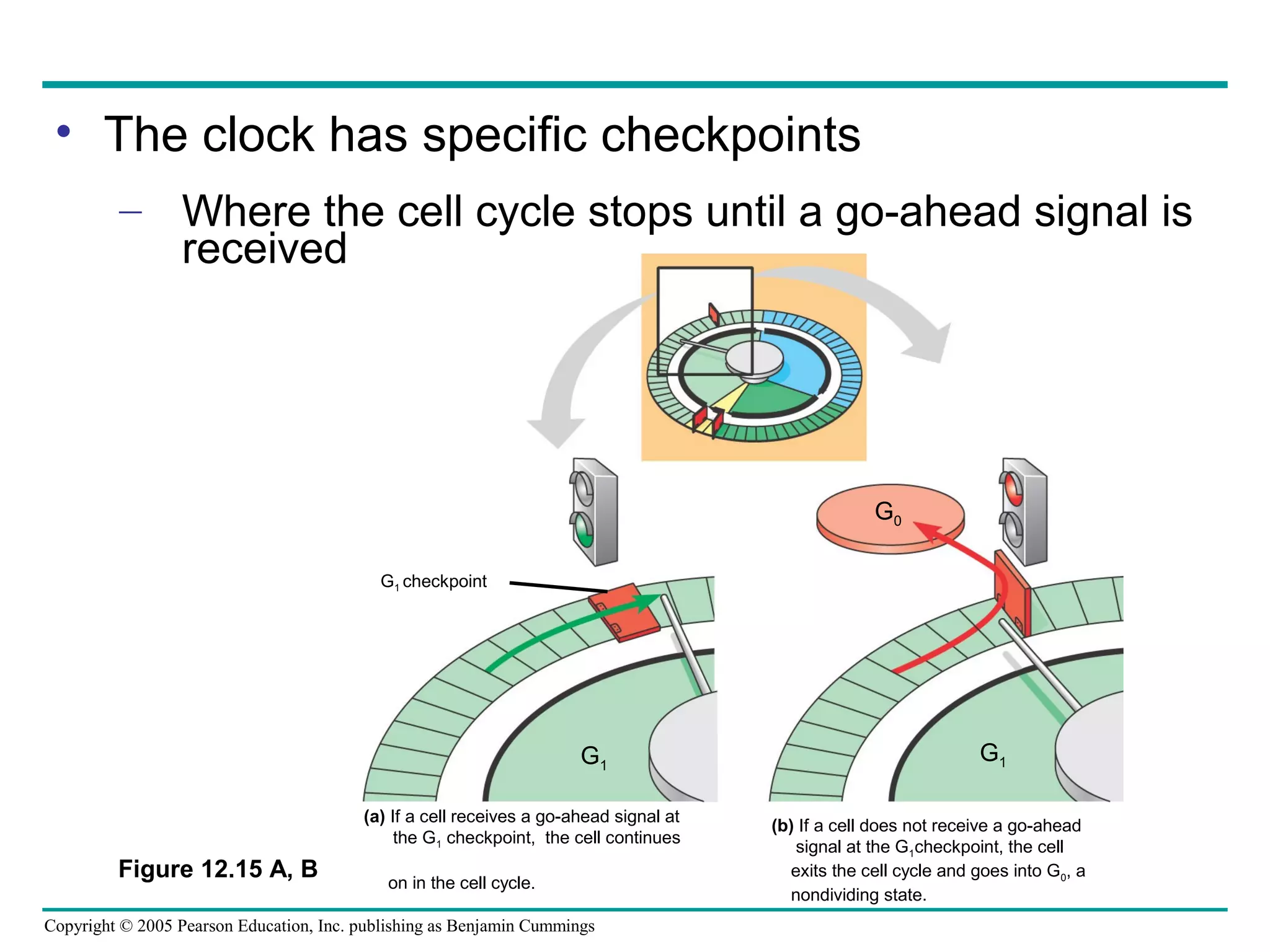
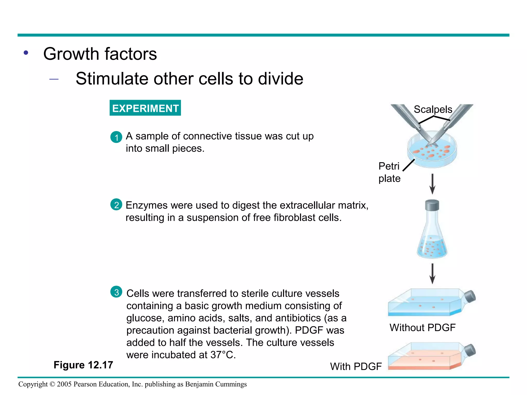
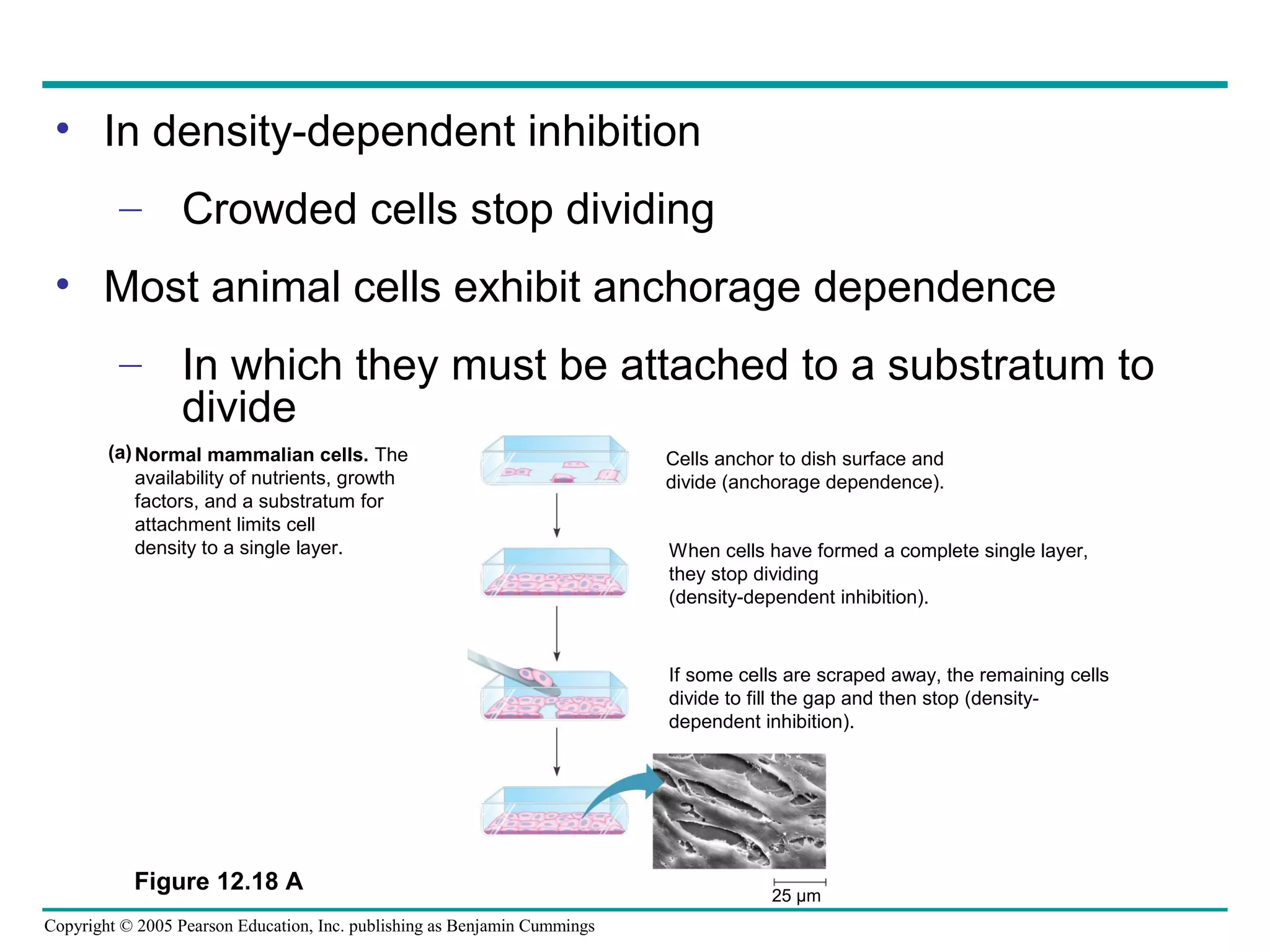
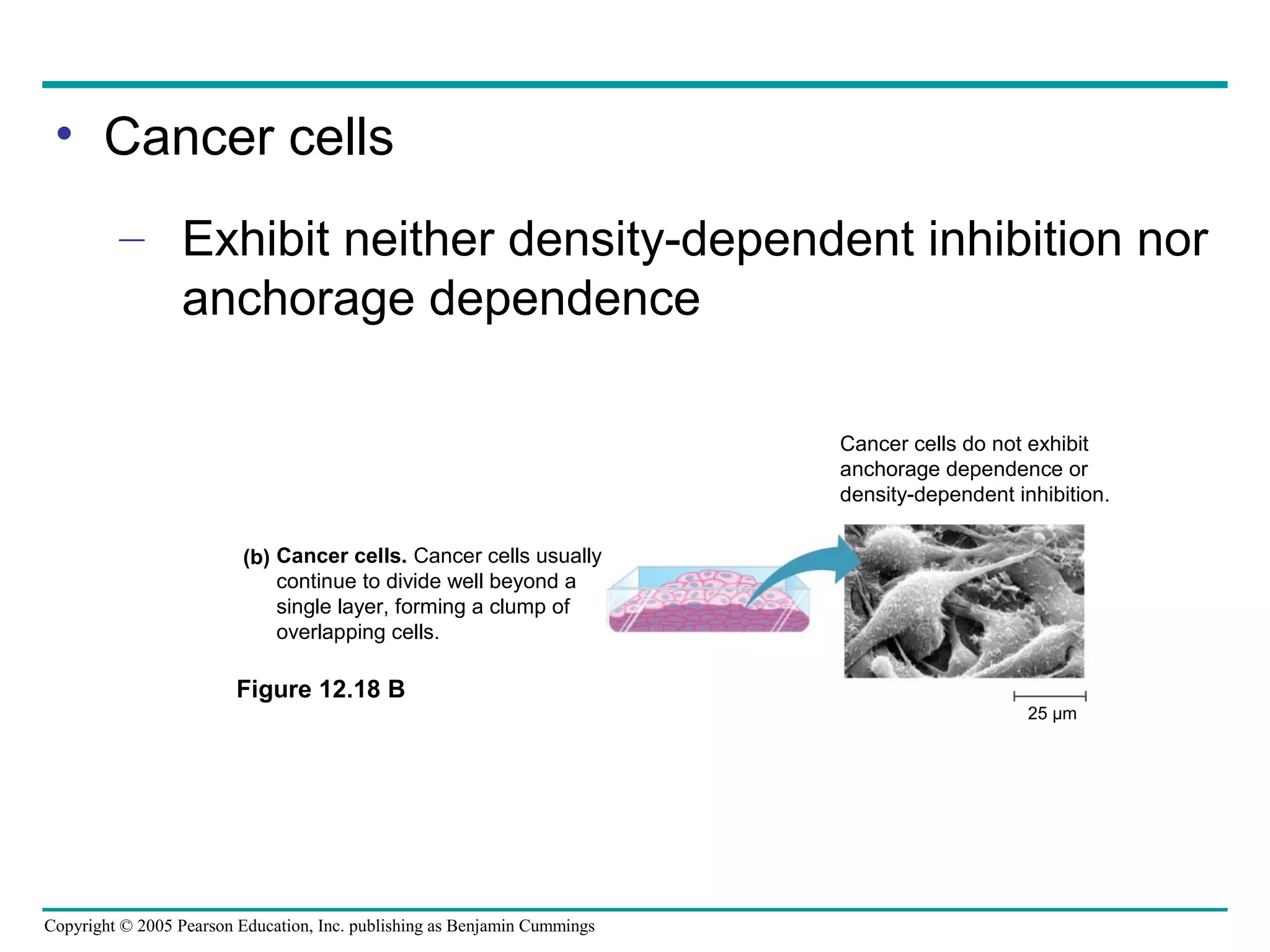
3) Both internal signals, like cyclin/CDK complexes, and external signals from growth factors control progression through the cell cycle checkpoints.


![25 lab algae quiz (25) [11 20-13]](https://cdn.slidesharecdn.com/ss_thumbnails/25labalgaequiz2511-20-13-131120211705-phpapp02-thumbnail.jpg?width=640&height=640&fit=bounds)

![Lecture on fungi [11 25-13 monday]](https://cdn.slidesharecdn.com/ss_thumbnails/lectureonfungi11-25-13monday-131126034938-phpapp01-thumbnail.jpg?width=640&height=640&fit=bounds)


![Lab examv questions [11 26-13]](https://cdn.slidesharecdn.com/ss_thumbnails/labexamvquestions11-26-13-131126152738-phpapp02-thumbnail.jpg?width=640&height=640&fit=bounds)
![Biology exam iv for dec 9-2013 monday [self quizzes] [all lecture notes]](https://cdn.slidesharecdn.com/ss_thumbnails/biologyexamivfordec-9-2013mondayselfquizzesalllecturenotes-131127174653-phpapp01-thumbnail.jpg?width=640&height=640&fit=bounds)


