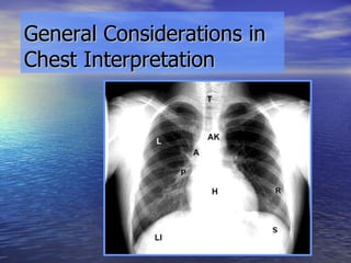
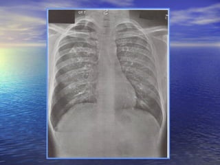
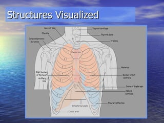
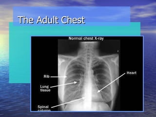
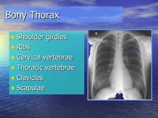
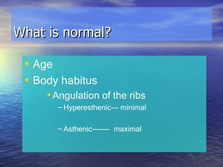
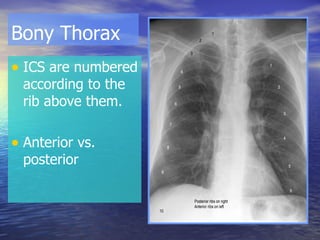
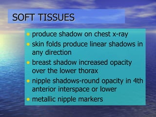
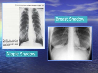
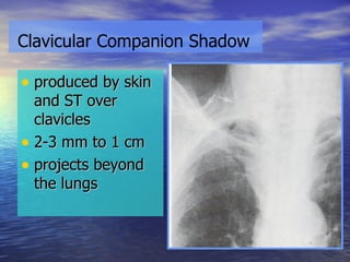
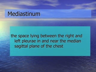
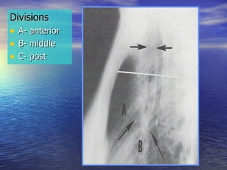
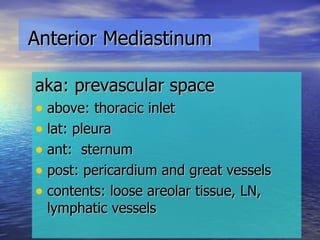
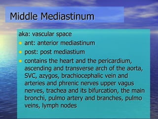
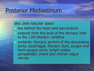
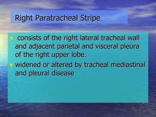
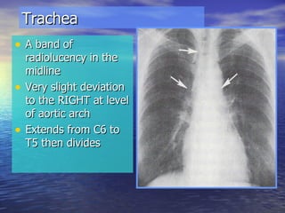
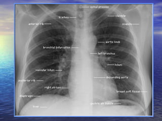
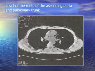
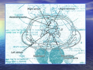
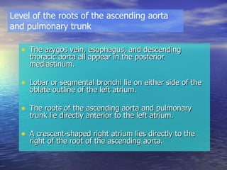
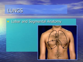
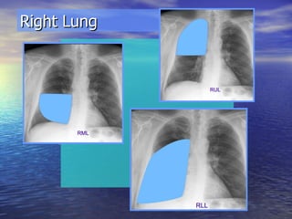
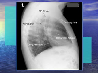
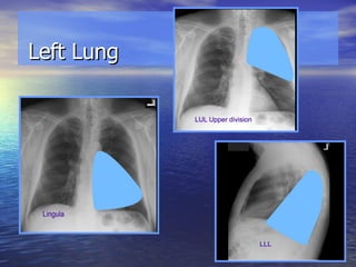
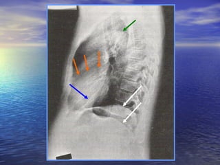
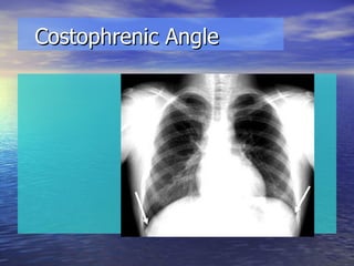
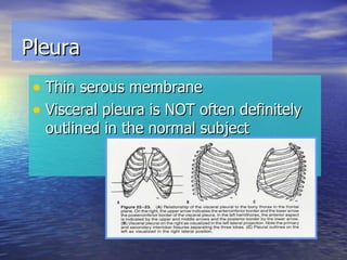
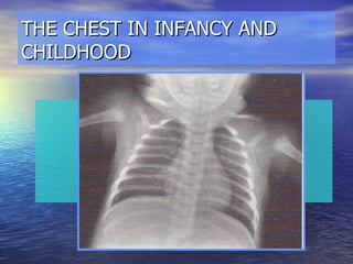
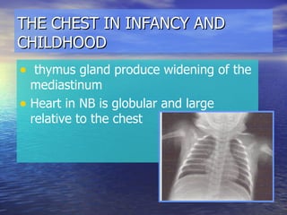
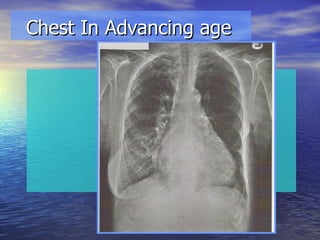
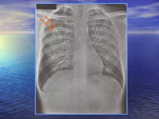
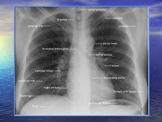
The Normal Chest http://crisbertcualteros.page.tl
General Considerations in Chest Interpretation
Developing the interpretative skills begins by looking at as many normal films as possible to develop an eye for the normal range…………
 
Objectives To be familiar with the normal chest roentgenogram Identify anatomical structures Know deviations from normal
Structures Visualized
Inspect mediastinum heart and great vessels trachea and central bronchi lungs and diaphragm bony thorax and soft tissue of thorax and neck
Note: compare lungs interspace by interspace
Remember make the initial examination of the film without knowledge of the clinical findings But before making a decision, xray observations must be correlated with  all available clinical findings
Patterns of Pulmonary Opacity Alveolar or Air Space Pattern char by homogenous opacity Interstitial Pattern  char by an increased prominence of the perivascular, interlobular and parenchymal interstitial spaces
First things first… Patient's name Date exam done  Check for position markers
Other things to check.. Patient’s position Type of film Technical quality of the film
INITIAL SURVEY General Body Size, Shape, and Symmetry  Male vs. Female  Is this an infant, child, young adult, elderly person?  Survey for foreign objects - tubes, IV lines, EKG leads, surgical drains, prosthesis, etc., as well as non-medical objects, bullets, shrapnel, glass, etc.
The Adult Chest
Bony Thorax Shoulder girdles Ribs Cervical vertebrae Thoracic vertebrae Clavicles Scapulae
What is normal? Age Body habitus Angulation of the ribs Hyperesthenic--- minimal Asthenic------- maximal
Bony Thorax ICS are numbered according to the rib above them. Anterior vs. posterior
Costal cartilage not calcified Calcification- mottled appearance men- peripheral women- central
Rhomboid Fossa anatomic variant of no clinical significance
SOFT TISSUES   produce shadow on chest x-ray skin folds produce linear shadows in any direction breast shadow increased opacity over the lower thorax  nipple shadows-round opacity in 4th anterior interspace or lower metallic nipple markers
Nipple Shadow Breast Shadow
produced by skin and ST over clavicles 2-3 mm to 1 cm projects beyond the lungs Clavicular Companion Shadow
Nodules and ST Masses may simulate pulmonary nodules subQ or deeper tissues of thoracic wall--->more sharply defined than intrapulmonary lesions CT scan if unsure
Mediastinum the space lying between the right and left pleurae in and near the median sagittal plane of the chest
Anatomic Division and Contents post aspect of the sternum to anterior aspect of thoracic vertebrae contains all thoracic viscera exept lungs
Divisions  A- anterior B- middle C- post
Anterior Mediastinum aka: prevascular space above: thoracic inlet lat: pleura ant:  sternum post: pericardium and great vessels contents: loose areolar tissue, LN, lymphatic vessels
Middle Mediastinum aka: vascular space ant: anterior mediastinum post: post mediastium contains the heart and the pericardium, ascending and transverse arch of the aorta, SVC, azygos, brachiocephalic vein and arteries and phrenic nerves upper vagus nerves, trachea and its bifurcation, the main bronchi, pulmo artery and branches, pulmo veins, lymph nodes
Posterior Mediastinum   aka: post vascular space lies behind the heart and pericardium extends from the level of the thoracic inlet to the 12th thoracic vertebra contents: thoracic portion of the descending aorta, esophagus, thoracic duct, azygos and hemi azygos veins, lymph nodes, sympathetic chains and inferior vagus nerves.
Mediastinal Pleural Reflections
Anterior Junction Line anterior to trachea, behind sternum from sternal angle downward and to the left represents the apposition of the visceral and parietal pleura of the upper lobes projected over the trachea as thin linear shadow, convex to the left
Posterior Junction Line post to trachea and esophagus from the thoracic inlet downward to level of the azygos and aortic arches represents the approximation of visceral and parietal pleura of the upper lobes posteriorly projects over the tracheal air shadow and is convex to the left
Azygoesophageal Recess extends from the inferior surface of the azygos arch downward and to the left to level of the diaphragm outlines medial aspect of the right lower lobe and right lateral aspect of the esophagus concave on its right
Right Paratracheal Stripe consists of the right lateral tracheal wall and adjacent parietal and visceral pleura of the right upper lobe. widened or altered by tracheal mediastinal and pleural disease
Paraspinal Interphase (pleural reflection) right side- posteriorly between parietal pleura and lateral aspect of thoracic vertebrae left side- left lateral wall of the descending aorta with lung
Lymph Nodes Anterior mediastinal (prevascular) Paratracheobronchial
Normal nodes are small and cannot be identified on normal chest radiographs
Anterior Mediastinal (prevascular) R: ant to SVC and R innominate vein L: anterior to aorta and carotid artery Ductus Nodes lie ant to ligamentum arteriosum in the aortopulmonary window
Paratracheobronchial Paratracheal Subcarinal (bifurcation) Hilar
Trachea A band of radiolucency in the midline Very slight deviation to the RIGHT at level of aortic arch Extends from C6 to T5 then divides
 
The angle formed by the bronchi with the sagittal plane is equal bilaterally until 15yo Tracheal deviation to the right increases the angle on the left Subcarinal angle = 60 0 Nice to know………….
Right Main Bronchi More vertical than left 2 primary branches Eparterial bronchus Hyparterial bronchus
Left Main Bronchi longer, deviates more laterally than right Divides into Lower lobe bronchus Left upper lobe bronchus
Aortic Nipple left superior intercostal, vein seen in cross section as it courses around the aorta
local concavity or notch between the aortic arch and the pulmonary artery Aortopulmonary Window
HILUM Contains the Pulmonary arteries and veins Bronchi Bronchial arteries and veins Lymph nodes
Hilar Height Ratio Useful in determining relative volume gain or loss in lower vs. upper lobes Height above hilum Height below hilum Right=1.3  Left= 0.84
CT of the Mediastinum
Relationships at the level of the origin of the three branches of the aortic arch
 
Origin of the three branches of the Aortic Arch The esophagus -anterior to and slightly to the left of the body of a thoracic vertebra. The trachea is anterior to and slightly to the right of the esophagus.  The brachiocephalic artery, left common carotid artery, and left subclavian artery each appears in transverse section. The thymus (if present) casts a triangular image directly anterior to the left brachiocephalic vein.
Level of the aortic-pulmonary window
 
Esophagus lies directly anterior to and to the left of the body of a thoracic vertebra. The main stem bronchi appear in oblique section just above or below the level of the carina. The azygos vein appears  at the level where it arches over the right main stem bronchus and ends via union with the superior vena cava  The region in the scan between the origin and end of the aortic arch represents the aortic pulmonary window. Level of the aortic-pulmonary window
Level of the roots of the ascending aorta and pulmonary trunk
 
The azygos vein, esophagus, and descending thoracic aorta all appear in the posterior mediastinum. Lobar or segmental bronchi lie on either side of the oblate outline of the left atrium. The roots of the ascending aorta and pulmonary trunk lie directly anterior to the left atrium.  A crescent-shaped right atrium lies directly to the right of the root of the ascending aorta. Level of the roots of the ascending aorta and pulmonary trunk
LUNGS Lobar and Segmental Anatomy
Right Lung
 
Left Lung
 
Bronchopulmonary  Segmental  Anatomy
These segments are the basic functional units of the lung Each pyramid-shaped segment is Enveloped by a connective tissue sheath Supplied by a single segmental bronchus and a single pulmonary arterial branch Oriented so that its apex projects toward the hilum of the lung Bronchopulmonary Segments of the Lungs
 
Right Lung Bronchogram
Left Lung Bronchogram
Surface projections of the lungs and the pleural cavities The apex of each lung projects above the medial third of the clavicle. Interlobar fissures The oblique fissure begins posteriorly in the upper thoracic region, crosses the 5th and 6th ribs, curves laterally to the midaxillary  line, and courses along the lower border of the 6 th  rib to the lateral border of the sternum. The right lung's horizontal fissure courses along the lower border of the 4th rib from the midaxillary line to the lateral border of the sternum
At midinspiration in quiet breathing, the lower margin of each lung lies at the level: The sixth rib anteriorly at the midclavicular line The eighth rib laterally at the midaxillary line The tenth rib posteriorly at the lateral border of the vertebral column Margins
The surface projection of the costodiaphragmatic margin of each pleural cavity passes through: The eighth rib anteriorly at the midclavicular line The tenth rib laterally at the midaxillary line The eleventh or twelfth rib posteriorly at the lateral border of the vertebral column Costodiaphragmatic Margin
Diaphragm The chief muscle of inspiration  Accounts for 2/3  of the lungs' volumetric increase during inspiration Most effective respiratory muscle when a person is supine
Diaphragm   higher R level of post arc of 10th rib 5th anterior rib or interspace on deep inspiration ribs below not clearly seen – opaque abdominal contents
 
Costophrenic Angle
Thin serous membrane Visceral pleura is NOT often definitely outlined in the normal subject Pleura
THE CHEST IN INFANCY AND CHILDHOOD
THE CHEST IN INFANCY AND CHILDHOOD AP diameter greater than transverse diameter Diaphragm is higher Ribs horizontal in infancy then angulate downwards with growth
THE CHEST IN INFANCY AND CHILDHOOD Sternum not completely ossified at birth- ossifies in segmental manner Two ossification centers in each segment Significance: Appear as small, rounded opacities, maybe mistaken for lesions
THE CHEST IN INFANCY AND CHILDHOOD thymus gland produce widening of the mediastinum Heart in NB is globular and large relative to the chest
THE CHEST IN INFANCY AND CHILDHOOD Lungs of infant and child more radiolucent Hilar shadow high at level of T3 Tracheal bifurcation reaches T5 at 10 yo Diaphragm higher, left maybe higher
Chest In Advancing age
Chest In Advancing age Increase dorsal kyphotic curve Increase AP diameter of chest Osteopenia Calcifications in costal cartilages Aorta becomes elongated, dilated and tortuous
Chest In Advancing age Emphysematous changes in lungs Apical scarring Senile emphysema Lower domes, less acute costophrenic angles
 
Review
Pls visit: http://crisbertcualteros.page.tl

BASIC RADIOLOGY

  • 1.
    The Normal Chesthttp://crisbertcualteros.page.tl
  • 2.
    General Considerations inChest Interpretation
  • 3.
    Developing the interpretativeskills begins by looking at as many normal films as possible to develop an eye for the normal range…………
  • 4.
  • 5.
    Objectives To befamiliar with the normal chest roentgenogram Identify anatomical structures Know deviations from normal
  • 6.
  • 7.
    Inspect mediastinum heartand great vessels trachea and central bronchi lungs and diaphragm bony thorax and soft tissue of thorax and neck
  • 8.
    Note: compare lungsinterspace by interspace
  • 9.
    Remember make theinitial examination of the film without knowledge of the clinical findings But before making a decision, xray observations must be correlated with all available clinical findings
  • 10.
    Patterns of PulmonaryOpacity Alveolar or Air Space Pattern char by homogenous opacity Interstitial Pattern char by an increased prominence of the perivascular, interlobular and parenchymal interstitial spaces
  • 11.
    First things first…Patient's name Date exam done Check for position markers
  • 12.
    Other things tocheck.. Patient’s position Type of film Technical quality of the film
  • 13.
    INITIAL SURVEY GeneralBody Size, Shape, and Symmetry Male vs. Female Is this an infant, child, young adult, elderly person? Survey for foreign objects - tubes, IV lines, EKG leads, surgical drains, prosthesis, etc., as well as non-medical objects, bullets, shrapnel, glass, etc.
  • 14.
  • 15.
    Bony Thorax Shouldergirdles Ribs Cervical vertebrae Thoracic vertebrae Clavicles Scapulae
  • 16.
    What is normal?Age Body habitus Angulation of the ribs Hyperesthenic--- minimal Asthenic------- maximal
  • 17.
    Bony Thorax ICSare numbered according to the rib above them. Anterior vs. posterior
  • 18.
    Costal cartilage notcalcified Calcification- mottled appearance men- peripheral women- central
  • 19.
    Rhomboid Fossa anatomicvariant of no clinical significance
  • 20.
    SOFT TISSUES produce shadow on chest x-ray skin folds produce linear shadows in any direction breast shadow increased opacity over the lower thorax nipple shadows-round opacity in 4th anterior interspace or lower metallic nipple markers
  • 21.
  • 22.
    produced by skinand ST over clavicles 2-3 mm to 1 cm projects beyond the lungs Clavicular Companion Shadow
  • 23.
    Nodules and STMasses may simulate pulmonary nodules subQ or deeper tissues of thoracic wall--->more sharply defined than intrapulmonary lesions CT scan if unsure
  • 24.
    Mediastinum the spacelying between the right and left pleurae in and near the median sagittal plane of the chest
  • 25.
    Anatomic Division andContents post aspect of the sternum to anterior aspect of thoracic vertebrae contains all thoracic viscera exept lungs
  • 26.
    Divisions A-anterior B- middle C- post
  • 27.
    Anterior Mediastinum aka:prevascular space above: thoracic inlet lat: pleura ant: sternum post: pericardium and great vessels contents: loose areolar tissue, LN, lymphatic vessels
  • 28.
    Middle Mediastinum aka:vascular space ant: anterior mediastinum post: post mediastium contains the heart and the pericardium, ascending and transverse arch of the aorta, SVC, azygos, brachiocephalic vein and arteries and phrenic nerves upper vagus nerves, trachea and its bifurcation, the main bronchi, pulmo artery and branches, pulmo veins, lymph nodes
  • 29.
    Posterior Mediastinum aka: post vascular space lies behind the heart and pericardium extends from the level of the thoracic inlet to the 12th thoracic vertebra contents: thoracic portion of the descending aorta, esophagus, thoracic duct, azygos and hemi azygos veins, lymph nodes, sympathetic chains and inferior vagus nerves.
  • 30.
  • 31.
    Anterior Junction Lineanterior to trachea, behind sternum from sternal angle downward and to the left represents the apposition of the visceral and parietal pleura of the upper lobes projected over the trachea as thin linear shadow, convex to the left
  • 32.
    Posterior Junction Linepost to trachea and esophagus from the thoracic inlet downward to level of the azygos and aortic arches represents the approximation of visceral and parietal pleura of the upper lobes posteriorly projects over the tracheal air shadow and is convex to the left
  • 33.
    Azygoesophageal Recess extendsfrom the inferior surface of the azygos arch downward and to the left to level of the diaphragm outlines medial aspect of the right lower lobe and right lateral aspect of the esophagus concave on its right
  • 34.
    Right Paratracheal Stripeconsists of the right lateral tracheal wall and adjacent parietal and visceral pleura of the right upper lobe. widened or altered by tracheal mediastinal and pleural disease
  • 35.
    Paraspinal Interphase (pleuralreflection) right side- posteriorly between parietal pleura and lateral aspect of thoracic vertebrae left side- left lateral wall of the descending aorta with lung
  • 36.
    Lymph Nodes Anteriormediastinal (prevascular) Paratracheobronchial
  • 37.
    Normal nodes aresmall and cannot be identified on normal chest radiographs
  • 38.
    Anterior Mediastinal (prevascular)R: ant to SVC and R innominate vein L: anterior to aorta and carotid artery Ductus Nodes lie ant to ligamentum arteriosum in the aortopulmonary window
  • 39.
  • 40.
    Trachea A bandof radiolucency in the midline Very slight deviation to the RIGHT at level of aortic arch Extends from C6 to T5 then divides
  • 41.
  • 42.
    The angle formedby the bronchi with the sagittal plane is equal bilaterally until 15yo Tracheal deviation to the right increases the angle on the left Subcarinal angle = 60 0 Nice to know………….
  • 43.
    Right Main BronchiMore vertical than left 2 primary branches Eparterial bronchus Hyparterial bronchus
  • 44.
    Left Main Bronchilonger, deviates more laterally than right Divides into Lower lobe bronchus Left upper lobe bronchus
  • 45.
    Aortic Nipple leftsuperior intercostal, vein seen in cross section as it courses around the aorta
  • 46.
    local concavity ornotch between the aortic arch and the pulmonary artery Aortopulmonary Window
  • 47.
    HILUM Contains thePulmonary arteries and veins Bronchi Bronchial arteries and veins Lymph nodes
  • 48.
    Hilar Height RatioUseful in determining relative volume gain or loss in lower vs. upper lobes Height above hilum Height below hilum Right=1.3 Left= 0.84
  • 49.
    CT of theMediastinum
  • 50.
    Relationships at thelevel of the origin of the three branches of the aortic arch
  • 51.
  • 52.
    Origin of thethree branches of the Aortic Arch The esophagus -anterior to and slightly to the left of the body of a thoracic vertebra. The trachea is anterior to and slightly to the right of the esophagus. The brachiocephalic artery, left common carotid artery, and left subclavian artery each appears in transverse section. The thymus (if present) casts a triangular image directly anterior to the left brachiocephalic vein.
  • 53.
    Level of theaortic-pulmonary window
  • 54.
  • 55.
    Esophagus lies directlyanterior to and to the left of the body of a thoracic vertebra. The main stem bronchi appear in oblique section just above or below the level of the carina. The azygos vein appears at the level where it arches over the right main stem bronchus and ends via union with the superior vena cava The region in the scan between the origin and end of the aortic arch represents the aortic pulmonary window. Level of the aortic-pulmonary window
  • 56.
    Level of theroots of the ascending aorta and pulmonary trunk
  • 57.
  • 58.
    The azygos vein,esophagus, and descending thoracic aorta all appear in the posterior mediastinum. Lobar or segmental bronchi lie on either side of the oblate outline of the left atrium. The roots of the ascending aorta and pulmonary trunk lie directly anterior to the left atrium. A crescent-shaped right atrium lies directly to the right of the root of the ascending aorta. Level of the roots of the ascending aorta and pulmonary trunk
  • 59.
    LUNGS Lobar andSegmental Anatomy
  • 60.
  • 61.
  • 62.
  • 63.
  • 64.
  • 65.
    These segments arethe basic functional units of the lung Each pyramid-shaped segment is Enveloped by a connective tissue sheath Supplied by a single segmental bronchus and a single pulmonary arterial branch Oriented so that its apex projects toward the hilum of the lung Bronchopulmonary Segments of the Lungs
  • 66.
  • 67.
  • 68.
  • 69.
    Surface projections ofthe lungs and the pleural cavities The apex of each lung projects above the medial third of the clavicle. Interlobar fissures The oblique fissure begins posteriorly in the upper thoracic region, crosses the 5th and 6th ribs, curves laterally to the midaxillary line, and courses along the lower border of the 6 th rib to the lateral border of the sternum. The right lung's horizontal fissure courses along the lower border of the 4th rib from the midaxillary line to the lateral border of the sternum
  • 70.
    At midinspiration inquiet breathing, the lower margin of each lung lies at the level: The sixth rib anteriorly at the midclavicular line The eighth rib laterally at the midaxillary line The tenth rib posteriorly at the lateral border of the vertebral column Margins
  • 71.
    The surface projectionof the costodiaphragmatic margin of each pleural cavity passes through: The eighth rib anteriorly at the midclavicular line The tenth rib laterally at the midaxillary line The eleventh or twelfth rib posteriorly at the lateral border of the vertebral column Costodiaphragmatic Margin
  • 72.
    Diaphragm The chiefmuscle of inspiration Accounts for 2/3 of the lungs' volumetric increase during inspiration Most effective respiratory muscle when a person is supine
  • 73.
    Diaphragm higher R level of post arc of 10th rib 5th anterior rib or interspace on deep inspiration ribs below not clearly seen – opaque abdominal contents
  • 74.
  • 75.
  • 76.
    Thin serous membraneVisceral pleura is NOT often definitely outlined in the normal subject Pleura
  • 77.
    THE CHEST ININFANCY AND CHILDHOOD
  • 78.
    THE CHEST ININFANCY AND CHILDHOOD AP diameter greater than transverse diameter Diaphragm is higher Ribs horizontal in infancy then angulate downwards with growth
  • 79.
    THE CHEST ININFANCY AND CHILDHOOD Sternum not completely ossified at birth- ossifies in segmental manner Two ossification centers in each segment Significance: Appear as small, rounded opacities, maybe mistaken for lesions
  • 80.
    THE CHEST ININFANCY AND CHILDHOOD thymus gland produce widening of the mediastinum Heart in NB is globular and large relative to the chest
  • 81.
    THE CHEST ININFANCY AND CHILDHOOD Lungs of infant and child more radiolucent Hilar shadow high at level of T3 Tracheal bifurcation reaches T5 at 10 yo Diaphragm higher, left maybe higher
  • 82.
  • 83.
    Chest In Advancingage Increase dorsal kyphotic curve Increase AP diameter of chest Osteopenia Calcifications in costal cartilages Aorta becomes elongated, dilated and tortuous
  • 84.
    Chest In Advancingage Emphysematous changes in lungs Apical scarring Senile emphysema Lower domes, less acute costophrenic angles
  • 85.
  • 86.
  • 87.