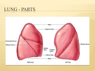
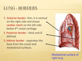
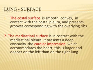
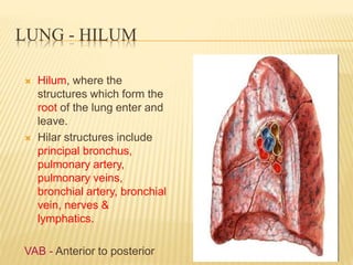
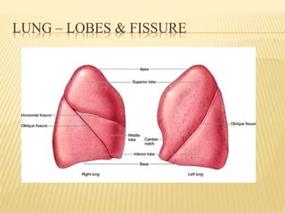
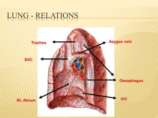
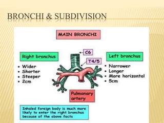
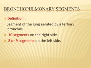
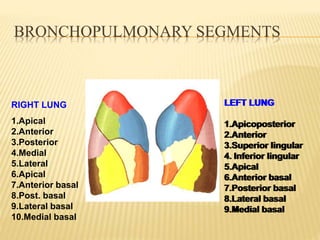
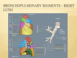
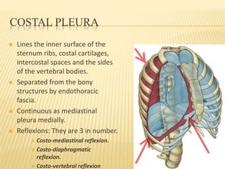
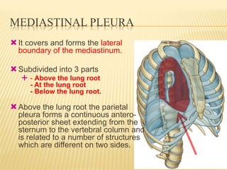
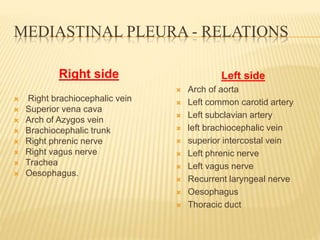
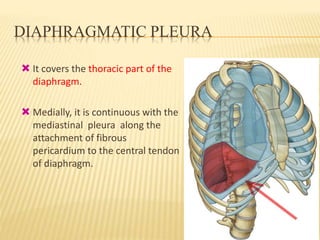
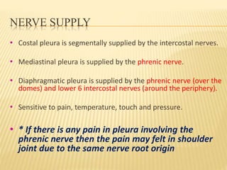
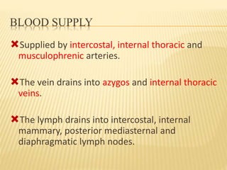
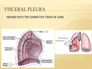
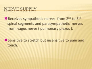
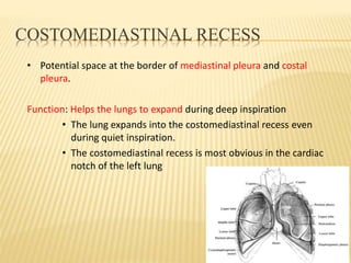
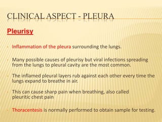
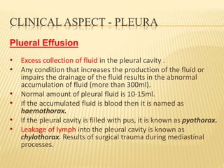
PLEURA
&
LUNG
Jekadeshnaidu Panirselvam
Faculty of Medicine,
AIMST University
INTRODUCTION - LUNG
 Pair of respiratory organs situated in the thoracic
cavity; separated by mediastinum.
 Each lung invaginates the corresponding pleural
cavity.
 Spongy in texture, brown or grey in colour. Gradually
they become mottled black due to deposition of
inhaled carbon particles.
 The surface is smooth, shining, and marked out into
numerous polyhedral areas, indicating the lobules of
the organ.
LUNG - PARTS
 Lung is conical in shape, and presents an apex, a
base, three borders, and two surfaces.
 The apex is rounded, and extends into the root of
the neck, 2.5 to 4 cm above the level of the sternal
end of first rib. Covered by cervical pleura and supra
pleural membrane.
 The base is broad, concave, and rests upon the
diaphragm, which separates the right lung from the
right lobe of the liver, and the left lung from the left
lobe of the liver, stomach, and spleen.
 As the diaphragm extends higher on the right than
on the left side, the concavity on the base of the
right lung is deeper than that on the left.
LUNG - PARTS
LUNG - BORDERS
1. Anterior border- thin, it is vertical
on the right side and shows
cardiac notch on the left side
below 4th costal cartilage.
2. Posterior border - thick and ill
defined.
3. Inferior border - separates the
base from the costal and
mediastinal surfaces.
Mediastinal surface of
right lung
LUNG - SURFACE
1. The costal surface is smooth, convex, in
contact with the costal pleura, and presents,
grooves corresponding with the overlying ribs.
2. The mediastinal surface is in contact with the
mediastinal pleura. It presents a deep
concavity, the cardiac impression, which
accommodates the heart; this is larger and
deeper on the left than on the right lung.
LUNG - HILUM
 Hilum, where the
structures which form the
root of the lung enter and
leave.
 Hilar structures include
principal bronchus,
pulmonary artery,
pulmonary veins,
bronchial artery, bronchial
vein, nerves &
lymphatics.
VAB - Anterior to posterior
LUNG – LOBES & FISSURE
 Right lung is divided into 3 lobes (superior,
middle & inferior) by 2 fissures, oblique &
horizontal.
 Left lung is divided into 2 lobes (superior &
inferior) by the oblique fissure.
 Left lung has tongue like projection below the
cardiac notch called lingula (corresponds to the
middle lobe of right lung).
LUNG – LOBES & FISSURE
LUNG - RELATIONS
Right lung – Medial surface
 Azygos vein - arched furrow above the hilum.
 Superior vena cava & right brachiocephalic vein - a wide groove
below the apex.
 Brachiocephalic artery - a furrow near the apex.
 Esophagus - vertical groove, behind the hilus.
 Inferior vena cava - in front and to the right of the lower part of the
esophageal groove.
 Large impression for the right atrium.
LUNG - RELATIONS
Azygos vein
Oesophagus
IVC
SVC
Rt. Atrium
Trachea
LUNG - RELATIONS
Left lung- Medial surface
 Aortic arch- furrow Immediately above the hilus.
 Left subclavian artery- groove in the apex.
 Left brachiocephalic vein - impression close to the anterior
border of the lung .
 Descending aorta- vertical furrow behind the hilus and
pulmonary ligament.
 Esophagus- impression near the base of the lung.
 Large impression for left ventricle.
LUNG - RELATIONS
Lt.Ventricle
Lt.Brachiocephalic
vein
Lt. Subclavian artery
Trachea & esophagus
Arch of Aorta
Descending
Aorta
Oesophagus
DIFFERENCES BETWEEN LEFT AND RIGHT
LUNG
Right Left
Larger,3 lobes & 2 fissures (oblique
& transverse).
2 lobes & 1 fissure (only oblique
fissure).
Hilum shows eparterial & hyparterial
bronchi.
Hilum shows only one principal
bronchus.
Absence of cardiac notch & lingula. Presence of cardiac notch & lingula.
10 bronchopulmonary segments 8 or 9 broncho pulmonary segments.
BRONCHI & SUBDIVISION
 Trachea divides into two primary bronchi.
 The primary bronchi divide into secondary bronchi – 3 on the
right and 2 on the left – supplying lobes.
 The secondary bronchi divide to become tertiary bronchi for
bronchopulmonary segments.
 Tertiary bronchi divide so on until 23 branching's occur. This
yields the bronchial tree.
 Passages with a diameter of <1mm are called bronchioles.
 Terminal bronchioles are the last portion of the conducting
zone.
BRONCHI & SUBDIVISION
BRONCHOPULMONARY SEGMENTS
 Definition :
Segment of the lung aerated by a tertiary
bronchus.
 10 segments on the right side
 8 or 9 segments on the left side.
BRONCHOPULMONARY SEGMENTS
RIGHT LUNG
1.Apical
2.Anterior
3.Posterior
4.Medial
5.Lateral
6.Apical
7.Anterior basal
8.Post. basal
9.Lateral basal
10.Medial basal
LEFT LUNG
1.Apicoposterior
2.Anterior
3.Superior lingular
4. Inferior lingular
5.Apical
6.Anterior basal
7.Posterior basal
8.Lateral basal
9.Medial basal
LEFT LUNG
1.Apicoposterior
2.Anterior
3.Superior lingular
4. Inferior lingular
5.Apical
6.Anterior basal
7.Posterior basal
8.Lateral basal
9.Medial basal
BRONCHOPULMONARY SEGMENTS – RIGHT
LUNG
BRONCHOPULMONARY SEGMENTS – LEFT
LUNG
BRONCHOPULMONARY SEGMENTS
Clinical importance:
 Usually infection of one segment restricted to that
segment but some infections like tuberculosis
spreads to other segments.
 Bronchogenic carcinoma spreads across segments.
 Knowledge of broncho pulmonary segments help in
the surgical resection ,drainage of infections by
postural drainage, visualizing the interior by
bronchoscope.
LUNG – BLOOD SUPPLY
• Arteries - Left side - 2 bronchial arteries arising from
descending thoracic aorta.
 - Right side - 1 bronchial artery arising from posterior
intercostal artery.
• Veins - 2 bronchial veins on both sides.
• Left side drains into left superior intercostal vein or hemiazygos
vein.
• Right side drains into azygos vein.
 Lymphatic drainage:
 Bronchopulmonary nodes-present in the hilum.
PLEURA
PLEURA
 Definition: The pleura is a delicate and closed
serous sac into which corresponding lung invaginates
from the medial side and reduces it into a potential
space.
 Layers: - Visceral Layer
- Parietal Layer
Both the layers are continuous around the lung root
and pulmonary ligament.
PARIETAL PLEURA
PARIETAL PLEURA
 The outer layer of the pleura is called parietal
pleura.
 Subdivided into 4 parts:
- Cervical
- Costal
- Mediastinal
- Diaphragmatic.
CERVICAL PLEURA
 Covered externally by the suprapleural membrane
(Sibson’s fascia).
 Extends from inner border of 1st rib to cover the apex of
the lung.
 Continuous downward and medially with the mediastinal
pleura.
 Summit is 3-4 cm above the 1st costal cartilage.
 2.5 cm above the sternal end of the clavicle.
SUPRAPLEURAL MEMBRANE
 Dome shaped musculo-fascial expansion.
 Muscular part- scalenus minimus muscle.
 Fascial part- endo thoracic fascia.
 Attachments:
In front- inner border of the 1st rib
Behind – Tip of the transverse process
of the 7th cervical vertebra.
Medially- Continuous with the
pretracheal fascia by the side of the
trachea.
 Protects the apex of the lung and
cervical pleura from the structures
of the root of the neck.
COSTAL PLEURA
 Lines the inner surface of the
sternum ribs, costal cartilages,
intercostal spaces and the sides
of the vertebral bodies.
 Separated from the bony
structures by endothoracic
fascia.
 Continuous as mediastinal
pleura medially.
 Reflexions: They are 3 in number.
 Costo-mediastinal reflexion.
 Costo-diaphragmatic
reflexion.
 Costo-vertebral reflexion
MEDIASTINAL PLEURA
It covers and forms the lateral
boundary of the mediastinum.
Subdivided into 3 parts
 - Above the lung root
- At the lung root
- Below the lung root.
Above the lung root the parietal
pleura forms a continuous antero-
posterior sheet extending from the
sternum to the vertebral column and
is related to a number of structures
which are different on two sides.
MEDIASTINAL PLEURA - RELATIONS
Right side
 Right brachiocephalic vein
 Superior vena cava
 Arch of Azygos vein
 Brachiocephalic trunk
 Right phrenic nerve
 Right vagus nerve
 Trachea
 Oesophagus.
Left side
 Arch of aorta
 Left common carotid artery
 Left subclavian artery
 left brachiocephalic vein
 superior intercostal vein
 Left phrenic nerve
 Left vagus nerve
 Recurrent laryngeal nerve
 Oesophagus
 Thoracic duct
DIAPHRAGMATIC PLEURA
 It covers the thoracic part of the
diaphragm.
 Medially, it is continuous with the
mediastinal pleura along the
attachment of fibrous
pericardium to the central tendon
of diaphragm.
NERVE SUPPLY
• Costal pleura is segmentally supplied by the intercostal nerves.
• Mediastinal pleura is supplied by the phrenic nerve.
• Diaphragmatic pleura is supplied by the phrenic nerve (over the
domes) and lower 6 intercostal nerves (around the periphery).
• Sensitive to pain, temperature, touch and pressure.
• * If there is any pain in pleura involving the
phrenic nerve then the pain may felt in shoulder
joint due to the same nerve root origin
BLOOD SUPPLY
Supplied by intercostal, internal thoracic and
musculophrenic arteries.
The vein drains into azygos and internal thoracic
veins.
The lymph drains into intercostal, internal
mammary, posterior mediasternal and
diaphragmatic lymph nodes.
VISCERAL PLEURA
•BLENDS WITH THE CONNECTIVE TISSUE OF LUNG
NERVE SUPPLY
Receives sympathetic nerves from 2nd to 5th
spinal segments and parasympathetic nerves
from vagus nerve ( pulmonary plexus ).
Sensitive to stretch but insensitive to pain and
touch.
BLOOD SUPPLY
Supplied by bronchial arteries.
The veins drain into bronchial veins.
The lymph drain into bronchopulmonary lymph
nodes.
PLEURAL CAVITY
 The pleural cavity is filled with a small amount of serous fluid
which forms a thin film of liquid between the pleural layers.
The normal amount is 10-15ml.
 It prevents separation of the two pleural layers. But facilitates
the sliding of two layers among them.
 lubricates the surface, so the lungs can move easily within the
thoracic cavity.
 The pleural fluid also provides surface tension, keeping the
lung suitably close to the wall of the thorax, despite the lungs
not being directly fixed to it.
COSTODIAPHRAGMATIC RECESS
Cleft like potential space between the lower limit of
pleural sac and lower border of the lung.
Extent:
The lower limit of the pleura along the 8th , 10th and 12th rib in
mid clavicular, mid-axillary and scapular line
Lower border of the lung along the 6th,8th and 10th rib.
COSTODIAPHRAGMATIC RECESS
 Function: - Allows expansion of lung during full
inspiration.
- Most dependant part so fluid first appear
in
this space.
 Relation: Right side – Right lobe and caudate lobe of
the
liver
- Upper part of the posterior
surface
of the right kidney.
Left side – Fundus of the stomach, spleen
- Upper part of posterior surface of
the
left kidney.
COSTOMEDIASTINAL RECESS
• Potential space at the border of mediastinal pleura and costal
pleura.
Function: Helps the lungs to expand during deep inspiration
• The lung expands into the costomediastinal recess even
during quiet inspiration.
• The costomediastinal recess is most obvious in the cardiac
notch of the left lung
CLINICAL ASPECT - PLEURA
Pleurisy
• Inflammation of the pleura surrounding the lungs.
• Many possible causes of pleurisy but viral infections spreading
from the lungs to pleural cavity are the most common.
• The inflamed pleural layers rub against each other every time the
lungs expand to breathe in air.
• This can cause sharp pain when breathing, also called
pleuritic chest pain
• Thoracentesis is normally performed to obtain sample for testing.
CLINICAL ASPECT - PLEURA
Plueral Effusion
• Excess collection of fluid in the pleural cavity .
• Any condition that increases the production of the fluid or
impairs the drainage of the fluid results in the abnormal
accumulation of fluid (more than 300ml).
• Normal amount of pleural fluid is 10-15ml.
• If the accumulated fluid is blood then it is named as
haemothorax.
• If the pleural cavity is filled with pus, it is known as pyothorax.
• Leakage of lymph into the pleural cavity is known as
chylothorax. Results of surgical trauma during mediastinal
processes.
CLINICALASPECT – PLEURA (PLEURAL
EFFUSION
CLINICAL ASPECT - PLEURA
Pneumothorax
Collection of free air or gas in the pleural cavity.
Types of pneumothorax:
- Open pneumothorax
- Spontaneous pneumothorax
- Tension pneumothorax
Collapse of ipsilateral lung due to pressure change & disruption
of surface tension.
Potential for mediastinal shifts.
Pleura and lung

Pleura and lung

  • 1.
  • 2.
    INTRODUCTION - LUNG Pair of respiratory organs situated in the thoracic cavity; separated by mediastinum.  Each lung invaginates the corresponding pleural cavity.  Spongy in texture, brown or grey in colour. Gradually they become mottled black due to deposition of inhaled carbon particles.  The surface is smooth, shining, and marked out into numerous polyhedral areas, indicating the lobules of the organ.
  • 3.
    LUNG - PARTS Lung is conical in shape, and presents an apex, a base, three borders, and two surfaces.  The apex is rounded, and extends into the root of the neck, 2.5 to 4 cm above the level of the sternal end of first rib. Covered by cervical pleura and supra pleural membrane.  The base is broad, concave, and rests upon the diaphragm, which separates the right lung from the right lobe of the liver, and the left lung from the left lobe of the liver, stomach, and spleen.  As the diaphragm extends higher on the right than on the left side, the concavity on the base of the right lung is deeper than that on the left.
  • 4.
  • 5.
    LUNG - BORDERS 1.Anterior border- thin, it is vertical on the right side and shows cardiac notch on the left side below 4th costal cartilage. 2. Posterior border - thick and ill defined. 3. Inferior border - separates the base from the costal and mediastinal surfaces. Mediastinal surface of right lung
  • 6.
    LUNG - SURFACE 1.The costal surface is smooth, convex, in contact with the costal pleura, and presents, grooves corresponding with the overlying ribs. 2. The mediastinal surface is in contact with the mediastinal pleura. It presents a deep concavity, the cardiac impression, which accommodates the heart; this is larger and deeper on the left than on the right lung.
  • 7.
    LUNG - HILUM Hilum, where the structures which form the root of the lung enter and leave.  Hilar structures include principal bronchus, pulmonary artery, pulmonary veins, bronchial artery, bronchial vein, nerves & lymphatics. VAB - Anterior to posterior
  • 8.
    LUNG – LOBES& FISSURE  Right lung is divided into 3 lobes (superior, middle & inferior) by 2 fissures, oblique & horizontal.  Left lung is divided into 2 lobes (superior & inferior) by the oblique fissure.  Left lung has tongue like projection below the cardiac notch called lingula (corresponds to the middle lobe of right lung).
  • 9.
    LUNG – LOBES& FISSURE
  • 10.
    LUNG - RELATIONS Rightlung – Medial surface  Azygos vein - arched furrow above the hilum.  Superior vena cava & right brachiocephalic vein - a wide groove below the apex.  Brachiocephalic artery - a furrow near the apex.  Esophagus - vertical groove, behind the hilus.  Inferior vena cava - in front and to the right of the lower part of the esophageal groove.  Large impression for the right atrium.
  • 11.
    LUNG - RELATIONS Azygosvein Oesophagus IVC SVC Rt. Atrium Trachea
  • 12.
    LUNG - RELATIONS Leftlung- Medial surface  Aortic arch- furrow Immediately above the hilus.  Left subclavian artery- groove in the apex.  Left brachiocephalic vein - impression close to the anterior border of the lung .  Descending aorta- vertical furrow behind the hilus and pulmonary ligament.  Esophagus- impression near the base of the lung.  Large impression for left ventricle.
  • 13.
    LUNG - RELATIONS Lt.Ventricle Lt.Brachiocephalic vein Lt.Subclavian artery Trachea & esophagus Arch of Aorta Descending Aorta Oesophagus
  • 14.
    DIFFERENCES BETWEEN LEFTAND RIGHT LUNG Right Left Larger,3 lobes & 2 fissures (oblique & transverse). 2 lobes & 1 fissure (only oblique fissure). Hilum shows eparterial & hyparterial bronchi. Hilum shows only one principal bronchus. Absence of cardiac notch & lingula. Presence of cardiac notch & lingula. 10 bronchopulmonary segments 8 or 9 broncho pulmonary segments.
  • 15.
    BRONCHI & SUBDIVISION Trachea divides into two primary bronchi.  The primary bronchi divide into secondary bronchi – 3 on the right and 2 on the left – supplying lobes.  The secondary bronchi divide to become tertiary bronchi for bronchopulmonary segments.  Tertiary bronchi divide so on until 23 branching's occur. This yields the bronchial tree.  Passages with a diameter of <1mm are called bronchioles.  Terminal bronchioles are the last portion of the conducting zone.
  • 16.
  • 17.
    BRONCHOPULMONARY SEGMENTS  Definition: Segment of the lung aerated by a tertiary bronchus.  10 segments on the right side  8 or 9 segments on the left side.
  • 18.
    BRONCHOPULMONARY SEGMENTS RIGHT LUNG 1.Apical 2.Anterior 3.Posterior 4.Medial 5.Lateral 6.Apical 7.Anteriorbasal 8.Post. basal 9.Lateral basal 10.Medial basal LEFT LUNG 1.Apicoposterior 2.Anterior 3.Superior lingular 4. Inferior lingular 5.Apical 6.Anterior basal 7.Posterior basal 8.Lateral basal 9.Medial basal LEFT LUNG 1.Apicoposterior 2.Anterior 3.Superior lingular 4. Inferior lingular 5.Apical 6.Anterior basal 7.Posterior basal 8.Lateral basal 9.Medial basal
  • 19.
  • 20.
  • 21.
    BRONCHOPULMONARY SEGMENTS Clinical importance: Usually infection of one segment restricted to that segment but some infections like tuberculosis spreads to other segments.  Bronchogenic carcinoma spreads across segments.  Knowledge of broncho pulmonary segments help in the surgical resection ,drainage of infections by postural drainage, visualizing the interior by bronchoscope.
  • 22.
    LUNG – BLOODSUPPLY • Arteries - Left side - 2 bronchial arteries arising from descending thoracic aorta.  - Right side - 1 bronchial artery arising from posterior intercostal artery. • Veins - 2 bronchial veins on both sides. • Left side drains into left superior intercostal vein or hemiazygos vein. • Right side drains into azygos vein.  Lymphatic drainage:  Bronchopulmonary nodes-present in the hilum.
  • 23.
  • 24.
    PLEURA  Definition: Thepleura is a delicate and closed serous sac into which corresponding lung invaginates from the medial side and reduces it into a potential space.  Layers: - Visceral Layer - Parietal Layer Both the layers are continuous around the lung root and pulmonary ligament.
  • 25.
  • 26.
    PARIETAL PLEURA  Theouter layer of the pleura is called parietal pleura.  Subdivided into 4 parts: - Cervical - Costal - Mediastinal - Diaphragmatic.
  • 27.
    CERVICAL PLEURA  Coveredexternally by the suprapleural membrane (Sibson’s fascia).  Extends from inner border of 1st rib to cover the apex of the lung.  Continuous downward and medially with the mediastinal pleura.  Summit is 3-4 cm above the 1st costal cartilage.  2.5 cm above the sternal end of the clavicle.
  • 28.
    SUPRAPLEURAL MEMBRANE  Domeshaped musculo-fascial expansion.  Muscular part- scalenus minimus muscle.  Fascial part- endo thoracic fascia.  Attachments: In front- inner border of the 1st rib Behind – Tip of the transverse process of the 7th cervical vertebra. Medially- Continuous with the pretracheal fascia by the side of the trachea.  Protects the apex of the lung and cervical pleura from the structures of the root of the neck.
  • 29.
    COSTAL PLEURA  Linesthe inner surface of the sternum ribs, costal cartilages, intercostal spaces and the sides of the vertebral bodies.  Separated from the bony structures by endothoracic fascia.  Continuous as mediastinal pleura medially.  Reflexions: They are 3 in number.  Costo-mediastinal reflexion.  Costo-diaphragmatic reflexion.  Costo-vertebral reflexion
  • 30.
    MEDIASTINAL PLEURA It coversand forms the lateral boundary of the mediastinum. Subdivided into 3 parts  - Above the lung root - At the lung root - Below the lung root. Above the lung root the parietal pleura forms a continuous antero- posterior sheet extending from the sternum to the vertebral column and is related to a number of structures which are different on two sides.
  • 31.
    MEDIASTINAL PLEURA -RELATIONS Right side  Right brachiocephalic vein  Superior vena cava  Arch of Azygos vein  Brachiocephalic trunk  Right phrenic nerve  Right vagus nerve  Trachea  Oesophagus. Left side  Arch of aorta  Left common carotid artery  Left subclavian artery  left brachiocephalic vein  superior intercostal vein  Left phrenic nerve  Left vagus nerve  Recurrent laryngeal nerve  Oesophagus  Thoracic duct
  • 32.
    DIAPHRAGMATIC PLEURA  Itcovers the thoracic part of the diaphragm.  Medially, it is continuous with the mediastinal pleura along the attachment of fibrous pericardium to the central tendon of diaphragm.
  • 33.
    NERVE SUPPLY • Costalpleura is segmentally supplied by the intercostal nerves. • Mediastinal pleura is supplied by the phrenic nerve. • Diaphragmatic pleura is supplied by the phrenic nerve (over the domes) and lower 6 intercostal nerves (around the periphery). • Sensitive to pain, temperature, touch and pressure. • * If there is any pain in pleura involving the phrenic nerve then the pain may felt in shoulder joint due to the same nerve root origin
  • 34.
    BLOOD SUPPLY Supplied byintercostal, internal thoracic and musculophrenic arteries. The vein drains into azygos and internal thoracic veins. The lymph drains into intercostal, internal mammary, posterior mediasternal and diaphragmatic lymph nodes.
  • 35.
    VISCERAL PLEURA •BLENDS WITHTHE CONNECTIVE TISSUE OF LUNG
  • 36.
    NERVE SUPPLY Receives sympatheticnerves from 2nd to 5th spinal segments and parasympathetic nerves from vagus nerve ( pulmonary plexus ). Sensitive to stretch but insensitive to pain and touch.
  • 37.
    BLOOD SUPPLY Supplied bybronchial arteries. The veins drain into bronchial veins. The lymph drain into bronchopulmonary lymph nodes.
  • 38.
    PLEURAL CAVITY  Thepleural cavity is filled with a small amount of serous fluid which forms a thin film of liquid between the pleural layers. The normal amount is 10-15ml.  It prevents separation of the two pleural layers. But facilitates the sliding of two layers among them.  lubricates the surface, so the lungs can move easily within the thoracic cavity.  The pleural fluid also provides surface tension, keeping the lung suitably close to the wall of the thorax, despite the lungs not being directly fixed to it.
  • 39.
    COSTODIAPHRAGMATIC RECESS Cleft likepotential space between the lower limit of pleural sac and lower border of the lung. Extent: The lower limit of the pleura along the 8th , 10th and 12th rib in mid clavicular, mid-axillary and scapular line Lower border of the lung along the 6th,8th and 10th rib.
  • 40.
    COSTODIAPHRAGMATIC RECESS  Function:- Allows expansion of lung during full inspiration. - Most dependant part so fluid first appear in this space.  Relation: Right side – Right lobe and caudate lobe of the liver - Upper part of the posterior surface of the right kidney. Left side – Fundus of the stomach, spleen - Upper part of posterior surface of the left kidney.
  • 41.
    COSTOMEDIASTINAL RECESS • Potentialspace at the border of mediastinal pleura and costal pleura. Function: Helps the lungs to expand during deep inspiration • The lung expands into the costomediastinal recess even during quiet inspiration. • The costomediastinal recess is most obvious in the cardiac notch of the left lung
  • 42.
    CLINICAL ASPECT -PLEURA Pleurisy • Inflammation of the pleura surrounding the lungs. • Many possible causes of pleurisy but viral infections spreading from the lungs to pleural cavity are the most common. • The inflamed pleural layers rub against each other every time the lungs expand to breathe in air. • This can cause sharp pain when breathing, also called pleuritic chest pain • Thoracentesis is normally performed to obtain sample for testing.
  • 43.
    CLINICAL ASPECT -PLEURA Plueral Effusion • Excess collection of fluid in the pleural cavity . • Any condition that increases the production of the fluid or impairs the drainage of the fluid results in the abnormal accumulation of fluid (more than 300ml). • Normal amount of pleural fluid is 10-15ml. • If the accumulated fluid is blood then it is named as haemothorax. • If the pleural cavity is filled with pus, it is known as pyothorax. • Leakage of lymph into the pleural cavity is known as chylothorax. Results of surgical trauma during mediastinal processes.
  • 44.
    CLINICALASPECT – PLEURA(PLEURAL EFFUSION
  • 45.
    CLINICAL ASPECT -PLEURA Pneumothorax Collection of free air or gas in the pleural cavity. Types of pneumothorax: - Open pneumothorax - Spontaneous pneumothorax - Tension pneumothorax Collapse of ipsilateral lung due to pressure change & disruption of surface tension. Potential for mediastinal shifts.