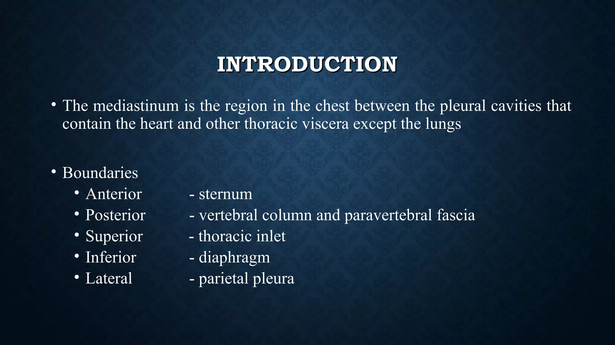
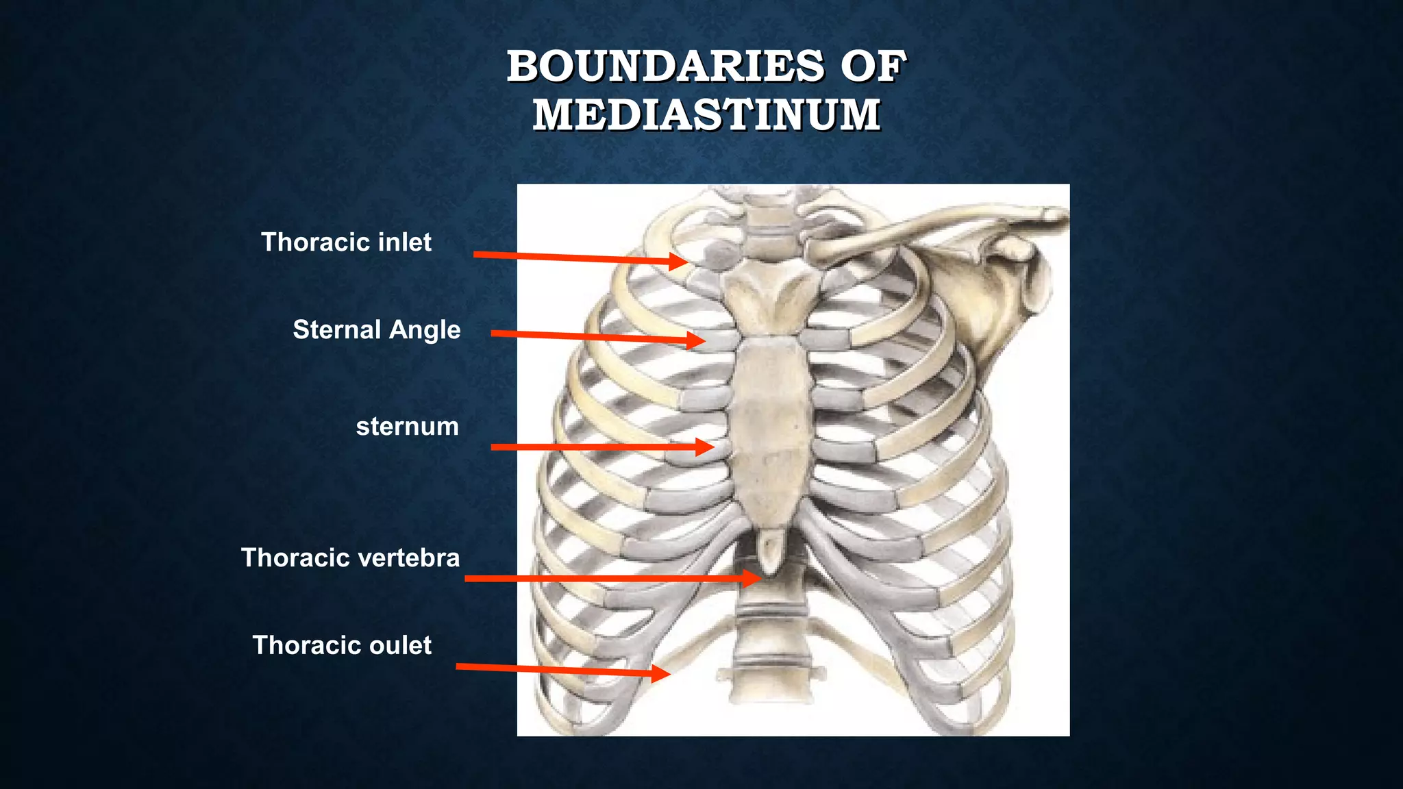
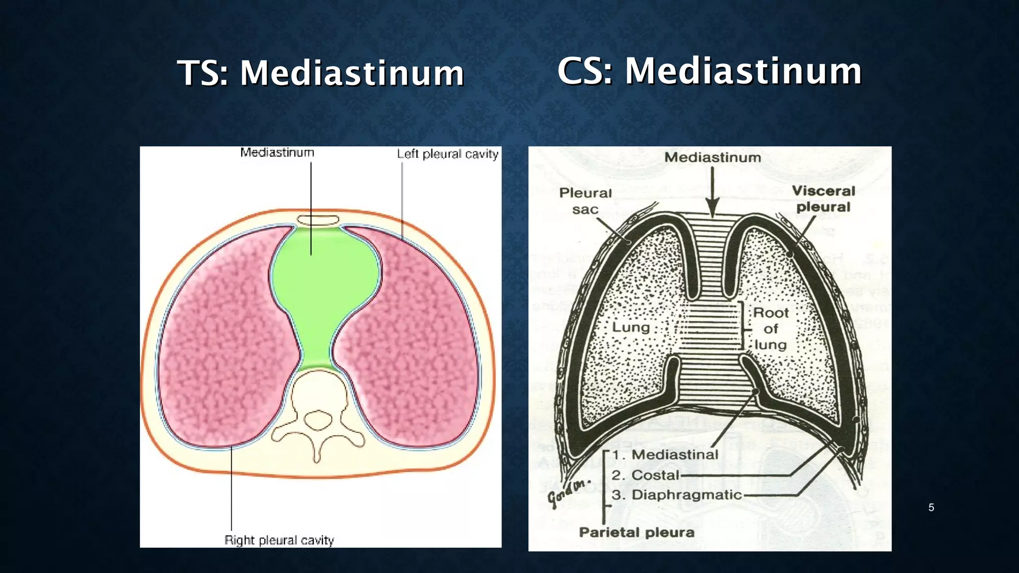
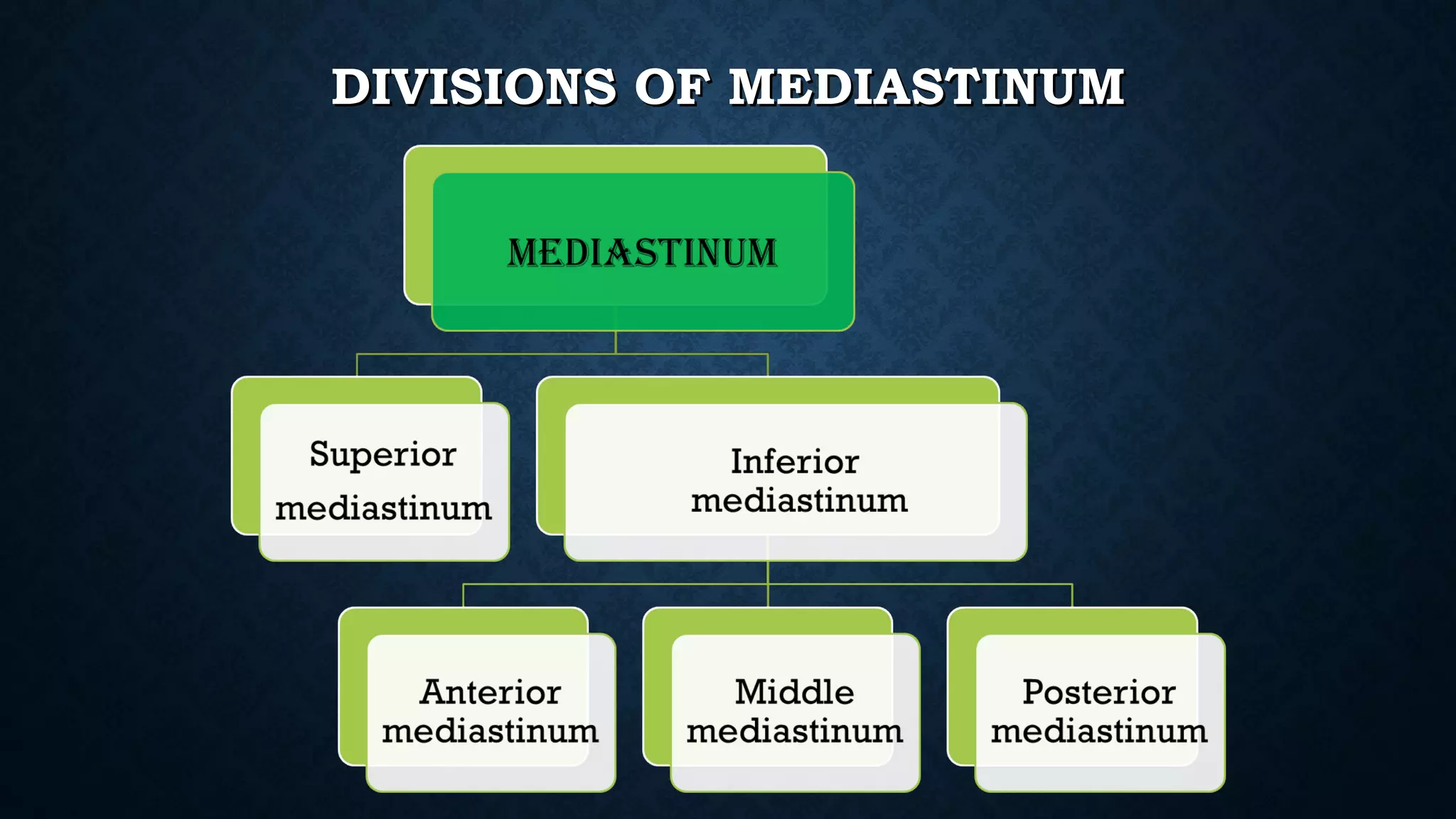
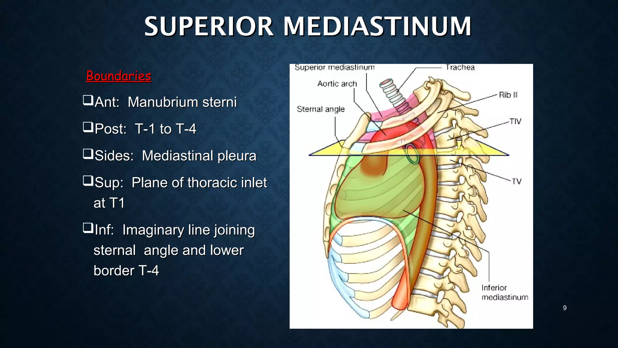
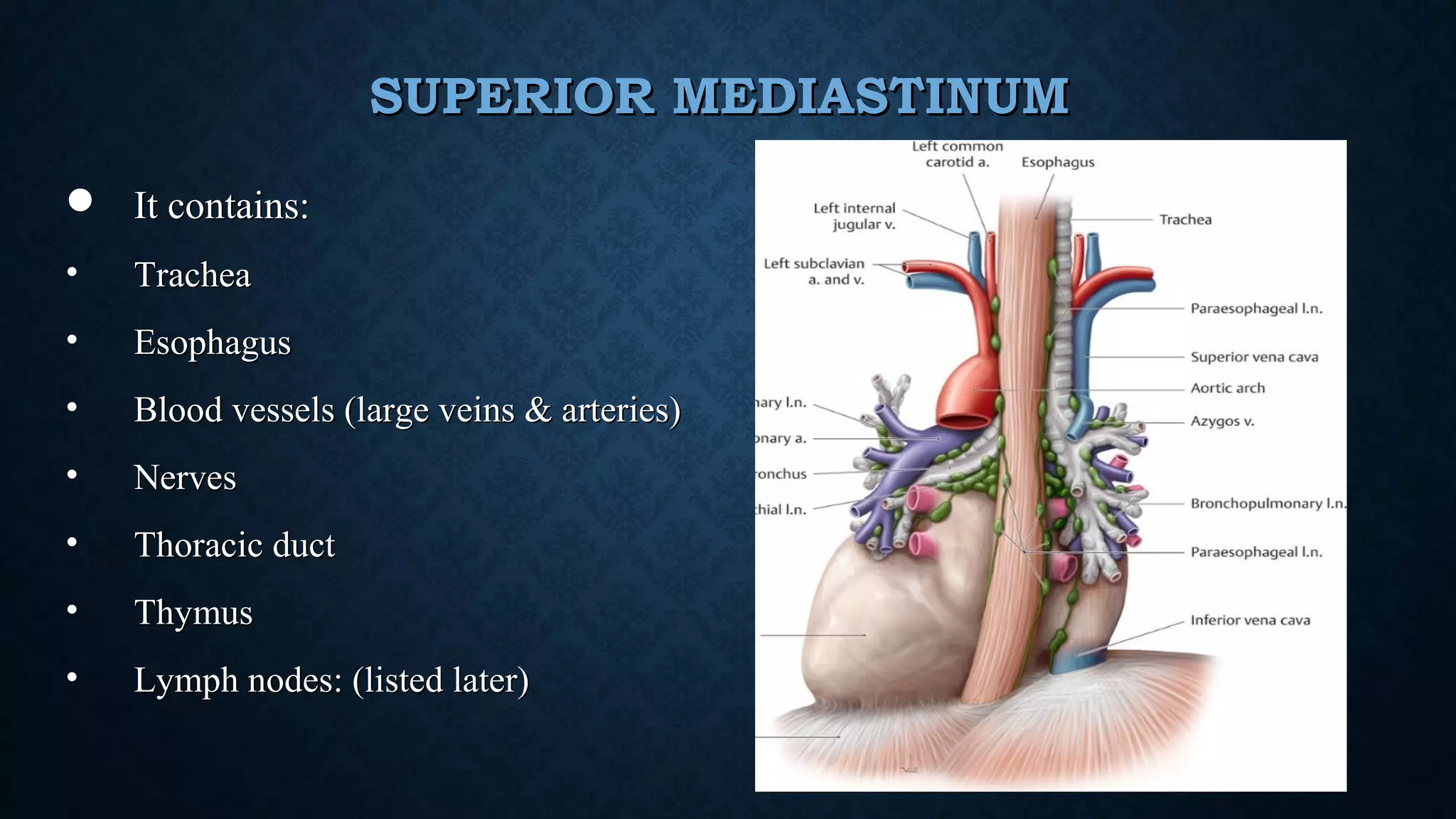
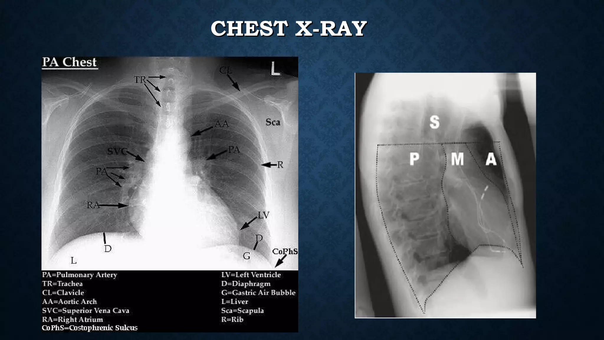
MEDIASTINUMMEDIASTINUM
Dr. Vibhay PareekDr. Vibhay Pareek
Radiation OncologyRadiation Oncology
Jupiter HospitalJupiter Hospital
INTRODUCTIONINTRODUCTION
• The mediastinum is the region in the chest between the pleural cavities that
contain the heart and other thoracic viscera except the lungs
• Boundaries
• Anterior - sternum
• Posterior - vertebral column and paravertebral fascia
• Superior - thoracic inlet
• Inferior - diaphragm
• Lateral - parietal pleura
Sternal Angle
Thoracic inlet
Thoracic oulet
BOUNDARIES OFBOUNDARIES OF
MEDIASTINUMMEDIASTINUM
sternum
Thoracic vertebra
TS: MediastinumTS: Mediastinum
5
CS: MediastinumCS: Mediastinum
DIVISIONS OF MEDIASTINUMDIVISIONS OF MEDIASTINUM
Superior
Mediastinum
Posterior
Mediastinum
Anterior
Mediastinum
Middle
Mediastinum
Sternal
Angle
T4
T5
divided into superior mediastinum and inferior mediastinum by an imaginary line passing
through sternal angle anteriorly lower border of 4th
thoracic vertebra posteriorly
Mediastinum divisionsMediastinum divisions
INFERIOR MEDIASTINUMINFERIOR MEDIASTINUM:: IS SUBDIVIDEDIS SUBDIVIDED INTOINTO
AnteriorAnterior
mediastinummediastinum
MiddleMiddle
mediastinummediastinum
PosteriorPosterior
mediastinummediastinum
SUPERIOR MEDIASTINUMSUPERIOR MEDIASTINUM
BoundariesBoundaries
Ant: Manubrium sterniAnt: Manubrium sterni
Post: T-1 to T-4Post: T-1 to T-4
Sides: Mediastinal pleuraSides: Mediastinal pleura
Sup: Plane of thoracic inletSup: Plane of thoracic inlet
at T1at T1
Inf: Imaginary line joiningInf: Imaginary line joining
sternal angle and lowersternal angle and lower
border T-4border T-4
9
SUPERIOR MEDIASTINUMSUPERIOR MEDIASTINUM
 It contains:It contains:
• TracheaTrachea
• EsophagusEsophagus
• Blood vessels (large veins & arteries)Blood vessels (large veins & arteries)
• NervesNerves
• Thoracic ductThoracic duct
• ThymusThymus
• Lymph nodes: (listed later)Lymph nodes: (listed later)
SUPERIOR MEDIASTINUM CONTENTSSUPERIOR MEDIASTINUM CONTENTS
Blood VesselsBlood Vessels
Veins:
SVC
Lt & Rt brachiocephalic
veins,
Arteries:
Arch of Aorta
Brachiocepalic artery
Lt Common carotid
Lt subclavian artery
SUPERIOR MEDIASTINUMSUPERIOR MEDIASTINUM
NervesNerves
1.1. Vagus nerveVagus nerve
2.2. Left RecurrentLeft Recurrent
Laryngeal nerve.Laryngeal nerve.
3.3. Phrenic nerve.Phrenic nerve.
SUPERIOR MEDIASTINUMSUPERIOR MEDIASTINUM
Lymph nodes:
 Highest mediastinal
 Paratracheal
 Prevascular
 retrotracheal
ANTERIOR MEDIASTINUMANTERIOR MEDIASTINUM
Lies ant. to pericardiumLies ant. to pericardium
Boundaries:Boundaries:
• Anterior: body of sternum
• Posterior: pericardium
• Superior: imaginary line separating sup. &
inf.mediastinum
• Infreior: diaphragm
• Lateral: mediastinal pleura
ANTERIOR MEDIASTINUM: CONTAINS:ANTERIOR MEDIASTINUM: CONTAINS:
a.a. Thymus glandThymus gland
b.b. Lymph NodesLymph Nodes
c.c. Fat.Fat.
MIDDLE MEDIASTINUMMIDDLE MEDIASTINUM
Boundaries:Boundaries:
• Anterior: posterior surface of sternum
• Posterior: oesophagus, desc. thoracic aorta,
azygous vein
• Superior: plane seperating sup.& inf
mediastinum
• Inferior: diaphragm
• Lateral: mediastinal pleura
MIDDLE MEDIASTINUMMIDDLE MEDIASTINUM
Contents:Contents:
HeartHeart enclosed in pericardiumenclosed in pericardium
Arteries:Arteries: Ascending Aorta,Ascending Aorta,
Pulmonary trunk with its Lt &Pulmonary trunk with its Lt &
Rt branchesRt branches
VeinsVeins: SVC,Pulmonary veins: SVC,Pulmonary veins
Nerves:Nerves: Phrenic, vagus nervePhrenic, vagus nerve
Bifurcation of Trachea withBifurcation of Trachea with
two principal bronchitwo principal bronchi
Tracheobronchial lymph nodesTracheobronchial lymph nodes 17
POSTERIOR MEDIASTINUMPOSTERIOR MEDIASTINUM
Boundaries:Boundaries:
Ant.Ant. Pericardium, Bifurcation of tracheaPericardium, Bifurcation of trachea
Post.Post. T5 to T12T5 to T12
sup.sup. Transverse thoracic planeTransverse thoracic plane
Inf.Inf. diaphragmdiaphragm
Sides:Sides: Mediastinal pleuraMediastinal pleura 18
19
POSTERIOR MEDIASTINUMPOSTERIOR MEDIASTINUM
Contents:Contents:
OesophagusOesophagus
ArteriesArteries
• Descending Aorta with its brsDescending Aorta with its brs
VeinsVeins
• AzygosAzygos
• HemizygosHemizygos
• Accessory hemizygosAccessory hemizygos
Nerves:Nerves:
• VagusVagus
• Splanchnic nervesSplanchnic nerves
Thoracic ductThoracic duct
lymph nodeslymph nodes
• Posterior mediastinalPosterior mediastinal
RADIOLOGICAL ANTOMYRADIOLOGICAL ANTOMY
CHEST X-RAYCHEST X-RAY
TRACHEOBRONCHIAL ANATOMYTRACHEOBRONCHIAL ANATOMY
23
Tracheal Displacement Due to Goiter
CLUES TO LOCATE MASS TO MEDIASTINUMCLUES TO LOCATE MASS TO MEDIASTINUM
Mediastinal Masses Masses In The Lung
 Not Contain Air
Bronchograms
 Mediastinal Mass Will
Create Obtuse Angles With
The Lung .
Mediastinal Lines Will Be
Disrupted
– May Contain Air
Bronchograms
– A Lung Mass Abutts The
Mediastinal Surface And
Creates Acute Angles With
The Lung
LEFT: A lung mass abutts the mediastinal surface and creates acute angles with the
lung.
RIGHT: A mediastinal mass will sit under the surface of the mediastinum, creating
obtuse angles with the lung
CERVICOTHORACIC SIGNCERVICOTHORACIC SIGN
• The anterior mediastinum ends at the level of the clavicles.The anterior mediastinum ends at the level of the clavicles.
• The posterior mediastinum extends much higher.The posterior mediastinum extends much higher.
• ThereforeTherefore
• any mass that remains sharply outlined in the apex of the thorax must beany mass that remains sharply outlined in the apex of the thorax must be
posterior and entirely within the chest, and posterior and entirely within the chest, and 
• any mass that disappears at the clavicles must be anterior and extendsany mass that disappears at the clavicles must be anterior and extends
into neckinto neck
See sharp
margin
above clavicle
Mass is in posterior mediastinum. because it remains sharply outlined in apex
of thorax, indicating that it is surrounded by lung.
This particular example is a ganglioneuroma
THORACOABDOMINAL SIGNTHORACOABDOMINAL SIGN
• A sharply marginated mediastinal mass seen through the diaphragmA sharply marginated mediastinal mass seen through the diaphragm
must lie entirely within the chest.must lie entirely within the chest.
• The posterior costophrenic sulcus extends far more caudally than theThe posterior costophrenic sulcus extends far more caudally than the
anterior aspect of the lunganterior aspect of the lung
• ThereforeTherefore
• Any mass that extends below the dome of the diaphragm and remainsAny mass that extends below the dome of the diaphragm and remains
sharply outlined must be in the posterior compartments andsharply outlined must be in the posterior compartments and
surrounded by lung, andsurrounded by lung, and
• Any mass that terminates at dome of diaphragm must be anteriorAny mass that terminates at dome of diaphragm must be anterior
Can you
see the
outline of the
mass below
the diaphragm?
Margin of mass is apparent and below diaphragm, therefore this must be in the middle or posterior
compartments where it is surrounded by lung
This example is a ‘Lipoma’
HILUM OVERLAY SIGNHILUM OVERLAY SIGN
• Principle of hilum overlayPrinciple of hilum overlay
• An anterior mediastinal mass will overlap the mainAn anterior mediastinal mass will overlap the main
pulmonary arteries, therefore they will be seen within thepulmonary arteries, therefore they will be seen within the
margins of the massmargins of the mass
Hilum can
be seen
through
mass
this must be an anterior
mediastinal mass
because it overlaps rather
than “pushes out” the
main pulmonary arteries
This particular example is a thymoma
VASCULAR ANATOMYVASCULAR ANATOMY
At T3 LevelAt T3 Level
At T4 LevelAt T4 Level
At T5 LevelAt T5 Level
At T6 LevelAt T6 Level
MEDIASTINAL TUMORS EPIDEMOLOGYMEDIASTINAL TUMORS EPIDEMOLOGY
MEDIASTINAL MASSESMEDIASTINAL MASSES
Compartment % Malignant
Anterosuperior 59
Middle 29
Posterior 16
Mediastinal
division
Most common
tumors
Anterior-
superior
thymoma
middle lymphoma
posterior Neurogenic tumors
Anterosuperior Masses
Thymus
• Thymoma
• Thymic carcinoma
• Thymic cyst
• Thymic carcinoid
• Thymolipoma
mediasTinal lymphoma
• Hodgkin’s Lymphoma
• Non-Hodgkin’s Lymphoma
mesenchymal Tumors
Germ cell Tumor
• Seminoma
• Non seminomatous Germ Cell
• Embryonal cell carcinoma
• Endodermal sinus tumor
• Choriocarcinoma
• Teratoma
• Mature
• Immature
endocrine Tumors
• Thyroid tumors
• Parathyroid adenoma
Middle mediastinal masses
mediasTinal lymphoma
• Hodgkin’s Lymphoma
• Non-Hodgkin’s Lymphoma
mesenchymal Tumors
cysT:
• Bronchogenic cyst
• Thoracic duct
• Meningoceles
cardiac & pericardial
Tumors
Tracheal Tumors
vascular Tumors
lymphadenopaThy
• Inflammatory
• Granulomatous
• sarcoidosis
Posterior mediastinal masses
mediasTinal lymphoma
• Hodgkin’s Lymphoma
• Non-Hodgkin’s Lymphoma
mesenchymal Tumors
neuroGenic Tumors
• Peripheral nerves
• Symphathetic ganglia
• paraganglia
endocrine Tumors
esophaGeal Tumors
& cysTs
TUMORS OF THYMUSTUMORS OF THYMUS
• ThymomasThymomas
• Thymic carcinomasThymic carcinomas
• Thymic lymphomasThymic lymphomas
• CarcinoidsCarcinoids
• ThymolipomasThymolipomas
• SecondariesSecondaries
THYMOMATHYMOMA
PresentationPresentation
• Most common primary anterior mediastinal tumorMost common primary anterior mediastinal tumor
• M=F, most >40M=F, most >40
• Most patients are asymptomaticMost patients are asymptomatic
• Half of patients suffer have associated parathymic syndromesHalf of patients suffer have associated parathymic syndromes
• myasthenia gravismyasthenia gravis
• hypogammaglobulinemiahypogammaglobulinemia
• pure red cell aplasiapure red cell aplasia
• 1/3 have chest pain, cough or dyspnea on presentation1/3 have chest pain, cough or dyspnea on presentation
• Myasthenia gravis occurs in 30-50% of pts with thymoma.Myasthenia gravis occurs in 30-50% of pts with thymoma.
Hypogammaglobulinemia occurs in 10% of pts with thymomaHypogammaglobulinemia occurs in 10% of pts with thymoma
• Pure red cell aplasia occurs in 5%, but thymoma occurs in 50% ofPure red cell aplasia occurs in 5%, but thymoma occurs in 50% of
pts with red cell aplasiapts with red cell aplasia
THYMOMATHYMOMA
• lobulated mass in the anterior mediastinum
THYMOMATHYMOMA
INVASIVE THYMOMAINVASIVE THYMOMA
• Encasement of mediastinal structures,
infiltration of fat planes, and an irregular
interface between the mass and lung
parenchyma, are highly suggestive of
invasion.
• Pleural thickening, nodularity, or effusion
generally indicates pleural invasion by
the thymoma
THYMIC CARCINOIDTHYMIC CARCINOID
carcinoid tumors (neuroendocrine tumors) of the thymus are very rare,carcinoid tumors (neuroendocrine tumors) of the thymus are very rare,
accounting for <5% of all neoplasms of the anterior mediastinum.accounting for <5% of all neoplasms of the anterior mediastinum.
They originate from the normal thymic Kulchitsky cells, which belong to the amine precursor uptakeThey originate from the normal thymic Kulchitsky cells, which belong to the amine precursor uptake
and decarboxylation (APUD) groupand decarboxylation (APUD) group
PresentationPresentation
• men aged 30 to 50 yearsmen aged 30 to 50 years
• (male/female ratio: 3:1)(male/female ratio: 3:1)
• Rarely associated with carcinoid syndromeRarely associated with carcinoid syndrome
• Associated endocrine abnormalities: Cushing’s syndrome due to ectopic ACTH or MENAssociated endocrine abnormalities: Cushing’s syndrome due to ectopic ACTH or MEN
• 73% have regional lymph node and/or distant osteoblastic bone mets73% have regional lymph node and/or distant osteoblastic bone mets
• Thymic carcinoid tumor in a 22-year-old man with a 3-month history of a persistent dry cough.Thymic carcinoid tumor in a 22-year-old man with a 3-month history of a persistent dry cough.
• Contrast-enhanced CT scan shows a heterogeneously enhancing thymic mass .Contrast-enhanced CT scan shows a heterogeneously enhancing thymic mass .
• PET image shows intense FDG uptake by the massPET image shows intense FDG uptake by the mass
THYMIC CARCINOMATHYMIC CARCINOMA
• Thymic carcinomas behave moreThymic carcinomas behave more
aggressively than invasive thymomas andaggressively than invasive thymomas and
are more likely to metastasize to distantare more likely to metastasize to distant
sitessites
THYMIC LYMPHOMASTHYMIC LYMPHOMAS
• Lymphoma is the most commonLymphoma is the most common
cause of an anterior mediastinalcause of an anterior mediastinal
mass in children and the secondmass in children and the second
most common cause of anmost common cause of an
anterior mediastinal mass inanterior mediastinal mass in
adults.adults.
CANCERS OF THE HEAD AND NECK, ABDOMEN, AND PELVIS CAN INVOLVE THECANCERS OF THE HEAD AND NECK, ABDOMEN, AND PELVIS CAN INVOLVE THE
THYMUS VIA LYMPHATIC PATHWAYSTHYMUS VIA LYMPHATIC PATHWAYS
• Metastatic disease to the thymusMetastatic disease to the thymus
in a 10-year-old boy 2 years afterin a 10-year-old boy 2 years after
diagnosis of alveolardiagnosis of alveolar
rhabdomyosarcoma of the thigh.rhabdomyosarcoma of the thigh.
Secondary Tumors of the Thymus
MEDIASTINAL LYMPHOMAMEDIASTINAL LYMPHOMA
PRIMARY MEDIASTINAL LYMPHOMAPRIMARY MEDIASTINAL LYMPHOMA
• 5-10% of patients with lymphoma present with primary5-10% of patients with lymphoma present with primary
mediastinal lesionsmediastinal lesions
• Primary mediastinal lymphoma represents 10-20% ofPrimary mediastinal lymphoma represents 10-20% of
primary mediastinal masses in adults and are usually inprimary mediastinal masses in adults and are usually in
the anterosuperior compartmentthe anterosuperior compartment
• Usually present with fever, weight loss and night sweatsUsually present with fever, weight loss and night sweats
• Pain, dyspnea, stridor, SVC syndrome due to mass effectsPain, dyspnea, stridor, SVC syndrome due to mass effects
are uncommonare uncommon
PRIMARY MEDIASTINAL LYMPHOMAPRIMARY MEDIASTINAL LYMPHOMA
Two TypesTwo Types
• Primary Mediastinal Hodgkin’s LymphomaPrimary Mediastinal Hodgkin’s Lymphoma
• Primary Mediastinal Non-Hodgkin’s LymphomaPrimary Mediastinal Non-Hodgkin’s Lymphoma
• Poorly differentiated lymphoblasticPoorly differentiated lymphoblastic
• Diffuse lymphocyticDiffuse lymphocytic
• Primary Mediastinal B-cell LymphomaPrimary Mediastinal B-cell Lymphoma
PRIMARY MEDIASTINAL HODGKIN’SPRIMARY MEDIASTINAL HODGKIN’S
LYMPHOMALYMPHOMA
PresentationPresentation
• Incidental mediastinal mass on chest xray is 2nd most commonIncidental mediastinal mass on chest xray is 2nd most common
presentation after asymptomatic lymphadenopathypresentation after asymptomatic lymphadenopathy
• Mass is usually large, rarely causes retrosternal chest pain, cough,Mass is usually large, rarely causes retrosternal chest pain, cough,
dyspnea, effusions or SVC syndromedyspnea, effusions or SVC syndrome
• Bimodal age distributionBimodal age distribution
• ““B” symptoms: fever, weight loss (>10% body wt in 6 months), nightB” symptoms: fever, weight loss (>10% body wt in 6 months), night
sweatssweats
• Generalized pruritus presentGeneralized pruritus present
A chest CT exam shows the mass to extend from the neck to the diaphragm, compressing the
tracheal and left mainstem bronchus leading to left lower lobe atelectasis. The chest wall mass is
partially eroding the sternum.
Two contiguous slices from an enhanced
chest CT exam show a homogenous, solid,
anterior mediastinal mass and a large right
pleural effusion.
Dx-Lymphoma
Non-Hodgkin,
Anterior Mediastinal
MEDIASTINAL GERM CELL TUMORSMEDIASTINAL GERM CELL TUMORS
• Primary extragonadal germ cell tumors comprise 2% to 5%Primary extragonadal germ cell tumors comprise 2% to 5%
of all germ cell tumorsof all germ cell tumors
• Approximately two thirds of these tumors occur in theApproximately two thirds of these tumors occur in the
mediastinummediastinum
• The mediastinum is the most common site of primaryThe mediastinum is the most common site of primary
extragonadal germ cell tumors in young adultsextragonadal germ cell tumors in young adults
• Represent 10-15% of adult anterosuperior mediastinalRepresent 10-15% of adult anterosuperior mediastinal
tumorstumors
MEDIASTINAL GERM CELL TUMORSMEDIASTINAL GERM CELL TUMORS
• they presumably arise from germ cells that migrate along thethey presumably arise from germ cells that migrate along the
urogenital ridge during embryonic development .urogenital ridge during embryonic development .
• The embryologic urogenital ridge extends from C6 to L4 and afterThe embryologic urogenital ridge extends from C6 to L4 and after
malignant transformation of displaced germ cells, explains themalignant transformation of displaced germ cells, explains the
development of primary germ cell tumors outside the gonadsdevelopment of primary germ cell tumors outside the gonads
MEDIASTINAL GERM CELL TUMORSMEDIASTINAL GERM CELL TUMORS
Three typesThree types
• TeratomaTeratoma
• SeminomaSeminoma
• Nonseminomatous Germ Cell TumorNonseminomatous Germ Cell Tumor
MEDIASTINAL TERATOMASMEDIASTINAL TERATOMAS
• Most common mediastinal germ cell tumorMost common mediastinal germ cell tumor
• Three types:Three types:
• Mature:Mature: benign, well-differentiatedbenign, well-differentiated
• Immature:Immature: contains >50% immature components, may recur orcontains >50% immature components, may recur or
metastasizemetastasize
• Malignant:Malignant: a mature teratoma that contains a focus of carcinoma,a mature teratoma that contains a focus of carcinoma,
sarcoma or malignant GCTsarcoma or malignant GCT
MATURE TERATOMAMATURE TERATOMA
• Occurs in children and young adultsOccurs in children and young adults
• Usually asymptomatic, but if large enough, may cause chest pain,Usually asymptomatic, but if large enough, may cause chest pain,
dyspnea, cough or other symptoms of mediastinal compressiondyspnea, cough or other symptoms of mediastinal compression
• Contains derivatives of all three primitive germ layers includingContains derivatives of all three primitive germ layers including
• Ectoderm: teeth, skin, hairEctoderm: teeth, skin, hair
• Mesoderm: cartilage and boneMesoderm: cartilage and bone
• Endoderm: bronchial, intestinal and pancreatic tissueEndoderm: bronchial, intestinal and pancreatic tissue
• Expectoration of hair (trichoptysis) is rare but pathognomonicExpectoration of hair (trichoptysis) is rare but pathognomonic
Dx Teratoma,
Anterior Mediastinal
CT exam show a low density mass
in the anterior mediastinum with
irregular walls with calcium in it.
MEDIASTINAL SEMINOMAMEDIASTINAL SEMINOMA
• Represents 40% of malignant mediastinal GCTsRepresents 40% of malignant mediastinal GCTs
• Afflicts Caucasian men in 20s-30sAfflicts Caucasian men in 20s-30s
• Only rarely represents a metastatic lesion from a testicular primaryOnly rarely represents a metastatic lesion from a testicular primary
tumor, but testicular USG is usually performed to rule this outtumor, but testicular USG is usually performed to rule this out
• If any other germ cell tumor histology is identified in the tumor, it isIf any other germ cell tumor histology is identified in the tumor, it is
treated as a mixed NSGCTtreated as a mixed NSGCT
• AFP normal,AFP normal, ββ-HCG may be elevated in 10%-HCG may be elevated in 10%
MEDIASTINAL SEMINOMAMEDIASTINAL SEMINOMA
PresentationPresentation
• Slow growing tumor, usually symptomatic at diagnosisSlow growing tumor, usually symptomatic at diagnosis
• Commonly presents with chest pain, dyspnea, cough, weightCommonly presents with chest pain, dyspnea, cough, weight
lossloss
• Presents infrequently with SVC syndromePresents infrequently with SVC syndrome
• Bulky, lobulated, homogeneous mass, no calcificationsBulky, lobulated, homogeneous mass, no calcifications
• Usually not invasive, but many have metastasized to regionalUsually not invasive, but many have metastasized to regional
lymph nodes, lung and/or bone by the time of diagnosislymph nodes, lung and/or bone by the time of diagnosis
MEDIASTINAL NONSEMINOMATOUS GERMMEDIASTINAL NONSEMINOMATOUS GERM
CELL TUMORSCELL TUMORS
• Five TypesFive Types
• Embryonal cell carcinomaEmbryonal cell carcinoma
• Endodermal sinus tumor: elevated AFPEndodermal sinus tumor: elevated AFP
• Choriocarcinoma: elevatedChoriocarcinoma: elevated ββ-HCG-HCG
• Malignant TeratomaMalignant Teratoma
• MixedMixed
MEDIASTINAL NONSEMINOMATOUS GERMMEDIASTINAL NONSEMINOMATOUS GERM
CELL TUMORSCELL TUMORS
• NSGCTs of the mediastinum have a worse prognosis thanNSGCTs of the mediastinum have a worse prognosis than
mediastinal seminomas or teratomasmediastinal seminomas or teratomas
• Occur in men in the 20-40 age groupOccur in men in the 20-40 age group
• 20% of patients also have Klinefelter’s syndrome20% of patients also have Klinefelter’s syndrome
TRACHEAL TUMORSTRACHEAL TUMORS
• Extremely rare tumors.Extremely rare tumors.
• Comprise of 0.1 to 0.4 %of all diagnosed malignanciesComprise of 0.1 to 0.4 %of all diagnosed malignancies
• Two types: squamous cell carcinoma M:F=3:1 Age:6Two types: squamous cell carcinoma M:F=3:1 Age:6thth
decadedecade
adenoid cystic carcinomas M:F=1:1 younger ageadenoid cystic carcinomas M:F=1:1 younger age
• Clinical feature: cough, dysnoea, dysphagia,stridor hemoptysis,Clinical feature: cough, dysnoea, dysphagia,stridor hemoptysis,
dysphoniadysphonia
• Middle mediastinum is the commonest site of intrathoracicMiddle mediastinum is the commonest site of intrathoracic
lymphadenopathy.lymphadenopathy.
• Gross lymphadenopathy is a feature ofGross lymphadenopathy is a feature of
1)Tuberculosis1)Tuberculosis
2)Histoplasmosis.2)Histoplasmosis.
3) Metastatic carcinoma3) Metastatic carcinoma
4) Lymphomas,4) Lymphomas,
5)Sarcoidosis.5)Sarcoidosis.
ENTERIC CYSTSENTERIC CYSTS
• Are located in the posterior mediastinumAre located in the posterior mediastinum
• Lined by gastric or intestinal epithelium.Lined by gastric or intestinal epithelium.
• All cysts may become1) InfectedAll cysts may become1) Infected
2) Bleed2) Bleed
3)Rupture3)Rupture
• Rupture into the Mediastinum.Rupture into the Mediastinum.
Pleural cavity.Pleural cavity.
NEUROGENIC TUMORSNEUROGENIC TUMORS
PNEUMOMEDIASTINUMPNEUMOMEDIASTINUM
CLINICAL PRESENTATION OF MEDIASTINAL MASSCLINICAL PRESENTATION OF MEDIASTINAL MASS
CLINICAL PRESENTATIONCLINICAL PRESENTATION
Asymptomatic massAsymptomatic mass
Incidental discovery – most commonIncidental discovery – most common
50% of all mediastinal mass are asymptomatic50% of all mediastinal mass are asymptomatic
80% of such mass are benign80% of such mass are benign
More than half are malignant if with symptomsMore than half are malignant if with symptoms
CLINICAL PRESENTATIONCLINICAL PRESENTATION
Effects on Compression or invasion of adjacent tissues
• Chest pain, from traction on mediastinal mass, tissue invasion, or
bone erosion is common
• Cough, because of extrinsic compression of the trachea or
bronchi, or erosion into the airway itself
• Hemoptysis, hoarseness or stridor
• Pleural effusion, invasion or irritation of pleural space
• Dysphagia, invasion or direct invasioin of the esophagus
• Pericarditis or pericardial tamponade
• Right ventricular outflow obstruction and cor pulmonale
CLINICAL PRESENTATIONCLINICAL PRESENTATION
Effects on Compression of nerves
• Hoarseness, invading or compressing the nerves recurrent laryngeal
nerve
• Horners syndrome, involvement of the sympathetic ganglia
• Dyspnea, from phrenic nerve involvement causing diaphragmatic
paralysis
• Tachycardia, secondary to vagus nerve involvement
CLINICAL PRESENTATIONCLINICAL PRESENTATION
• Superior vena cava
• Vulnerable to extrinsic compression and obstruction because it is thin walled and
its intravascular pressure is low.
• Superior vena cava syndrome
• Results from the increase venous pressure in the upper thorax , head and neck
• characterized by dilation of the collateral veins in the upper portion of the head
and thorax and edema and phlethora of the face, neck and upper torso, suffusion
and edema of the conjunctiva and cerebral symptoms such as headache,
disturbance of consciousness and visual distortion
• Bronchogenic carcinoma and lymphoma are the most common etiologies
MEDIASTINAL MASS: PRE TREATMENTMEDIASTINAL MASS: PRE TREATMENT
EVALUATIONEVALUATION
LYMPH NODESLYMPH NODES
SUPRACLAVICULAR AND UPPERSUPRACLAVICULAR AND UPPER
PARATRACHEALPARATRACHEAL
PREVERTEBRAL AND PREVASCULARPREVERTEBRAL AND PREVASCULAR
LOWER PARATRACHEALLOWER PARATRACHEAL
SUBAORTIC AND PARAAORTICSUBAORTIC AND PARAAORTIC
CARINAL, PARAESOPHAGEAL AND HILARCARINAL, PARAESOPHAGEAL AND HILAR
THANK YOUTHANK YOU

Radiology day 3 mediastinal anatomy

  • 1.
    MEDIASTINUMMEDIASTINUM Dr. Vibhay PareekDr.Vibhay Pareek Radiation OncologyRadiation Oncology Jupiter HospitalJupiter Hospital
  • 2.
    INTRODUCTIONINTRODUCTION • The mediastinumis the region in the chest between the pleural cavities that contain the heart and other thoracic viscera except the lungs • Boundaries • Anterior - sternum • Posterior - vertebral column and paravertebral fascia • Superior - thoracic inlet • Inferior - diaphragm • Lateral - parietal pleura
  • 3.
    Sternal Angle Thoracic inlet Thoracicoulet BOUNDARIES OFBOUNDARIES OF MEDIASTINUMMEDIASTINUM sternum Thoracic vertebra
  • 4.
    TS: MediastinumTS: Mediastinum 5 CS:MediastinumCS: Mediastinum
  • 5.
  • 6.
    Superior Mediastinum Posterior Mediastinum Anterior Mediastinum Middle Mediastinum Sternal Angle T4 T5 divided into superiormediastinum and inferior mediastinum by an imaginary line passing through sternal angle anteriorly lower border of 4th thoracic vertebra posteriorly Mediastinum divisionsMediastinum divisions
  • 7.
    INFERIOR MEDIASTINUMINFERIOR MEDIASTINUM::IS SUBDIVIDEDIS SUBDIVIDED INTOINTO AnteriorAnterior mediastinummediastinum MiddleMiddle mediastinummediastinum PosteriorPosterior mediastinummediastinum
  • 8.
    SUPERIOR MEDIASTINUMSUPERIOR MEDIASTINUM BoundariesBoundaries Ant:Manubrium sterniAnt: Manubrium sterni Post: T-1 to T-4Post: T-1 to T-4 Sides: Mediastinal pleuraSides: Mediastinal pleura Sup: Plane of thoracic inletSup: Plane of thoracic inlet at T1at T1 Inf: Imaginary line joiningInf: Imaginary line joining sternal angle and lowersternal angle and lower border T-4border T-4 9
  • 9.
    SUPERIOR MEDIASTINUMSUPERIOR MEDIASTINUM It contains:It contains: • TracheaTrachea • EsophagusEsophagus • Blood vessels (large veins & arteries)Blood vessels (large veins & arteries) • NervesNerves • Thoracic ductThoracic duct • ThymusThymus • Lymph nodes: (listed later)Lymph nodes: (listed later)
  • 10.
    SUPERIOR MEDIASTINUM CONTENTSSUPERIORMEDIASTINUM CONTENTS Blood VesselsBlood Vessels Veins: SVC Lt & Rt brachiocephalic veins, Arteries: Arch of Aorta Brachiocepalic artery Lt Common carotid Lt subclavian artery
  • 11.
    SUPERIOR MEDIASTINUMSUPERIOR MEDIASTINUM NervesNerves 1.1.Vagus nerveVagus nerve 2.2. Left RecurrentLeft Recurrent Laryngeal nerve.Laryngeal nerve. 3.3. Phrenic nerve.Phrenic nerve.
  • 12.
    SUPERIOR MEDIASTINUMSUPERIOR MEDIASTINUM Lymphnodes:  Highest mediastinal  Paratracheal  Prevascular  retrotracheal
  • 13.
    ANTERIOR MEDIASTINUMANTERIOR MEDIASTINUM Liesant. to pericardiumLies ant. to pericardium Boundaries:Boundaries: • Anterior: body of sternum • Posterior: pericardium • Superior: imaginary line separating sup. & inf.mediastinum • Infreior: diaphragm • Lateral: mediastinal pleura
  • 14.
    ANTERIOR MEDIASTINUM: CONTAINS:ANTERIORMEDIASTINUM: CONTAINS: a.a. Thymus glandThymus gland b.b. Lymph NodesLymph Nodes c.c. Fat.Fat.
  • 15.
    MIDDLE MEDIASTINUMMIDDLE MEDIASTINUM Boundaries:Boundaries: •Anterior: posterior surface of sternum • Posterior: oesophagus, desc. thoracic aorta, azygous vein • Superior: plane seperating sup.& inf mediastinum • Inferior: diaphragm • Lateral: mediastinal pleura
  • 16.
    MIDDLE MEDIASTINUMMIDDLE MEDIASTINUM Contents:Contents: HeartHeartenclosed in pericardiumenclosed in pericardium Arteries:Arteries: Ascending Aorta,Ascending Aorta, Pulmonary trunk with its Lt &Pulmonary trunk with its Lt & Rt branchesRt branches VeinsVeins: SVC,Pulmonary veins: SVC,Pulmonary veins Nerves:Nerves: Phrenic, vagus nervePhrenic, vagus nerve Bifurcation of Trachea withBifurcation of Trachea with two principal bronchitwo principal bronchi Tracheobronchial lymph nodesTracheobronchial lymph nodes 17
  • 17.
    POSTERIOR MEDIASTINUMPOSTERIOR MEDIASTINUM Boundaries:Boundaries: Ant.Ant.Pericardium, Bifurcation of tracheaPericardium, Bifurcation of trachea Post.Post. T5 to T12T5 to T12 sup.sup. Transverse thoracic planeTransverse thoracic plane Inf.Inf. diaphragmdiaphragm Sides:Sides: Mediastinal pleuraMediastinal pleura 18
  • 18.
    19 POSTERIOR MEDIASTINUMPOSTERIOR MEDIASTINUM Contents:Contents: OesophagusOesophagus ArteriesArteries •Descending Aorta with its brsDescending Aorta with its brs VeinsVeins • AzygosAzygos • HemizygosHemizygos • Accessory hemizygosAccessory hemizygos Nerves:Nerves: • VagusVagus • Splanchnic nervesSplanchnic nerves Thoracic ductThoracic duct lymph nodeslymph nodes • Posterior mediastinalPosterior mediastinal
  • 19.
  • 20.
  • 22.
  • 23.
    CLUES TO LOCATEMASS TO MEDIASTINUMCLUES TO LOCATE MASS TO MEDIASTINUM Mediastinal Masses Masses In The Lung  Not Contain Air Bronchograms  Mediastinal Mass Will Create Obtuse Angles With The Lung . Mediastinal Lines Will Be Disrupted – May Contain Air Bronchograms – A Lung Mass Abutts The Mediastinal Surface And Creates Acute Angles With The Lung
  • 24.
    LEFT: A lungmass abutts the mediastinal surface and creates acute angles with the lung. RIGHT: A mediastinal mass will sit under the surface of the mediastinum, creating obtuse angles with the lung
  • 25.
    CERVICOTHORACIC SIGNCERVICOTHORACIC SIGN •The anterior mediastinum ends at the level of the clavicles.The anterior mediastinum ends at the level of the clavicles. • The posterior mediastinum extends much higher.The posterior mediastinum extends much higher. • ThereforeTherefore • any mass that remains sharply outlined in the apex of the thorax must beany mass that remains sharply outlined in the apex of the thorax must be posterior and entirely within the chest, and posterior and entirely within the chest, and  • any mass that disappears at the clavicles must be anterior and extendsany mass that disappears at the clavicles must be anterior and extends into neckinto neck
  • 26.
    See sharp margin above clavicle Massis in posterior mediastinum. because it remains sharply outlined in apex of thorax, indicating that it is surrounded by lung. This particular example is a ganglioneuroma
  • 27.
    THORACOABDOMINAL SIGNTHORACOABDOMINAL SIGN •A sharply marginated mediastinal mass seen through the diaphragmA sharply marginated mediastinal mass seen through the diaphragm must lie entirely within the chest.must lie entirely within the chest. • The posterior costophrenic sulcus extends far more caudally than theThe posterior costophrenic sulcus extends far more caudally than the anterior aspect of the lunganterior aspect of the lung • ThereforeTherefore • Any mass that extends below the dome of the diaphragm and remainsAny mass that extends below the dome of the diaphragm and remains sharply outlined must be in the posterior compartments andsharply outlined must be in the posterior compartments and surrounded by lung, andsurrounded by lung, and • Any mass that terminates at dome of diaphragm must be anteriorAny mass that terminates at dome of diaphragm must be anterior
  • 28.
    Can you see the outlineof the mass below the diaphragm? Margin of mass is apparent and below diaphragm, therefore this must be in the middle or posterior compartments where it is surrounded by lung This example is a ‘Lipoma’
  • 29.
    HILUM OVERLAY SIGNHILUMOVERLAY SIGN • Principle of hilum overlayPrinciple of hilum overlay • An anterior mediastinal mass will overlap the mainAn anterior mediastinal mass will overlap the main pulmonary arteries, therefore they will be seen within thepulmonary arteries, therefore they will be seen within the margins of the massmargins of the mass
  • 30.
    Hilum can be seen through mass thismust be an anterior mediastinal mass because it overlaps rather than “pushes out” the main pulmonary arteries This particular example is a thymoma
  • 31.
  • 33.
    At T3 LevelAtT3 Level
  • 34.
    At T4 LevelAtT4 Level
  • 35.
    At T5 LevelAtT5 Level
  • 36.
    At T6 LevelAtT6 Level
  • 37.
  • 38.
    MEDIASTINAL MASSESMEDIASTINAL MASSES Compartment% Malignant Anterosuperior 59 Middle 29 Posterior 16 Mediastinal division Most common tumors Anterior- superior thymoma middle lymphoma posterior Neurogenic tumors
  • 39.
    Anterosuperior Masses Thymus • Thymoma •Thymic carcinoma • Thymic cyst • Thymic carcinoid • Thymolipoma mediasTinal lymphoma • Hodgkin’s Lymphoma • Non-Hodgkin’s Lymphoma mesenchymal Tumors Germ cell Tumor • Seminoma • Non seminomatous Germ Cell • Embryonal cell carcinoma • Endodermal sinus tumor • Choriocarcinoma • Teratoma • Mature • Immature endocrine Tumors • Thyroid tumors • Parathyroid adenoma
  • 40.
    Middle mediastinal masses mediasTinallymphoma • Hodgkin’s Lymphoma • Non-Hodgkin’s Lymphoma mesenchymal Tumors cysT: • Bronchogenic cyst • Thoracic duct • Meningoceles cardiac & pericardial Tumors Tracheal Tumors vascular Tumors lymphadenopaThy • Inflammatory • Granulomatous • sarcoidosis
  • 41.
    Posterior mediastinal masses mediasTinallymphoma • Hodgkin’s Lymphoma • Non-Hodgkin’s Lymphoma mesenchymal Tumors neuroGenic Tumors • Peripheral nerves • Symphathetic ganglia • paraganglia endocrine Tumors esophaGeal Tumors & cysTs
  • 42.
    TUMORS OF THYMUSTUMORSOF THYMUS • ThymomasThymomas • Thymic carcinomasThymic carcinomas • Thymic lymphomasThymic lymphomas • CarcinoidsCarcinoids • ThymolipomasThymolipomas • SecondariesSecondaries
  • 43.
    THYMOMATHYMOMA PresentationPresentation • Most commonprimary anterior mediastinal tumorMost common primary anterior mediastinal tumor • M=F, most >40M=F, most >40 • Most patients are asymptomaticMost patients are asymptomatic • Half of patients suffer have associated parathymic syndromesHalf of patients suffer have associated parathymic syndromes • myasthenia gravismyasthenia gravis • hypogammaglobulinemiahypogammaglobulinemia • pure red cell aplasiapure red cell aplasia
  • 44.
    • 1/3 havechest pain, cough or dyspnea on presentation1/3 have chest pain, cough or dyspnea on presentation • Myasthenia gravis occurs in 30-50% of pts with thymoma.Myasthenia gravis occurs in 30-50% of pts with thymoma. Hypogammaglobulinemia occurs in 10% of pts with thymomaHypogammaglobulinemia occurs in 10% of pts with thymoma • Pure red cell aplasia occurs in 5%, but thymoma occurs in 50% ofPure red cell aplasia occurs in 5%, but thymoma occurs in 50% of pts with red cell aplasiapts with red cell aplasia
  • 45.
    THYMOMATHYMOMA • lobulated massin the anterior mediastinum
  • 46.
  • 47.
    INVASIVE THYMOMAINVASIVE THYMOMA •Encasement of mediastinal structures, infiltration of fat planes, and an irregular interface between the mass and lung parenchyma, are highly suggestive of invasion. • Pleural thickening, nodularity, or effusion generally indicates pleural invasion by the thymoma
  • 48.
    THYMIC CARCINOIDTHYMIC CARCINOID carcinoidtumors (neuroendocrine tumors) of the thymus are very rare,carcinoid tumors (neuroendocrine tumors) of the thymus are very rare, accounting for <5% of all neoplasms of the anterior mediastinum.accounting for <5% of all neoplasms of the anterior mediastinum. They originate from the normal thymic Kulchitsky cells, which belong to the amine precursor uptakeThey originate from the normal thymic Kulchitsky cells, which belong to the amine precursor uptake and decarboxylation (APUD) groupand decarboxylation (APUD) group PresentationPresentation • men aged 30 to 50 yearsmen aged 30 to 50 years • (male/female ratio: 3:1)(male/female ratio: 3:1) • Rarely associated with carcinoid syndromeRarely associated with carcinoid syndrome • Associated endocrine abnormalities: Cushing’s syndrome due to ectopic ACTH or MENAssociated endocrine abnormalities: Cushing’s syndrome due to ectopic ACTH or MEN • 73% have regional lymph node and/or distant osteoblastic bone mets73% have regional lymph node and/or distant osteoblastic bone mets
  • 49.
    • Thymic carcinoidtumor in a 22-year-old man with a 3-month history of a persistent dry cough.Thymic carcinoid tumor in a 22-year-old man with a 3-month history of a persistent dry cough. • Contrast-enhanced CT scan shows a heterogeneously enhancing thymic mass .Contrast-enhanced CT scan shows a heterogeneously enhancing thymic mass . • PET image shows intense FDG uptake by the massPET image shows intense FDG uptake by the mass
  • 50.
    THYMIC CARCINOMATHYMIC CARCINOMA •Thymic carcinomas behave moreThymic carcinomas behave more aggressively than invasive thymomas andaggressively than invasive thymomas and are more likely to metastasize to distantare more likely to metastasize to distant sitessites
  • 51.
    THYMIC LYMPHOMASTHYMIC LYMPHOMAS •Lymphoma is the most commonLymphoma is the most common cause of an anterior mediastinalcause of an anterior mediastinal mass in children and the secondmass in children and the second most common cause of anmost common cause of an anterior mediastinal mass inanterior mediastinal mass in adults.adults.
  • 52.
    CANCERS OF THEHEAD AND NECK, ABDOMEN, AND PELVIS CAN INVOLVE THECANCERS OF THE HEAD AND NECK, ABDOMEN, AND PELVIS CAN INVOLVE THE THYMUS VIA LYMPHATIC PATHWAYSTHYMUS VIA LYMPHATIC PATHWAYS • Metastatic disease to the thymusMetastatic disease to the thymus in a 10-year-old boy 2 years afterin a 10-year-old boy 2 years after diagnosis of alveolardiagnosis of alveolar rhabdomyosarcoma of the thigh.rhabdomyosarcoma of the thigh. Secondary Tumors of the Thymus
  • 53.
  • 54.
    PRIMARY MEDIASTINAL LYMPHOMAPRIMARYMEDIASTINAL LYMPHOMA • 5-10% of patients with lymphoma present with primary5-10% of patients with lymphoma present with primary mediastinal lesionsmediastinal lesions • Primary mediastinal lymphoma represents 10-20% ofPrimary mediastinal lymphoma represents 10-20% of primary mediastinal masses in adults and are usually inprimary mediastinal masses in adults and are usually in the anterosuperior compartmentthe anterosuperior compartment • Usually present with fever, weight loss and night sweatsUsually present with fever, weight loss and night sweats • Pain, dyspnea, stridor, SVC syndrome due to mass effectsPain, dyspnea, stridor, SVC syndrome due to mass effects are uncommonare uncommon
  • 55.
    PRIMARY MEDIASTINAL LYMPHOMAPRIMARYMEDIASTINAL LYMPHOMA Two TypesTwo Types • Primary Mediastinal Hodgkin’s LymphomaPrimary Mediastinal Hodgkin’s Lymphoma • Primary Mediastinal Non-Hodgkin’s LymphomaPrimary Mediastinal Non-Hodgkin’s Lymphoma • Poorly differentiated lymphoblasticPoorly differentiated lymphoblastic • Diffuse lymphocyticDiffuse lymphocytic • Primary Mediastinal B-cell LymphomaPrimary Mediastinal B-cell Lymphoma
  • 56.
    PRIMARY MEDIASTINAL HODGKIN’SPRIMARYMEDIASTINAL HODGKIN’S LYMPHOMALYMPHOMA PresentationPresentation • Incidental mediastinal mass on chest xray is 2nd most commonIncidental mediastinal mass on chest xray is 2nd most common presentation after asymptomatic lymphadenopathypresentation after asymptomatic lymphadenopathy • Mass is usually large, rarely causes retrosternal chest pain, cough,Mass is usually large, rarely causes retrosternal chest pain, cough, dyspnea, effusions or SVC syndromedyspnea, effusions or SVC syndrome • Bimodal age distributionBimodal age distribution • ““B” symptoms: fever, weight loss (>10% body wt in 6 months), nightB” symptoms: fever, weight loss (>10% body wt in 6 months), night sweatssweats • Generalized pruritus presentGeneralized pruritus present
  • 57.
    A chest CTexam shows the mass to extend from the neck to the diaphragm, compressing the tracheal and left mainstem bronchus leading to left lower lobe atelectasis. The chest wall mass is partially eroding the sternum.
  • 58.
    Two contiguous slicesfrom an enhanced chest CT exam show a homogenous, solid, anterior mediastinal mass and a large right pleural effusion. Dx-Lymphoma Non-Hodgkin, Anterior Mediastinal
  • 59.
    MEDIASTINAL GERM CELLTUMORSMEDIASTINAL GERM CELL TUMORS • Primary extragonadal germ cell tumors comprise 2% to 5%Primary extragonadal germ cell tumors comprise 2% to 5% of all germ cell tumorsof all germ cell tumors • Approximately two thirds of these tumors occur in theApproximately two thirds of these tumors occur in the mediastinummediastinum • The mediastinum is the most common site of primaryThe mediastinum is the most common site of primary extragonadal germ cell tumors in young adultsextragonadal germ cell tumors in young adults • Represent 10-15% of adult anterosuperior mediastinalRepresent 10-15% of adult anterosuperior mediastinal tumorstumors
  • 60.
    MEDIASTINAL GERM CELLTUMORSMEDIASTINAL GERM CELL TUMORS • they presumably arise from germ cells that migrate along thethey presumably arise from germ cells that migrate along the urogenital ridge during embryonic development .urogenital ridge during embryonic development . • The embryologic urogenital ridge extends from C6 to L4 and afterThe embryologic urogenital ridge extends from C6 to L4 and after malignant transformation of displaced germ cells, explains themalignant transformation of displaced germ cells, explains the development of primary germ cell tumors outside the gonadsdevelopment of primary germ cell tumors outside the gonads
  • 61.
    MEDIASTINAL GERM CELLTUMORSMEDIASTINAL GERM CELL TUMORS Three typesThree types • TeratomaTeratoma • SeminomaSeminoma • Nonseminomatous Germ Cell TumorNonseminomatous Germ Cell Tumor
  • 62.
    MEDIASTINAL TERATOMASMEDIASTINAL TERATOMAS •Most common mediastinal germ cell tumorMost common mediastinal germ cell tumor • Three types:Three types: • Mature:Mature: benign, well-differentiatedbenign, well-differentiated • Immature:Immature: contains >50% immature components, may recur orcontains >50% immature components, may recur or metastasizemetastasize • Malignant:Malignant: a mature teratoma that contains a focus of carcinoma,a mature teratoma that contains a focus of carcinoma, sarcoma or malignant GCTsarcoma or malignant GCT
  • 63.
    MATURE TERATOMAMATURE TERATOMA •Occurs in children and young adultsOccurs in children and young adults • Usually asymptomatic, but if large enough, may cause chest pain,Usually asymptomatic, but if large enough, may cause chest pain, dyspnea, cough or other symptoms of mediastinal compressiondyspnea, cough or other symptoms of mediastinal compression • Contains derivatives of all three primitive germ layers includingContains derivatives of all three primitive germ layers including • Ectoderm: teeth, skin, hairEctoderm: teeth, skin, hair • Mesoderm: cartilage and boneMesoderm: cartilage and bone • Endoderm: bronchial, intestinal and pancreatic tissueEndoderm: bronchial, intestinal and pancreatic tissue • Expectoration of hair (trichoptysis) is rare but pathognomonicExpectoration of hair (trichoptysis) is rare but pathognomonic
  • 65.
    Dx Teratoma, Anterior Mediastinal CTexam show a low density mass in the anterior mediastinum with irregular walls with calcium in it.
  • 66.
    MEDIASTINAL SEMINOMAMEDIASTINAL SEMINOMA •Represents 40% of malignant mediastinal GCTsRepresents 40% of malignant mediastinal GCTs • Afflicts Caucasian men in 20s-30sAfflicts Caucasian men in 20s-30s • Only rarely represents a metastatic lesion from a testicular primaryOnly rarely represents a metastatic lesion from a testicular primary tumor, but testicular USG is usually performed to rule this outtumor, but testicular USG is usually performed to rule this out • If any other germ cell tumor histology is identified in the tumor, it isIf any other germ cell tumor histology is identified in the tumor, it is treated as a mixed NSGCTtreated as a mixed NSGCT • AFP normal,AFP normal, ββ-HCG may be elevated in 10%-HCG may be elevated in 10%
  • 67.
    MEDIASTINAL SEMINOMAMEDIASTINAL SEMINOMA PresentationPresentation •Slow growing tumor, usually symptomatic at diagnosisSlow growing tumor, usually symptomatic at diagnosis • Commonly presents with chest pain, dyspnea, cough, weightCommonly presents with chest pain, dyspnea, cough, weight lossloss • Presents infrequently with SVC syndromePresents infrequently with SVC syndrome • Bulky, lobulated, homogeneous mass, no calcificationsBulky, lobulated, homogeneous mass, no calcifications • Usually not invasive, but many have metastasized to regionalUsually not invasive, but many have metastasized to regional lymph nodes, lung and/or bone by the time of diagnosislymph nodes, lung and/or bone by the time of diagnosis
  • 68.
    MEDIASTINAL NONSEMINOMATOUS GERMMEDIASTINALNONSEMINOMATOUS GERM CELL TUMORSCELL TUMORS • Five TypesFive Types • Embryonal cell carcinomaEmbryonal cell carcinoma • Endodermal sinus tumor: elevated AFPEndodermal sinus tumor: elevated AFP • Choriocarcinoma: elevatedChoriocarcinoma: elevated ββ-HCG-HCG • Malignant TeratomaMalignant Teratoma • MixedMixed
  • 69.
    MEDIASTINAL NONSEMINOMATOUS GERMMEDIASTINALNONSEMINOMATOUS GERM CELL TUMORSCELL TUMORS • NSGCTs of the mediastinum have a worse prognosis thanNSGCTs of the mediastinum have a worse prognosis than mediastinal seminomas or teratomasmediastinal seminomas or teratomas • Occur in men in the 20-40 age groupOccur in men in the 20-40 age group • 20% of patients also have Klinefelter’s syndrome20% of patients also have Klinefelter’s syndrome
  • 70.
    TRACHEAL TUMORSTRACHEAL TUMORS •Extremely rare tumors.Extremely rare tumors. • Comprise of 0.1 to 0.4 %of all diagnosed malignanciesComprise of 0.1 to 0.4 %of all diagnosed malignancies • Two types: squamous cell carcinoma M:F=3:1 Age:6Two types: squamous cell carcinoma M:F=3:1 Age:6thth decadedecade adenoid cystic carcinomas M:F=1:1 younger ageadenoid cystic carcinomas M:F=1:1 younger age • Clinical feature: cough, dysnoea, dysphagia,stridor hemoptysis,Clinical feature: cough, dysnoea, dysphagia,stridor hemoptysis, dysphoniadysphonia
  • 71.
    • Middle mediastinumis the commonest site of intrathoracicMiddle mediastinum is the commonest site of intrathoracic lymphadenopathy.lymphadenopathy. • Gross lymphadenopathy is a feature ofGross lymphadenopathy is a feature of 1)Tuberculosis1)Tuberculosis 2)Histoplasmosis.2)Histoplasmosis. 3) Metastatic carcinoma3) Metastatic carcinoma 4) Lymphomas,4) Lymphomas, 5)Sarcoidosis.5)Sarcoidosis.
  • 72.
    ENTERIC CYSTSENTERIC CYSTS •Are located in the posterior mediastinumAre located in the posterior mediastinum • Lined by gastric or intestinal epithelium.Lined by gastric or intestinal epithelium. • All cysts may become1) InfectedAll cysts may become1) Infected 2) Bleed2) Bleed 3)Rupture3)Rupture • Rupture into the Mediastinum.Rupture into the Mediastinum. Pleural cavity.Pleural cavity.
  • 73.
  • 76.
  • 77.
    CLINICAL PRESENTATION OFMEDIASTINAL MASSCLINICAL PRESENTATION OF MEDIASTINAL MASS
  • 78.
    CLINICAL PRESENTATIONCLINICAL PRESENTATION AsymptomaticmassAsymptomatic mass Incidental discovery – most commonIncidental discovery – most common 50% of all mediastinal mass are asymptomatic50% of all mediastinal mass are asymptomatic 80% of such mass are benign80% of such mass are benign More than half are malignant if with symptomsMore than half are malignant if with symptoms
  • 79.
    CLINICAL PRESENTATIONCLINICAL PRESENTATION Effectson Compression or invasion of adjacent tissues • Chest pain, from traction on mediastinal mass, tissue invasion, or bone erosion is common • Cough, because of extrinsic compression of the trachea or bronchi, or erosion into the airway itself • Hemoptysis, hoarseness or stridor • Pleural effusion, invasion or irritation of pleural space • Dysphagia, invasion or direct invasioin of the esophagus • Pericarditis or pericardial tamponade • Right ventricular outflow obstruction and cor pulmonale
  • 80.
    CLINICAL PRESENTATIONCLINICAL PRESENTATION Effectson Compression of nerves • Hoarseness, invading or compressing the nerves recurrent laryngeal nerve • Horners syndrome, involvement of the sympathetic ganglia • Dyspnea, from phrenic nerve involvement causing diaphragmatic paralysis • Tachycardia, secondary to vagus nerve involvement
  • 81.
    CLINICAL PRESENTATIONCLINICAL PRESENTATION •Superior vena cava • Vulnerable to extrinsic compression and obstruction because it is thin walled and its intravascular pressure is low. • Superior vena cava syndrome • Results from the increase venous pressure in the upper thorax , head and neck • characterized by dilation of the collateral veins in the upper portion of the head and thorax and edema and phlethora of the face, neck and upper torso, suffusion and edema of the conjunctiva and cerebral symptoms such as headache, disturbance of consciousness and visual distortion • Bronchogenic carcinoma and lymphoma are the most common etiologies
  • 82.
    MEDIASTINAL MASS: PRETREATMENTMEDIASTINAL MASS: PRE TREATMENT EVALUATIONEVALUATION
  • 86.
  • 88.
    SUPRACLAVICULAR AND UPPERSUPRACLAVICULARAND UPPER PARATRACHEALPARATRACHEAL
  • 89.
  • 90.
  • 91.
  • 92.
    CARINAL, PARAESOPHAGEAL ANDHILARCARINAL, PARAESOPHAGEAL AND HILAR
  • 110.

Editor's Notes

  • #45 1/3 have chest pain, cough or dyspnea on presentation Myasthenia gravis occurs in 30-50% of pts with thymoma. All pts should have antiacetylcholine receptor antibodies measured prior to surgery and subsequently Hypogammaglobulinemia occurs in 10% of pts with thymoma Pure red cell aplasia occurs in 5%, but thymoma occurs in 50% of pts with red cell aplasia