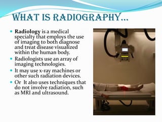
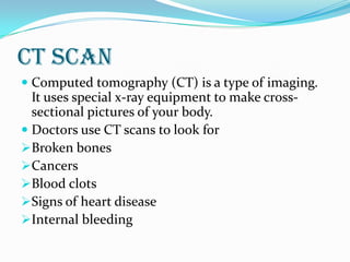
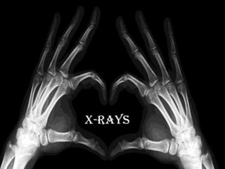
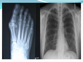
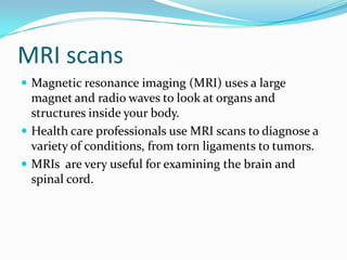
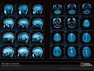
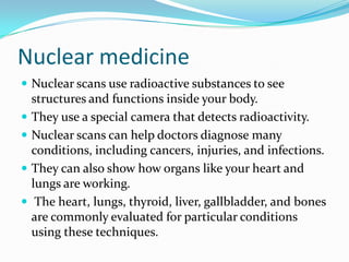
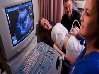
Medical imaging is the technique and process of creating visual representations of the interior of a body for clinical analysis and medical intervention, as well as visual representation of the function of some organs or tissues (physiology).
Nuclear Medicine.................
Radioactivity………………
Gamma camera………………
PET scan and SPECT scan…...........
Nuclear Medicine Studies…………..
Nuclear Medicine Team……………
Safety in Nuclear Medicine…………
Brief explanation of what is PET, the main components for a PET system along with their basic functions. The principle behind PET inclusive of positron emission and emission detection. Acquisition and reconstruction of the collected data to produce the final image. Finally the pros and cons of Positron emission tomography.
Computed tomography (CT scan) is a medical imaging procedure that uses computer-processed X-rays to produce tomographic images or 'slices' of specific areas of the body. These cross-sectional images are used for diagnostic and therapeutic purposes in various medical disciplines.
Overview of radiology basics, scan types and pros and cons of each, presented by David J. Eschelman, MD, FSIR, Professor of Radiology, Sidney Kimmel Medical College of Thomas Jefferson University, Co-Director of Interventional Radiology, Thomas Jefferson University Hospital.
Wind Turbines and their Potential for Cost ReductionsJeffrey Funk
These slides show how that long-term reductions in the cost of electricity from wind turbines have primarily come more from increasing the scale (rotor diameter and tower height) of wind turbines. See my other slides for details on concepts, methodology, and other new industries..
Indian Dental Academy: will be one of the most relevant and exciting training center with best faculty and flexible training programs for dental professionals who wish to advance in their dental practice,Offers certified courses in Dental implants,Orthodontics,Endodontics,Cosmetic Dentistry, Prosthetic Dentistry, Periodontics and General Dentistry.
In nuclear medicine, molecular imaging is applied. It employs various methods to picture biological processes that are reacting in the cells. Radiopharmaceuticals are used in molecular imaging. It is used in small quantities called radioactive markers. It offers detailed information about the biological processes that are occurring in the body especially at molecular and cellular levels. It can show the disease in the early stages. Other kinds of medical imaging are ultrasound imaging, CT and magnetic resonance imaging. The best feature of all these imaging systems is they function without ionizing radiation. Visit here https://www.alliedbusiness.co.in/ to know more about the medical imaging systems.
Pulmonary Thromboembolism - etilogy, types, medical- Surgical and nursing man...VarunMahajani
Disruption of blood supply to lung alveoli due to blockage of one or more pulmonary blood vessels is called as Pulmonary thromboembolism. In this presentation we will discuss its causes, types and its management in depth.
New Drug Discovery and Development .....NEHA GUPTA
The "New Drug Discovery and Development" process involves the identification, design, testing, and manufacturing of novel pharmaceutical compounds with the aim of introducing new and improved treatments for various medical conditions. This comprehensive endeavor encompasses various stages, including target identification, preclinical studies, clinical trials, regulatory approval, and post-market surveillance. It involves multidisciplinary collaboration among scientists, researchers, clinicians, regulatory experts, and pharmaceutical companies to bring innovative therapies to market and address unmet medical needs.
These simplified slides by Dr. Sidra Arshad present an overview of the non-respiratory functions of the respiratory tract.
Learning objectives:
1. Enlist the non-respiratory functions of the respiratory tract
2. Briefly explain how these functions are carried out
3. Discuss the significance of dead space
4. Differentiate between minute ventilation and alveolar ventilation
5. Describe the cough and sneeze reflexes
Study Resources:
1. Chapter 39, Guyton and Hall Textbook of Medical Physiology, 14th edition
2. Chapter 34, Ganong’s Review of Medical Physiology, 26th edition
3. Chapter 17, Human Physiology by Lauralee Sherwood, 9th edition
4. Non-respiratory functions of the lungs https://academic.oup.com/bjaed/article/13/3/98/278874
NVBDCP.pptx Nation vector borne disease control programSapna Thakur
NVBDCP was launched in 2003-2004 . Vector-Borne Disease: Disease that results from an infection transmitted to humans and other animals by blood-feeding arthropods, such as mosquitoes, ticks, and fleas. Examples of vector-borne diseases include Dengue fever, West Nile Virus, Lyme disease, and malaria.
Knee anatomy and clinical tests 2024.pdfvimalpl1234
This includes all relevant anatomy and clinical tests compiled from standard textbooks, Campbell,netter etc..It is comprehensive and best suited for orthopaedicians and orthopaedic residents.
HOT NEW PRODUCT! BIG SALES FAST SHIPPING NOW FROM CHINA!! EU KU DB BK substit...GL Anaacs
Contact us if you are interested:
Email / Skype : kefaya1771@gmail.com
Threema: PXHY5PDH
New BATCH Ku !!! MUCH IN DEMAND FAST SALE EVERY BATCH HAPPY GOOD EFFECT BIG BATCH !
Contact me on Threema or skype to start big business!!
Hot-sale products:
NEW HOT EUTYLONE WHITE CRYSTAL!!
5cl-adba precursor (semi finished )
5cl-adba raw materials
ADBB precursor (semi finished )
ADBB raw materials
APVP powder
5fadb/4f-adb
Jwh018 / Jwh210
Eutylone crystal
Protonitazene (hydrochloride) CAS: 119276-01-6
Flubrotizolam CAS: 57801-95-3
Metonitazene CAS: 14680-51-4
Payment terms: Western Union,MoneyGram,Bitcoin or USDT.
Deliver Time: Usually 7-15days
Shipping method: FedEx, TNT, DHL,UPS etc.Our deliveries are 100% safe, fast, reliable and discreet.
Samples will be sent for your evaluation!If you are interested in, please contact me, let's talk details.
We specializes in exporting high quality Research chemical, medical intermediate, Pharmaceutical chemicals and so on. Products are exported to USA, Canada, France, Korea, Japan,Russia, Southeast Asia and other countries.
These lecture slides, by Dr Sidra Arshad, offer a quick overview of physiological basis of a normal electrocardiogram.
Learning objectives:
1. Define an electrocardiogram (ECG) and electrocardiography
2. Describe how dipoles generated by the heart produce the waveforms of the ECG
3. Describe the components of a normal electrocardiogram of a typical bipolar leads (limb II)
4. Differentiate between intervals and segments
5. Enlist some common indications for obtaining an ECG
Study Resources:
1. Chapter 11, Guyton and Hall Textbook of Medical Physiology, 14th edition
2. Chapter 9, Human Physiology - From Cells to Systems, Lauralee Sherwood, 9th edition
3. Chapter 29, Ganong’s Review of Medical Physiology, 26th edition
4. Electrocardiogram, StatPearls - https://www.ncbi.nlm.nih.gov/books/NBK549803/
5. ECG in Medical Practice by ABM Abdullah, 4th edition
6. ECG Basics, http://www.nataliescasebook.com/tag/e-c-g-basics
The prostate is an exocrine gland of the male mammalian reproductive system
It is a walnut-sized gland that forms part of the male reproductive system and is located in front of the rectum and just below the urinary bladder
Function is to store and secrete a clear, slightly alkaline fluid that constitutes 10-30% of the volume of the seminal fluid that along with the spermatozoa, constitutes semen
A healthy human prostate measures (4cm-vertical, by 3cm-horizontal, 2cm ant-post ).
It surrounds the urethra just below the urinary bladder. It has anterior, median, posterior and two lateral lobes
It’s work is regulated by androgens which are responsible for male sex characteristics
Generalised disease of the prostate due to hormonal derangement which leads to non malignant enlargement of the gland (increase in the number of epithelial cells and stromal tissue)to cause compression of the urethra leading to symptoms (LUTS
Ethanol (CH3CH2OH), or beverage alcohol, is a two-carbon alcohol
that is rapidly distributed in the body and brain. Ethanol alters many
neurochemical systems and has rewarding and addictive properties. It
is the oldest recreational drug and likely contributes to more morbidity,
mortality, and public health costs than all illicit drugs combined. The
5th edition of the Diagnostic and Statistical Manual of Mental Disorders
(DSM-5) integrates alcohol abuse and alcohol dependence into a single
disorder called alcohol use disorder (AUD), with mild, moderate,
and severe subclassifications (American Psychiatric Association, 2013).
In the DSM-5, all types of substance abuse and dependence have been
combined into a single substance use disorder (SUD) on a continuum
from mild to severe. A diagnosis of AUD requires that at least two of
the 11 DSM-5 behaviors be present within a 12-month period (mild
AUD: 2–3 criteria; moderate AUD: 4–5 criteria; severe AUD: 6–11 criteria).
The four main behavioral effects of AUD are impaired control over
drinking, negative social consequences, risky use, and altered physiological
effects (tolerance, withdrawal). This chapter presents an overview
of the prevalence and harmful consequences of AUD in the U.S.,
the systemic nature of the disease, neurocircuitry and stages of AUD,
comorbidities, fetal alcohol spectrum disorders, genetic risk factors, and
pharmacotherapies for AUD.
Report Back from SGO 2024: What’s the Latest in Cervical Cancer?bkling
Are you curious about what’s new in cervical cancer research or unsure what the findings mean? Join Dr. Emily Ko, a gynecologic oncologist at Penn Medicine, to learn about the latest updates from the Society of Gynecologic Oncology (SGO) 2024 Annual Meeting on Women’s Cancer. Dr. Ko will discuss what the research presented at the conference means for you and answer your questions about the new developments.
2. What is radiography…
Radiology is a medical
specialty that employs the use
of imaging to both diagnose
and treat disease visualized
within the human body.
Radiologists use an array of
imaging technologies.
It may use x-ray machines or
other such radiation devices.
Or It also uses techniques that
do not involve radiation, such
as MRI and ultrasound.
3. types…
Radiology can refer to two sub-fields, diagnostic
radiology and therapeutic radiology.
Diagnostic radiology is concerned with the use of
various imaging modalities to aid in the diagnosis
of disease.
Therapeutic radiology—or, as it is now called,
radiation oncology uses radiation to treat diseases
such as cancer using a form of treatment called
radiation therapy.
4. Commonly used techniques for diagnostic radiology
includes
Computed tomography (CT)
Magnetic resonance imaging (MRI)
Ultrasound
X-rays
Nuclear imaging techniques
6. CT scan
Computed tomography (CT) is a type of imaging.
It uses special x-ray equipment to make cross-
sectional pictures of your body.
Doctors use CT scans to look for
Broken bones
Cancers
Blood clots
Signs of heart disease
Internal bleeding
8. X-rays
X-rays are basically electromagnetic radiations which
are used to create images of inside your body.
The images show the parts of your body in different
shades of black and white due to different level of
absorption of x-rays by different tissues
Calcium in bones absorbs x-rays the most, so bones
look white. Fat and other soft tissues absorb less, and
look gray.
Air absorbs the least, so lungs look black.
9.
10. The most familiar use of x-rays is checking for broken
bones
However, x-rays are also used in other ways.
For example, chest x-rays can spot pneumonia.
Mammograms use x-rays to look for breast cancer.
13. MRI scans
Magnetic resonance imaging (MRI) uses a large
magnet and radio waves to look at organs and
structures inside your body.
Health care professionals use MRI scans to diagnose a
variety of conditions, from torn ligaments to tumors.
MRIs are very useful for examining the brain and
spinal cord.
14.
15. Nuclear medicine
Nuclear scans use radioactive substances to see
structures and functions inside your body.
They use a special camera that detects radioactivity.
Nuclear scans can help doctors diagnose many
conditions, including cancers, injuries, and infections.
They can also show how organs like your heart and
lungs are working.
The heart, lungs, thyroid, liver, gallbladder, and bones
are commonly evaluated for particular conditions
using these techniques.
16.
17. Ultrasound
Ultrasound is a type of imaging. It uses high-frequency
sound waves to look at organs and structures inside
the body.
Health care professionals use it to view the heart,
blood vessels, kidneys, liver, and other organs.
During pregnancy, doctors use ultrasound to view the
fetus. Unlike x-rays, ultrasound does not expose you to
radiation.