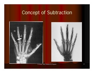
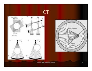
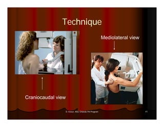
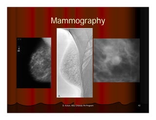
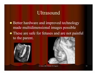
This document provides an introduction to various medical imaging modalities including X-ray, CT, mammography, MRI, PET, SPECT, and ultrasound. It discusses the principles, techniques, and indications for each modality. Key terms are defined. Images demonstrate examples of each type of imaging. The objectives are to recognize imaging study types, discuss how images are produced, list common indications, and describe imaging precaution considerations.
4. Objectives
Recognize various types of imaging
studies
Discuss the mechanism for producing
images with each modality
List th
Li t the common indications for different
i di ti f diff t
imaging modalities
Describe the precautions for ordering
imaging studies
4
D. Kotun, NSU Orlando PA Program
5. Pre – Test
I am looking forward to this test
because:
I want to learn all I can
I am demented
It i a good time to catch a nap
is di h
I heard that Prof. Kotun is a wonderful
lecturer
5
D. Kotun, NSU Orlando PA Program
7. Terms
Tomography: a cross-sectional image formed from a set of projection
cross-
images. The Greek word tomo means cut.
CT: Computed (or Computerized) Tomography
MR, or MRI: Magnetic Resonance Imaging. This was first called nuclear
magnetic resonance (NMR), but the mention of anything nuclear scared
patients, so the “N” was dropped.
PET: Positron Emission Tomography. Understanding this phenomenon
requires acceptance of the theory that there is antimatter in the universe,
and when antimatter meets matter, then both kinds of matter are
annihilated, and pure energy is formed.
SPECT: Single Photon Emission Tomography
Ultrasound: Sonar in the body
OCT: O ti l C h
OCT Optical Coherent Tomography – th use of infrared light to image
tT h the fi f d li ht t i
(particularity) the walls of an artery.
7
D. Kotun, NSU Orlando PA Program
9. The First X-Ray Exam
X-
William Conrad Roentgen
89 Nov 1895
Took a picture of his wife s hand and
wife’s
developed it on photographic film
Ferdinand and Mazwell predicted X-Rays
X-
from Maxwell’s EM radiation theory
9
D. Kotun, NSU Orlando PA Program
20. In an X-ray tube, the photons are
X-
generated by what?
db h?
A high density nuclear source
1.
1
25%
2. An electron stream onto a metal
25%
target
3. A focused heated metal plate
25%
4. Magic pixies in a glass tube (hence
25%
the name pixels)
20
D. Kotun, NSU Orlando PA Program
28. Planes
(Frontal)
28
D. Kotun, NSU Orlando PA Program
29. The left lateral view would mean:
The beam goes from front to back
1.
1
25%
2. The beam goes from left to right
25%
3. Th patient’s left side is closest to the
The ti t’ l ft id i l t t th
beam
25%
4. The patient’s left side is closest to the
25%
film
29
D. Kotun, NSU Orlando PA Program
30. Putting It On Film
Figure 7-7 Scattered X-ray photons can be
removed from the image by positioning a grid
bet ee the patient and the detector (or
between t e pat e t a d t e detecto (o film).
)
30
D. Kotun, NSU Orlando PA Program
31. Putting It On Film
X-Ray Photons are not very good at
exposing film
There is some type of amplification system
Photostimulable cassette
Phospors coat a screen and emit light when struck
by X-Ray photons
X-
These can be catured on film or amplified with an
electronic amplification system
l lf
31
D. Kotun, NSU Orlando PA Program
36. Helical CT
Instead of going
back and forth,
this corkscrews
itself down your
body.
36
D. Kotun, NSU Orlando PA Program
37. CAT Scan
Now multiple heads allow faster imaging
with more resolution
Chest in < 30 sec. with .5cm resolution
sec 5cm
Pictures are reconstructed using a digital
reconstruction algorithm
These must be digitalized
Can be placed on film
37
D. Kotun, NSU Orlando PA Program
39. Mammography
Specific to a woman’s or man’s breasts
woman s man s
Lower dose
Used as cancer screening
Two types
Radiomammography
d h
Uses X-Ray technique
X-
Xeromammography
X h
Electrostatic image production
39
D. Kotun, NSU Orlando PA Program
40. VIEWS
Cranio-caudal (CC) Medio lateral (ML)
()
Latero medial (LM)
()
view and mediolateral
i d di l t l mammographic view mammographic view
oblique (MLO)
mammographic view
Spot compression and cone down views
40
D. Kotun, NSU Orlando PA Program
41. Technique
Mediolateral view
Craniocaudal view
41
D. Kotun, NSU Orlando PA Program
42. Mammography
42
D. Kotun, NSU Orlando PA Program
43. (N)MRI
Uses high strength magnets on hydrogen
bonds (in water)
Magnet strength is in Tesla Units (0.3 – 3)
g g ( )
The magnetic field excites the protons and
as it relaxes, it emits a radiofrequency
, q y
signal
Non-
Non-ionizing (very important)
Fields are aligned in 3 planes but can be
adjusted
43
D. Kotun, NSU Orlando PA Program
45. ()
(N)MRI
High Quality three dimensional pictures
Uses Voxels vs Pixels (What is the
vs.
difference?)
Good for soft tissue water filled tissue
tissue,
45
D. Kotun, NSU Orlando PA Program
46. Positron Emission Tomography) PET
Uses a prepared positron emitting nuclide
Positrons are emitted at 180° apart
180°
46
D. Kotun, NSU Orlando PA Program
48. PET
Uptake is where the tissue has the most
activity
Increased uptake shows increased tissue
function
f ti
Great for mapping brain activity
Nuclide is usually injected
20 y.o. vs. 80 y.o.
48
D. Kotun, NSU Orlando PA Program
49. SPEC/Nuclear Medicine
Uses a gamma emitting radionuclide to take a
2 dimensional picture
This is gathered on a scintillation camera
picking up the photons
Planar images can be combined from different
angles to reconstruct a 3 – D image
The nuclides are usually injected and are
designed to go to the organ or system in
q
question
49
D. Kotun, NSU Orlando PA Program
51. Ultrasound
Uses sound waves higher than detectable by
humans
Non-
Non-invasive and non-painful
non-
Patients aren’t usually afraid of U/S
51
D. Kotun, NSU Orlando PA Program
53. Ultrasound
2 – D ultrasounds
became more diagnostic
because the transducers
could produce a planar
image
53
D. Kotun, NSU Orlando PA Program
54. Ultrasound
Better hardware and improved technology
made multidimensional images possible
These are safe for fetuses and are not painful
to the parent.
54
D. Kotun, NSU Orlando PA Program
55. If I wanted to see organ function I
would use this type of imaging.
ld hi fi i
Plane X-ray
X-
1.
1
25%
2. Nuclear medicine scan
25%
3. C
Computerized tomography
t i dt h
25%
4. Magnetic resonance tomography
25%
55
D. Kotun, NSU Orlando PA Program
56. To image an unborn fetus, the safest
imaging is:
i ii
Ultrasound
1.
1
25%
2. Nuclear medicine
25%
3. P it
Positron emission
ii
25%
4. Computerized tomography
25%
56
D. Kotun, NSU Orlando PA Program
57. Did Ms Roentgen have a ring on?
Ms.
Yes
1.
1
33%
2. No
33%
3. M b
Maybe
33%
57
D. Kotun, NSU Orlando PA Program
58. In the Future
Cellular imaging uses a combination of MRT,
MRT
CT, and U/S
Mouse done with
cellular imaging
58
D. Kotun, NSU Orlando PA Program
59. Funny Bone…
Bone
59
D. Kotun, NSU Orlando PA Program
60. Acknowledgements
Dove, E. L., 2003, Physics of Medical Imaging – An Introduction. U.
of Iowa, 53pp. Sep. 22, 2003
60
D. Kotun, NSU Orlando PA Program
61. Questions?
61
D. Kotun, NSU Orlando PA Program