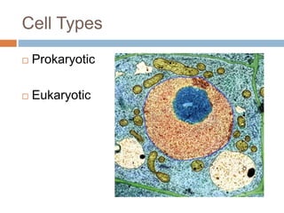
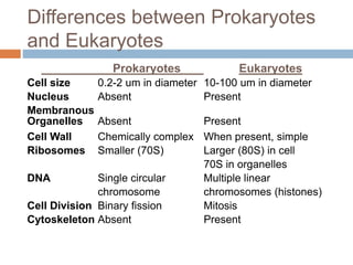
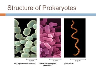
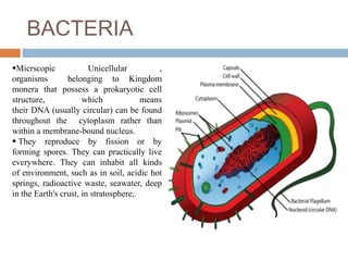
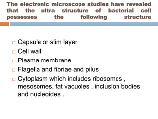
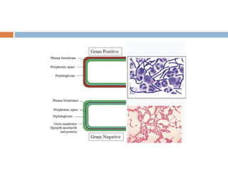
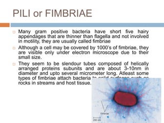
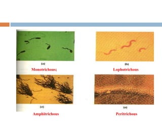
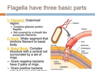
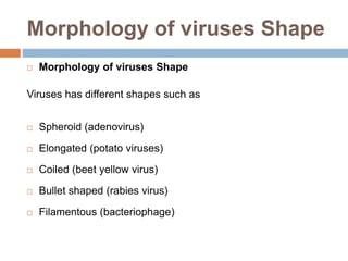
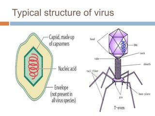
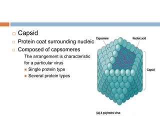
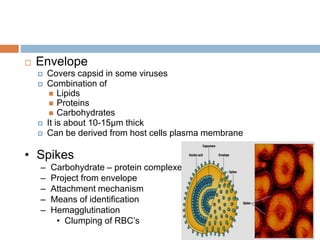
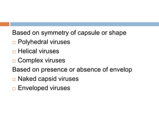
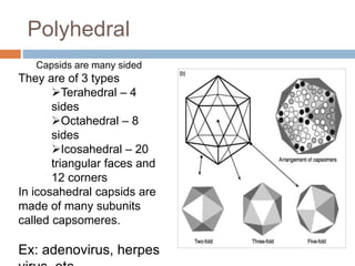
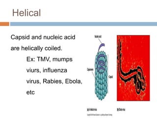
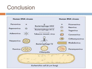
By :-
Vikas C J
ARCHITECTURE OF
MICROBIAL CELL
Contents
 Introduction
 Classification of bacteria
 Structure of bacteria
 Conclusion
 Reference
Cells
 Smallest living unit
 Most are microscopic
Cell size:
 Dimensions of most bacterial cells:
 Diameter: 0.2 to 2.0 mm.
 Human red blood cell is about 7.5-10 mm in
diameter.
 Length: 2 to 8 mm.
 Some cyanobacteria are up to 60 mm long.
 Bacterial cells have large surface to volume
ratios. Therefore all parts of the cell:
 Are close to the surface.
 Can be quickly reached by nutrients
Cell Types
 Prokaryotic
 Eukaryotic
Distinguishing Features of
Prokaryotic Cells:
1. DNA is:
 Not enclosed within a nuclear membrane.
 A single circular chromosome.
 Not associated with histone proteins.
2. Lack membrane-enclosed organelles like
mitochondria, chloroplasts, Golgi, etc.
3. Cell walls usually contain peptidoglycan, a
complex polysaccharide.
4. Divide by binary fission
Distinguishing Features of
Eukaryotic Cells:
1. DNA is:
 Enclosed within a nuclear membrane.
 Several linear chromosomes.
 Associated with histones and other proteins.
2. Have membrane-enclosed organelles like
mitochondria, chloroplasts, Golgi, endoplasmic
reticulum, etc.
3. Divide by mitosis.
Differences between Prokaryotes
and Eukaryotes
Prokaryotes Eukaryotes
Cell size 0.2-2 um in diameter 10-100 um in diameter
Nucleus Absent Present
Membranous
Organelles Absent Present
Cell Wall Chemically complex When present, simple
Ribosomes Smaller (70S) Larger (80S) in cell
70S in organelles
DNA Single circular Multiple linear
chromosome chromosomes (histones)
Cell Division Binary fission Mitosis
Cytoskeleton Absent Present
Structure of Prokaryotes
BACTERIA
Micrscopic Unicellular ,
organisms belonging to Kingdom
monera that possess a prokaryotic cell
structure, which means
their DNA (usually circular) can be found
throughout the cytoplasm rather than
within a membrane-bound nucleus.
 They reproduce by fission or by
forming spores. They can practically live
everywhere. They can inhabit all kinds
of environment, such as in soil, acidic hot
springs, radioactive waste, seawater, deep
in the Earth's crust, in stratosphere,.
The electronic microscope studies have revealed
that the ultra structure of bacterial cell
possesses the following structure
 Capsule or slim layer
 Cell wall
 Plasma membrane
 Flagella and fibriae and pilus
 Cytoplasm which includes ribosomes ,
mesosomes, fat vacuoles , inclusion bodies
and nucleoides .
CAPSULE
 Certain bacteria like diplococcus and pneumococcal are
externally covered by a layer of that layer is called
capsule.
 Glycocalyx - Polysaccharide on external surface
 Forms an envelope around the cell wall and can be observed under
light microscope.
 Chemically capsules are made up of proteins, polysaccharide and
lipids
 The bacterial capsule is species specific and can be used for
immunological detections. amount these polymers vary with bacterial
species.
 It is sticky in nature ,secreted by the cell which gets firmly attached to
the cell wall.
 If the substances are unorganized and loosely attached to cell wall, -
slime layer.
 Many gram positive and gram negative bacteria have a regular
structured layer called an S-layer on their surface. These are very
common among Archea. where they may be the only wall structure
Functions of Capsule
 Prevent attachment of bacteiophages.
 Prevents bacterial cell against desiccation.
 Survive in natural environment –sticky
property.
 Prevents from phagocytosis.
 It may protect the cell against ion and ph
fluctuation.
CELL WALL
 A typical bacterial cell possesses a rigid structure
called cell wall that lies outside the plasma
membrane
 It is about 10 -25 nm thick and accounts for about
20 -30% of the dry weight of the cell
 It is responsible for maintaining the shape of the
bacterium and prevents the cell from osmotic
lysise
 The chemical subsatance that comfers rigidity to
the cell wall is peptidoglycon.
it is a strong polymer found only in prokaryotes.
 peptidoglycon is a polymer of N-acetyl
glucosomine and N-acetyle muramic acid .
Bacteria are classified into gram positive
and gram negative depending on the nature
of the cell wall
 Gram’s positive bacteria will have thick
peptidoglycon layer where as gram negative
bacteria are having thin peptidoglycon layer.
Gram positive bacteria also contains teichoic
acid which is absent in gram negative bacteria.
 Gram negative bacteria contain an outer
membrane that surrounds the peptidoglycon
layer which is made up of lipopoly
saccharides. The important function of this
outer membrane is to serve as protective
barrier and prevents or slows the entry of toxic
substance that might kill or injure the bacteria.
In addition this membrane is anchored to the
peptidoglucon by Braun’s lipoprotein.
PLASMA MEMBRANE
 The contents of the bacterial cell with in the boundries of the cell wall
form the protoplast. It consists of outer plasma membrane, cytoplasm
, and nucleoids.
 The plasma membrane of bacterial cell is similar to that of eukaryotic
cell. The most widely accepted current model for membrane structure
is the fluid mosaic model of Singer and Nicolson.
 According this membrane structurally is
a lipid bilayer consisting of two types
of proteins peripheral proteins are
loosely connected to the membrane and
can be easily removed and make
20-30% of the total membrane proteins.
about 70-80% of the membrane
protein are integral proteins, they are
not easily extracted from membrane
Functions of plasma membrane
 Organic and inorganic nutrients are transported
through plasma membrane.
 Consists of enzymes of biosynthetic pathways.
 The inner membrane invaginates to form
mesosomes.
 It has selective permiability.
PILI or FIMBRIAE
 Many gram positive bacteria have short five hairy
appendages that are thinner than flagella and not involved
in motility, they are usually called fimbriae
 Although a cell may be covered by 1000’s of fimbriae, they
are visible only under electron microscope due to their
small size.
 They seem to be slendour tubes composed of helically
arranged proteins subunits and are about 3-10nm in
diameter and upto several micrometer long. Atleast some
types of fimbriae attach bacteria to solid surfaces such as
rocks in streams and host tissue.
Flagella
 About half of all known bacteria are motile, most
use flagella.
 Long, thin, helical appendages.
 A bacterium may have one or several flagella,
which can be in the following arrangements:
 Monotrichous: Single polar flagellum at one end.
 Amphitrichous: Two polar flagella, one at each end.
 Lophotrichous: Two or more flagella at one or both
ends.
 Peritrichous: Many flagella over entire cell surface.
Monotrichous; Lophotrichous
Amphitrichous Peritrichous
Flagella have three basic parts
1. Filament: Outermost
region.
 Contains globular protein
flagellin.
 Not covered by a sheath like
eucaryotic filaments.
2. Hook: Wider segment that
anchors filament to basal
body.
3. Basal Body: Complex
structure with a central rod
surrounded by a set of
rings.
 Gram negative bacteria
have 2 pairs of rings.
 Gram positive bacteria
Cytoskeleton
 These are Filaments & fibers
 Made of 3 fiber types
 Microfilaments
 Microtubules
 Intermediate filaments
 3 functions:
 mechanical support
 anchor organelles
 help move substances
VIRUSES
 WHAT ARE VIRUSES????
 Viruses are considered to be the smallest ‘living
units’.
 Viruses may be defined as acellular , Sub
microscopic entity consisting of a single nucleic
acid surrounded by a protein coat and capable of
replication only within the host cells using host
metabolic machinery.
Morphology of viruses Shape
 Morphology of viruses Shape
Viruses has different shapes such as
 Spheroid (adenovirus)
 Elongated (potato viruses)
 Coiled (beet yellow virus)
 Bullet shaped (rabies virus)
 Filamentous (bacteriophage)
Typical structure of virus
 Capsid
 Protein coat surrounding nucleic acid
 Composed of capsomeres
The arrangement is characteristic
for a particular virus
 Single protein type
 Several protein types
 Envelope
 Covers capsid in some viruses
 Combination of
 Lipids
 Proteins
 Carbohydrates
 It is about 10-15µm thick
 Can be derived from host cells plasma membrane
• Spikes
– Carbohydrate – protein complexes
– Project from envelope
– Attachment mechanism
– Means of identification
– Hemagglutination
• Clumping of RBC’s
Based on symmetry of capsule or shape
 Polyhedral viruses
 Helical viruses
 Complex viruses
Based on presence or absence of envelop
 Naked capsid viruses
 Enveloped viruses
Polyhedral
Capsids are many sided
They are of 3 types
Terahedral – 4
sides
Octahedral – 8
sides
Icosahedral – 20
triangular faces and
12 corners
In icosahedral capsids are
made of many subunits
called capsomeres.
Ex: adenovirus, herpes
Helical
Capsid and nucleic acid
are helically coiled.
Ex: TMV, mumps
viurs, influenza
virus, Rabies, Ebola,
etc
Complex
 Capsid is attached with
additional structures
Ex: vaccinia virus,
phages of
T-even series
 Bacteriophages - Capsid is
polyhedral in shape where
as sheath is helical
Conclusion
Reference
 Dr.R.C Dubey and Dr.D.K Maheshwari. A text
book of microbiology.
 https://scholar.google.co.in/scholar?q=architec
ture+of+microbial+cell
 https://www.google.co.in/webhp?sourceid=chr
ome-instant&ion=1&espv=2&ie=UTF-
8#q=characteristics+of+anaerobes
Architechture of microbial cell

Architechture of microbial cell

  • 1.
    By :- Vikas CJ ARCHITECTURE OF MICROBIAL CELL
  • 2.
    Contents  Introduction  Classificationof bacteria  Structure of bacteria  Conclusion  Reference
  • 3.
    Cells  Smallest livingunit  Most are microscopic
  • 5.
    Cell size:  Dimensionsof most bacterial cells:  Diameter: 0.2 to 2.0 mm.  Human red blood cell is about 7.5-10 mm in diameter.  Length: 2 to 8 mm.  Some cyanobacteria are up to 60 mm long.  Bacterial cells have large surface to volume ratios. Therefore all parts of the cell:  Are close to the surface.  Can be quickly reached by nutrients
  • 6.
  • 7.
    Distinguishing Features of ProkaryoticCells: 1. DNA is:  Not enclosed within a nuclear membrane.  A single circular chromosome.  Not associated with histone proteins. 2. Lack membrane-enclosed organelles like mitochondria, chloroplasts, Golgi, etc. 3. Cell walls usually contain peptidoglycan, a complex polysaccharide. 4. Divide by binary fission
  • 8.
    Distinguishing Features of EukaryoticCells: 1. DNA is:  Enclosed within a nuclear membrane.  Several linear chromosomes.  Associated with histones and other proteins. 2. Have membrane-enclosed organelles like mitochondria, chloroplasts, Golgi, endoplasmic reticulum, etc. 3. Divide by mitosis.
  • 9.
    Differences between Prokaryotes andEukaryotes Prokaryotes Eukaryotes Cell size 0.2-2 um in diameter 10-100 um in diameter Nucleus Absent Present Membranous Organelles Absent Present Cell Wall Chemically complex When present, simple Ribosomes Smaller (70S) Larger (80S) in cell 70S in organelles DNA Single circular Multiple linear chromosome chromosomes (histones) Cell Division Binary fission Mitosis Cytoskeleton Absent Present
  • 10.
  • 11.
    BACTERIA Micrscopic Unicellular , organismsbelonging to Kingdom monera that possess a prokaryotic cell structure, which means their DNA (usually circular) can be found throughout the cytoplasm rather than within a membrane-bound nucleus.  They reproduce by fission or by forming spores. They can practically live everywhere. They can inhabit all kinds of environment, such as in soil, acidic hot springs, radioactive waste, seawater, deep in the Earth's crust, in stratosphere,.
  • 12.
    The electronic microscopestudies have revealed that the ultra structure of bacterial cell possesses the following structure  Capsule or slim layer  Cell wall  Plasma membrane  Flagella and fibriae and pilus  Cytoplasm which includes ribosomes , mesosomes, fat vacuoles , inclusion bodies and nucleoides .
  • 13.
    CAPSULE  Certain bacterialike diplococcus and pneumococcal are externally covered by a layer of that layer is called capsule.  Glycocalyx - Polysaccharide on external surface  Forms an envelope around the cell wall and can be observed under light microscope.  Chemically capsules are made up of proteins, polysaccharide and lipids  The bacterial capsule is species specific and can be used for immunological detections. amount these polymers vary with bacterial species.  It is sticky in nature ,secreted by the cell which gets firmly attached to the cell wall.  If the substances are unorganized and loosely attached to cell wall, - slime layer.  Many gram positive and gram negative bacteria have a regular structured layer called an S-layer on their surface. These are very common among Archea. where they may be the only wall structure
  • 14.
    Functions of Capsule Prevent attachment of bacteiophages.  Prevents bacterial cell against desiccation.  Survive in natural environment –sticky property.  Prevents from phagocytosis.  It may protect the cell against ion and ph fluctuation.
  • 15.
    CELL WALL  Atypical bacterial cell possesses a rigid structure called cell wall that lies outside the plasma membrane  It is about 10 -25 nm thick and accounts for about 20 -30% of the dry weight of the cell  It is responsible for maintaining the shape of the bacterium and prevents the cell from osmotic lysise  The chemical subsatance that comfers rigidity to the cell wall is peptidoglycon. it is a strong polymer found only in prokaryotes.  peptidoglycon is a polymer of N-acetyl glucosomine and N-acetyle muramic acid .
  • 16.
    Bacteria are classifiedinto gram positive and gram negative depending on the nature of the cell wall  Gram’s positive bacteria will have thick peptidoglycon layer where as gram negative bacteria are having thin peptidoglycon layer. Gram positive bacteria also contains teichoic acid which is absent in gram negative bacteria.
  • 17.
     Gram negativebacteria contain an outer membrane that surrounds the peptidoglycon layer which is made up of lipopoly saccharides. The important function of this outer membrane is to serve as protective barrier and prevents or slows the entry of toxic substance that might kill or injure the bacteria. In addition this membrane is anchored to the peptidoglucon by Braun’s lipoprotein.
  • 19.
    PLASMA MEMBRANE  Thecontents of the bacterial cell with in the boundries of the cell wall form the protoplast. It consists of outer plasma membrane, cytoplasm , and nucleoids.  The plasma membrane of bacterial cell is similar to that of eukaryotic cell. The most widely accepted current model for membrane structure is the fluid mosaic model of Singer and Nicolson.  According this membrane structurally is a lipid bilayer consisting of two types of proteins peripheral proteins are loosely connected to the membrane and can be easily removed and make 20-30% of the total membrane proteins. about 70-80% of the membrane protein are integral proteins, they are not easily extracted from membrane
  • 20.
    Functions of plasmamembrane  Organic and inorganic nutrients are transported through plasma membrane.  Consists of enzymes of biosynthetic pathways.  The inner membrane invaginates to form mesosomes.  It has selective permiability.
  • 21.
    PILI or FIMBRIAE Many gram positive bacteria have short five hairy appendages that are thinner than flagella and not involved in motility, they are usually called fimbriae  Although a cell may be covered by 1000’s of fimbriae, they are visible only under electron microscope due to their small size.  They seem to be slendour tubes composed of helically arranged proteins subunits and are about 3-10nm in diameter and upto several micrometer long. Atleast some types of fimbriae attach bacteria to solid surfaces such as rocks in streams and host tissue.
  • 22.
    Flagella  About halfof all known bacteria are motile, most use flagella.  Long, thin, helical appendages.  A bacterium may have one or several flagella, which can be in the following arrangements:  Monotrichous: Single polar flagellum at one end.  Amphitrichous: Two polar flagella, one at each end.  Lophotrichous: Two or more flagella at one or both ends.  Peritrichous: Many flagella over entire cell surface.
  • 23.
  • 24.
    Flagella have threebasic parts 1. Filament: Outermost region.  Contains globular protein flagellin.  Not covered by a sheath like eucaryotic filaments. 2. Hook: Wider segment that anchors filament to basal body. 3. Basal Body: Complex structure with a central rod surrounded by a set of rings.  Gram negative bacteria have 2 pairs of rings.  Gram positive bacteria
  • 25.
    Cytoskeleton  These areFilaments & fibers  Made of 3 fiber types  Microfilaments  Microtubules  Intermediate filaments  3 functions:  mechanical support  anchor organelles  help move substances
  • 26.
    VIRUSES  WHAT AREVIRUSES????  Viruses are considered to be the smallest ‘living units’.  Viruses may be defined as acellular , Sub microscopic entity consisting of a single nucleic acid surrounded by a protein coat and capable of replication only within the host cells using host metabolic machinery.
  • 27.
    Morphology of virusesShape  Morphology of viruses Shape Viruses has different shapes such as  Spheroid (adenovirus)  Elongated (potato viruses)  Coiled (beet yellow virus)  Bullet shaped (rabies virus)  Filamentous (bacteriophage)
  • 28.
  • 29.
     Capsid  Proteincoat surrounding nucleic acid  Composed of capsomeres The arrangement is characteristic for a particular virus  Single protein type  Several protein types
  • 30.
     Envelope  Coverscapsid in some viruses  Combination of  Lipids  Proteins  Carbohydrates  It is about 10-15µm thick  Can be derived from host cells plasma membrane • Spikes – Carbohydrate – protein complexes – Project from envelope – Attachment mechanism – Means of identification – Hemagglutination • Clumping of RBC’s
  • 31.
    Based on symmetryof capsule or shape  Polyhedral viruses  Helical viruses  Complex viruses Based on presence or absence of envelop  Naked capsid viruses  Enveloped viruses
  • 32.
    Polyhedral Capsids are manysided They are of 3 types Terahedral – 4 sides Octahedral – 8 sides Icosahedral – 20 triangular faces and 12 corners In icosahedral capsids are made of many subunits called capsomeres. Ex: adenovirus, herpes
  • 33.
    Helical Capsid and nucleicacid are helically coiled. Ex: TMV, mumps viurs, influenza virus, Rabies, Ebola, etc
  • 34.
    Complex  Capsid isattached with additional structures Ex: vaccinia virus, phages of T-even series  Bacteriophages - Capsid is polyhedral in shape where as sheath is helical
  • 35.
  • 36.
    Reference  Dr.R.C Dubeyand Dr.D.K Maheshwari. A text book of microbiology.  https://scholar.google.co.in/scholar?q=architec ture+of+microbial+cell  https://www.google.co.in/webhp?sourceid=chr ome-instant&ion=1&espv=2&ie=UTF- 8#q=characteristics+of+anaerobes