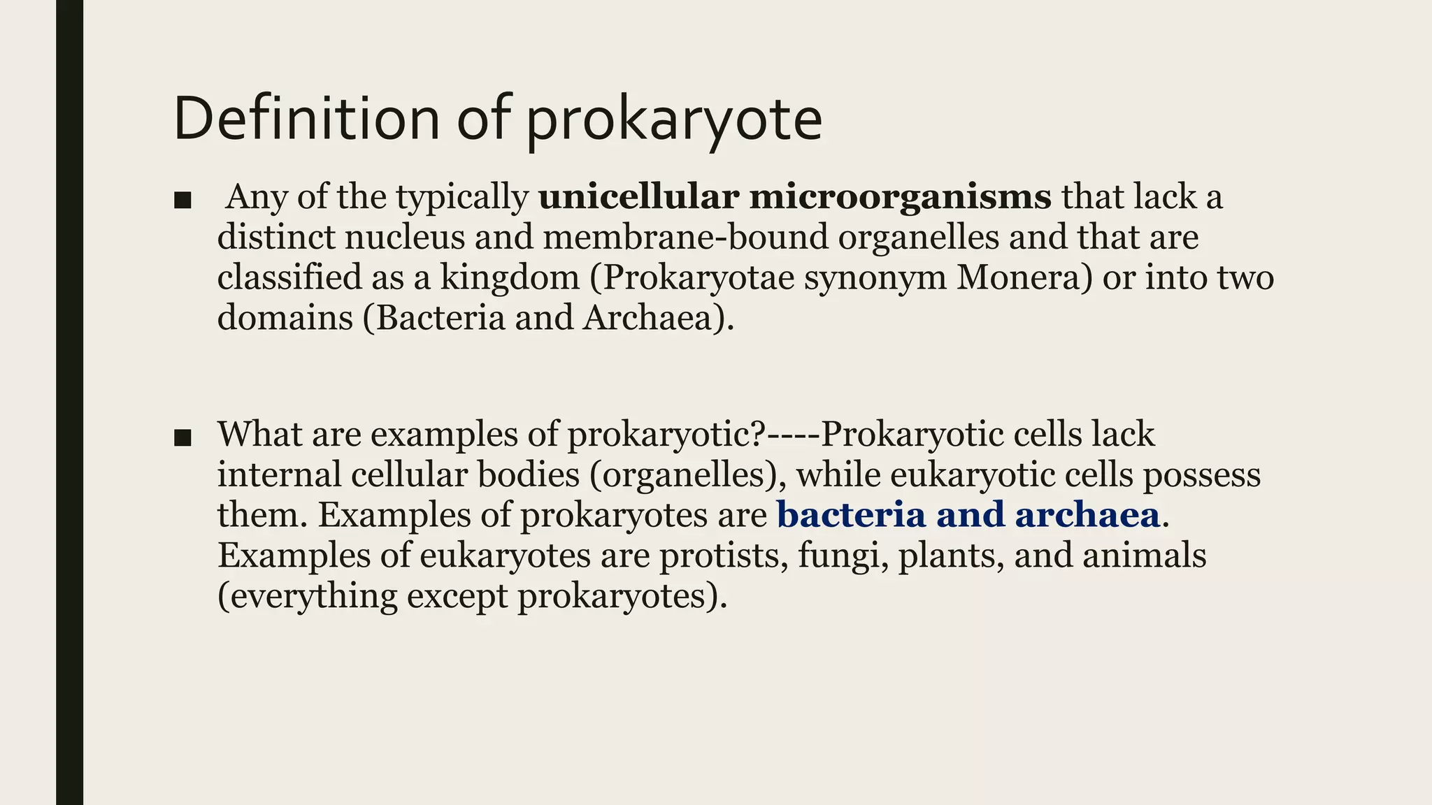
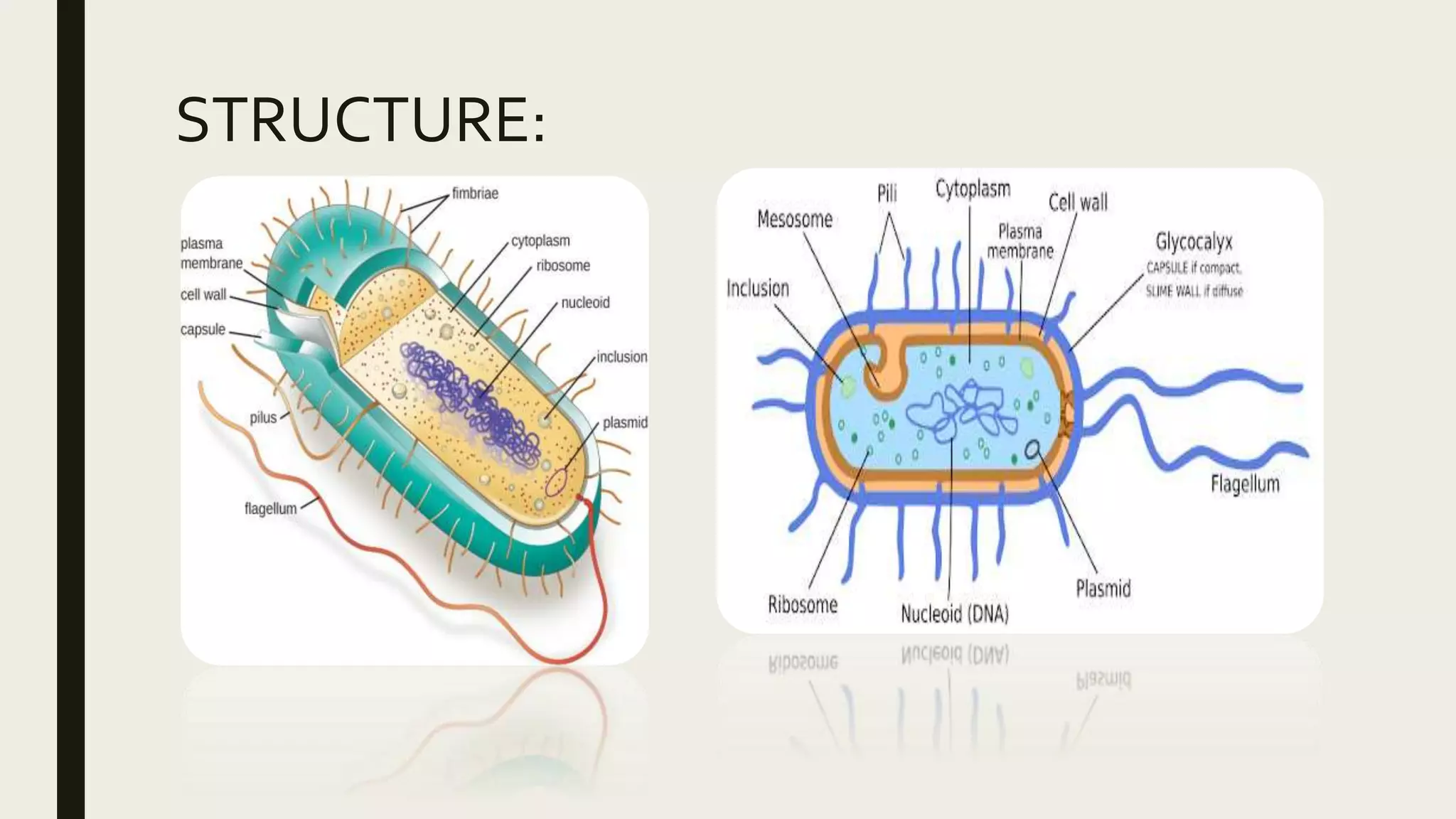
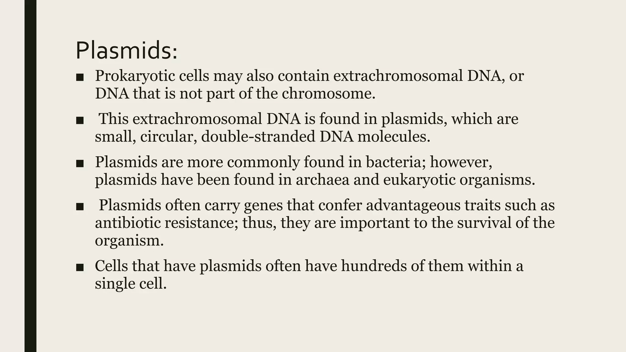
Prokaryotic cells lack membrane-bound organelles and have a circular chromosome located in an irregular region called the nucleoid. They may contain extrachromosomal DNA in plasmids. Prokaryotic cells share components like the plasma membrane, ribosomes, and DNA. Some prokaryotes perform photosynthesis using thylakoid or chromatophore membranes. Prokaryotic cells may also contain mesosomes, cell walls, glycocalyces, S-layers, and filamentous appendages like flagella, fimbriae, and pili that allow interaction with the environment.


![What are the fundamental components of a prokaryotic cell?
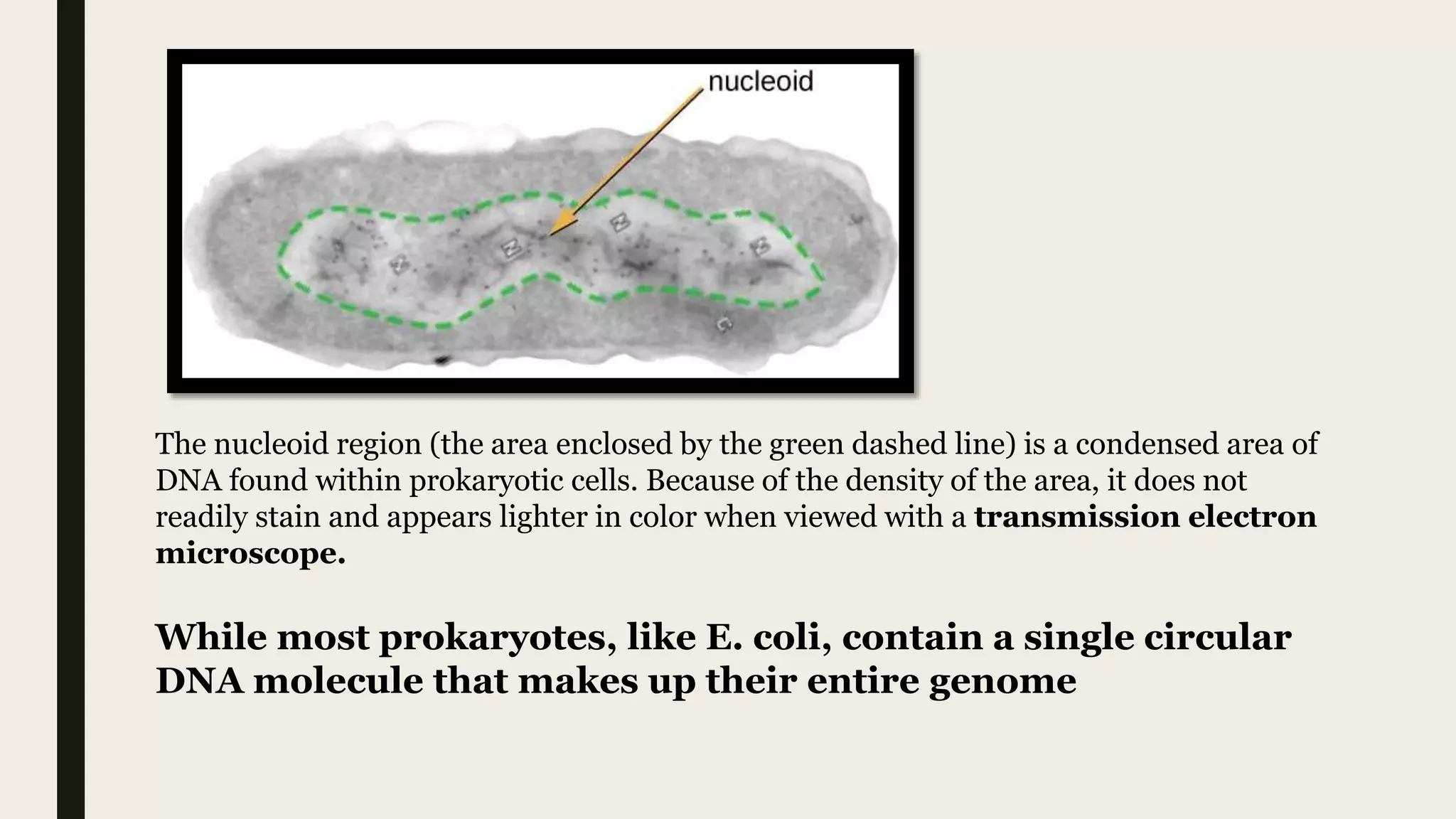
The Nucleoid----The nucleoid (meaning nucleus-like) is an
irregularly shaped region within the prokaryotic cell that contains all
or most of the genetic material.[While most prokaryotes, like
E. coli, contain a single circular DNA molecule that makes up
their entire genome]
■ In contrast to the nucleus of a eukaryotic cell, it is not surrounded
by a nuclear membrane.
■ Prokaryotic DNA and DNA-associated proteins are
concentrated within the nucleoid region of the cell .
■ In general, prokaryotic DNA interacts with nucleoid-associated
proteins (NAPs) that assist in the organization and packaging of the
chromosome.
■ NAPs function similar to histones, which are the DNA-organizing
proteins found in eukaryotic cells.](https://image.slidesharecdn.com/prokaryoticcell-200905105746/75/Prokaryotic-cell-6-2048.jpg)


![■ These phospholipids and proteins have the ability to move laterally
within the plane of the membranes as well as between the two
phospholipid layers.
[figure]The bacterial plasma membrane is a phospholipid bilayer with a variety of embedded proteins
that perform various functions for the cell. Note the presence of glycoproteins and glycolipids, whose
carbohydrate components extend out from the surface of the cell.The abundance and arrangement of
these proteins and lipids can vary greatly between species.](https://image.slidesharecdn.com/prokaryoticcell-200905105746/75/Prokaryotic-cell-14-2048.jpg)

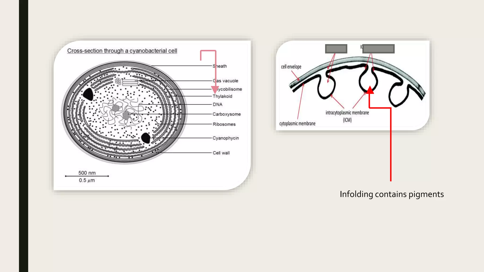
![Photosynthetic Membrane Structures [cell
membrane modification]
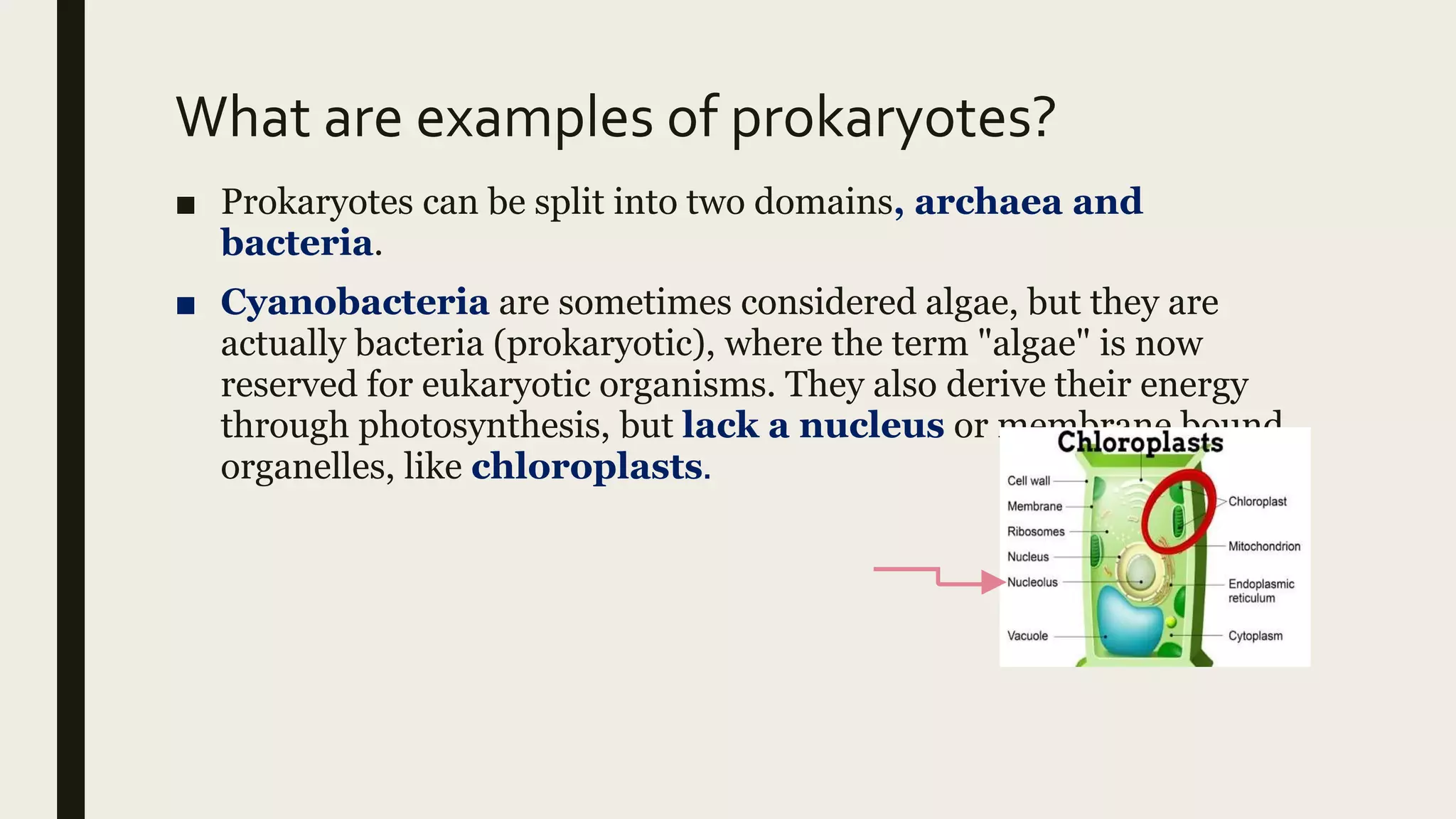
■ Some prokaryotic cells, namely cyanobacteria and photosynthetic
bacteria, have membrane structures that enable them to perform
photosynthesis.
■ These structures consist of an infolding of the plasma membrane
that encloses photosynthetic pigments such as green chlorophylls
and bacteriochlorophylls.
■ In cyanobacteria, these membrane structures are called thylakoids;
in photosynthetic bacteria, they are called chromatophores,
lamellae, or chlorosomes.](https://image.slidesharecdn.com/prokaryoticcell-200905105746/75/Prokaryotic-cell-16-2048.jpg)
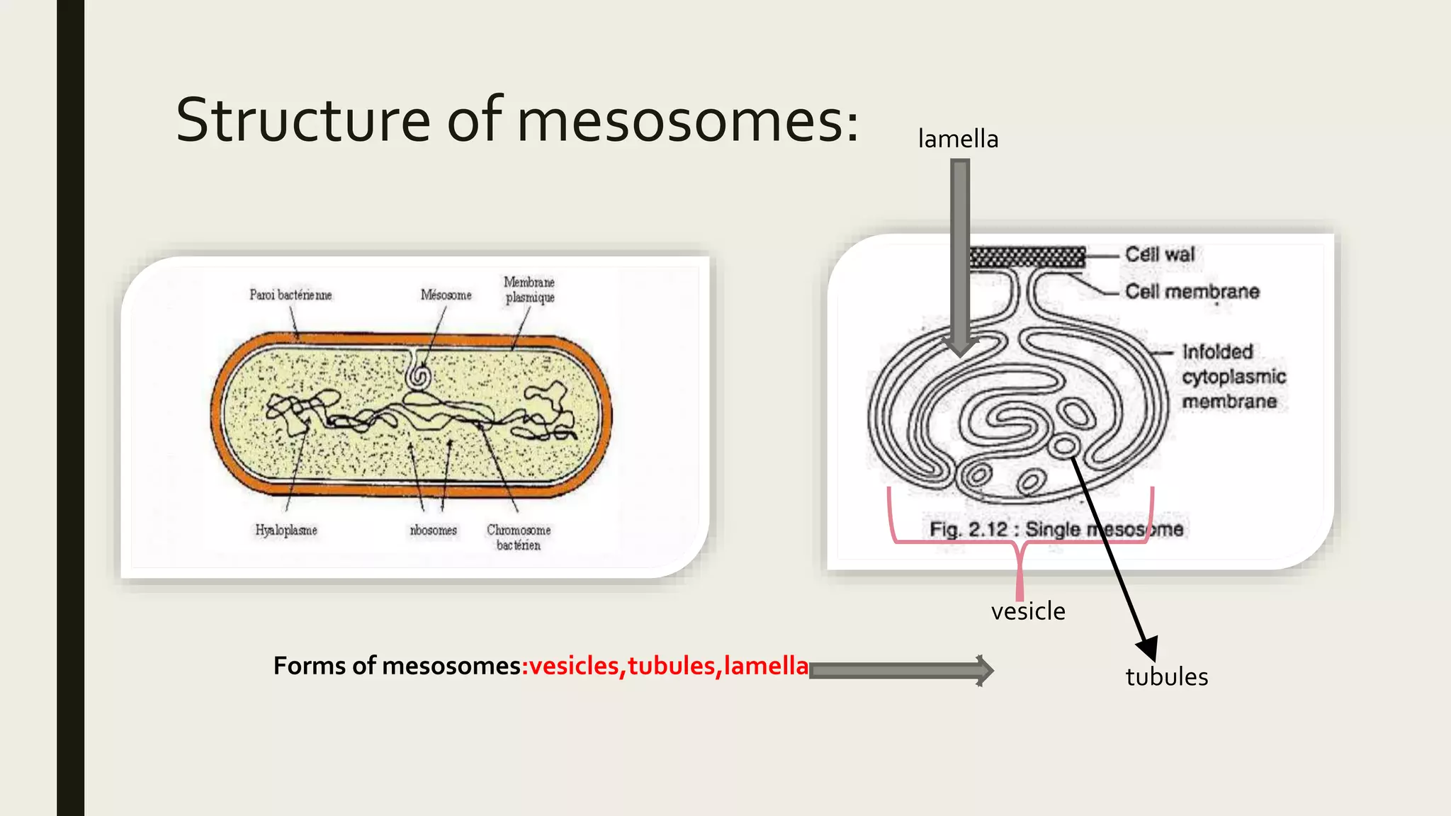
![MESOSOMES [CELL MEMBRANE
MODIFICATION]■ Mesosomes or chondrioids are folded invaginations in the
plasma membrane of bacteria.
■ A specialized membraneous structure is a mesosome which is
formed by an extension of plasma membrane into the cell. These
extensions are in the form of vesicle, tubules, and lamellae.
■ FUNCTIONS:(1) These extensions help in the synthesis of the cell
wall and replication of DNA. They also help in the equal
distribution of chromosomes into the daughter cells.
■ (2) It also increases the surface area of the plasma membrane to
carry out various enzymatic activities.
■ (3) It helps in secretion processes as well as in bacterial
respiration.](https://image.slidesharecdn.com/prokaryoticcell-200905105746/75/Prokaryotic-cell-18-2048.jpg)
![TYPES OF MESOSOMES:
■ 1]septal or central mesosome.
■ Help in DNA replication and transfer
of replicated DNA to daughter cell.
■ Help in formation of septa / Cross
wall during binary fission.
■ 2]peripheral or lateral mesosome
■ Also called chondroid.
■ Analogous to mitochondria of
eukaryotic cell.
■ Site for respiration bacteria.
■ Has enzyme for electron transport
chain[ETC]
■ Provide large surface area for
enzyme activity.](https://image.slidesharecdn.com/prokaryoticcell-200905105746/75/Prokaryotic-cell-19-2048.jpg)


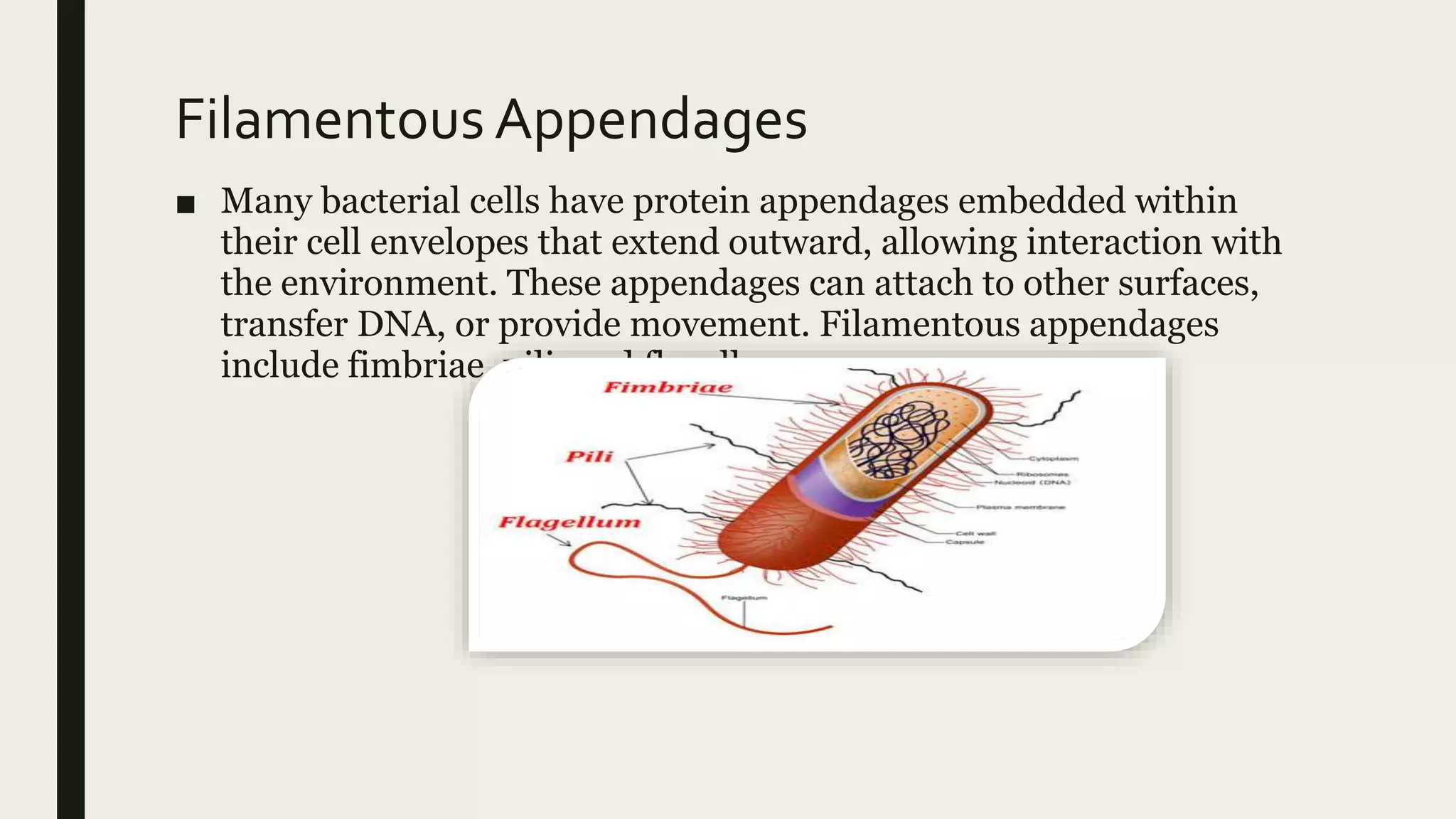


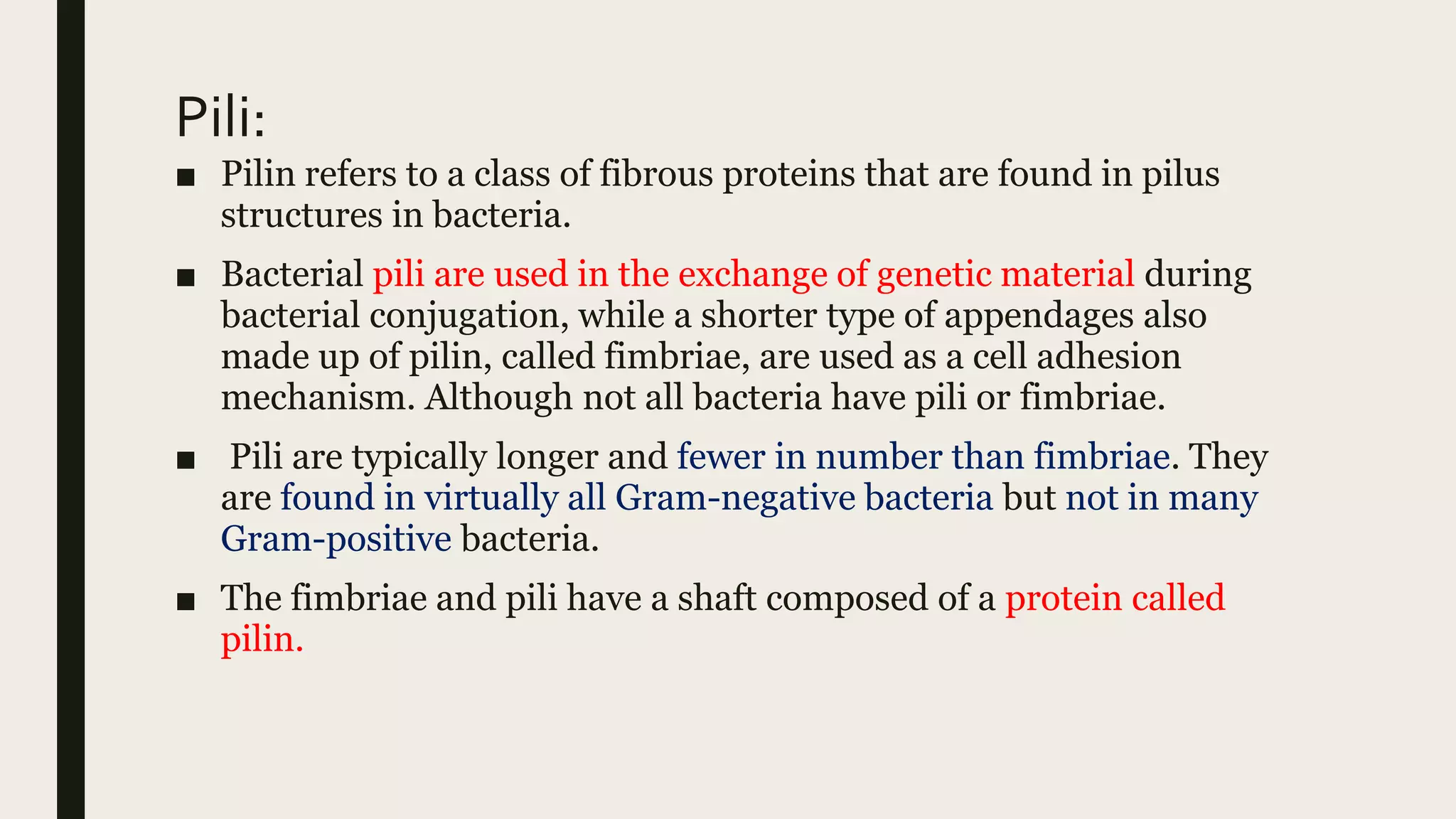
![Fimbriae [short attachment pili]
■ In bacteriology, a fimbria (Latin for 'fringe', plural fimbriae), also
referred to as an "attachment pilus" by some scientists, is a type of
appendage that is found on many Gram-negative and some Gram-
positive bacteria, and that is thinner and shorter than a flagellum
■ Fimbriae are used by bacteria to adhere to one another and to
adhere to animal cells and some inanimate objects. A bacterium
can have as many as 1,000 fimbriae. Fimbriae are only visible with
the use of an electron microscope. They may be straight or flexible.
■ Made up of fimbrillin protein.
■ Comparatively shorter in length than pili and flagella.
■ No role in bacterial motility and conjugation.](https://image.slidesharecdn.com/prokaryoticcell-200905105746/75/Prokaryotic-cell-35-2048.jpg)


![ATHLETE'S FOOT [TENEA PEDIS] FUNGI](https://cdn.slidesharecdn.com/ss_thumbnails/fungi-200817091534-thumbnail.jpg?width=640&height=640&fit=bounds)


