Download as PDF, PPTX
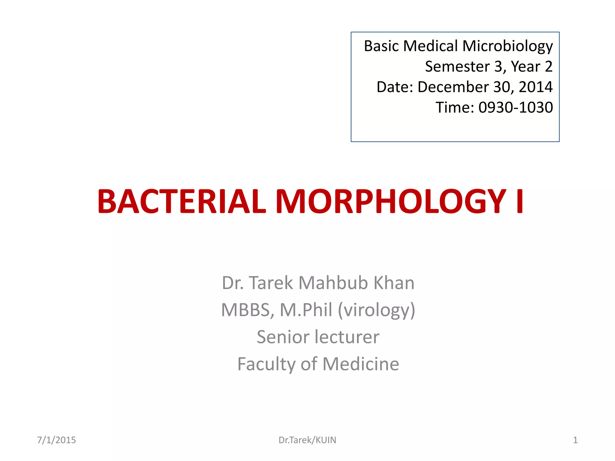

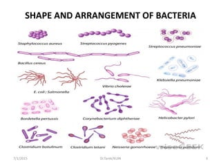
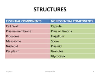
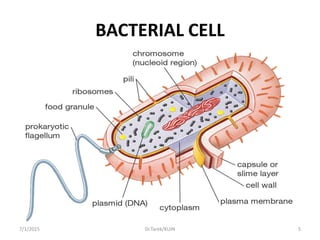
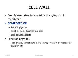
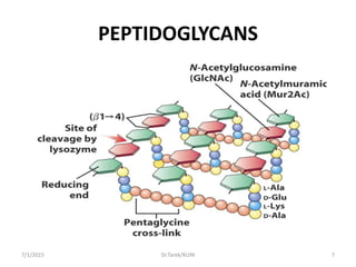
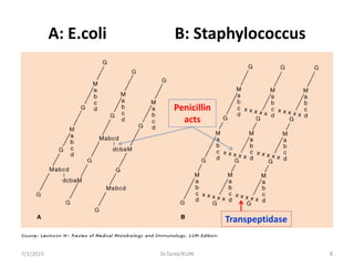
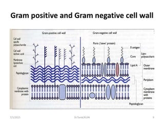
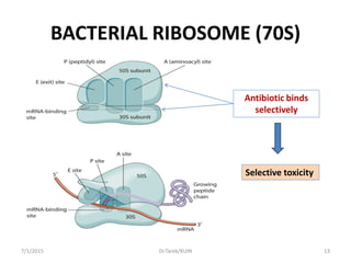
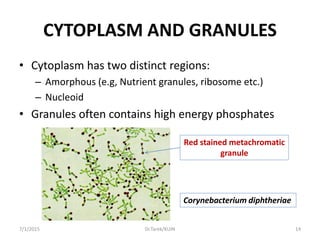
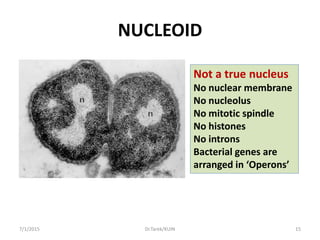
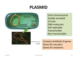
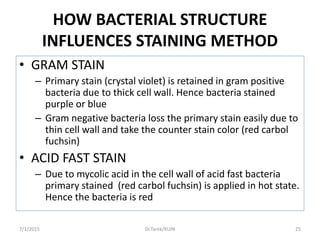


This document discusses the morphology and structures of bacterial cells. It begins by outlining the learning objectives which are to describe different bacterial shapes and arrangements, basic bacterial cell structures and their functions, staining characteristics based on structures, and how structure relates to laboratory identification. It then details the various bacterial cell components such as the cell wall, plasma membrane, ribosomes, and other internal structures. It explains how these structures determine staining properties and relates morphology to identification. The document uses diagrams to illustrate the different bacterial forms, components, and arrangements.


![PERI-PROSTHETIC FRACTURE NAIL-PLATE CONSTRUCT [NPC].pptx](https://cdn.slidesharecdn.com/ss_thumbnails/drarunkumardrmohamedashrafperiprostheticfrasturenail-plateconstructnpc-260209164459-7e9d15a1-thumbnail.jpg?width=640&height=640&fit=bounds)


![CTEV [ clubfoot] DR ARUN LAL ,DR MOHAMED ASHRAF travancore medical college k...](https://cdn.slidesharecdn.com/ss_thumbnails/ctevclubfootdrarunlaldrmohamedashraftravancoremedicalcollegekollamkeralaindia-260208063247-18fc466c-thumbnail.jpg?width=640&height=640&fit=bounds)


