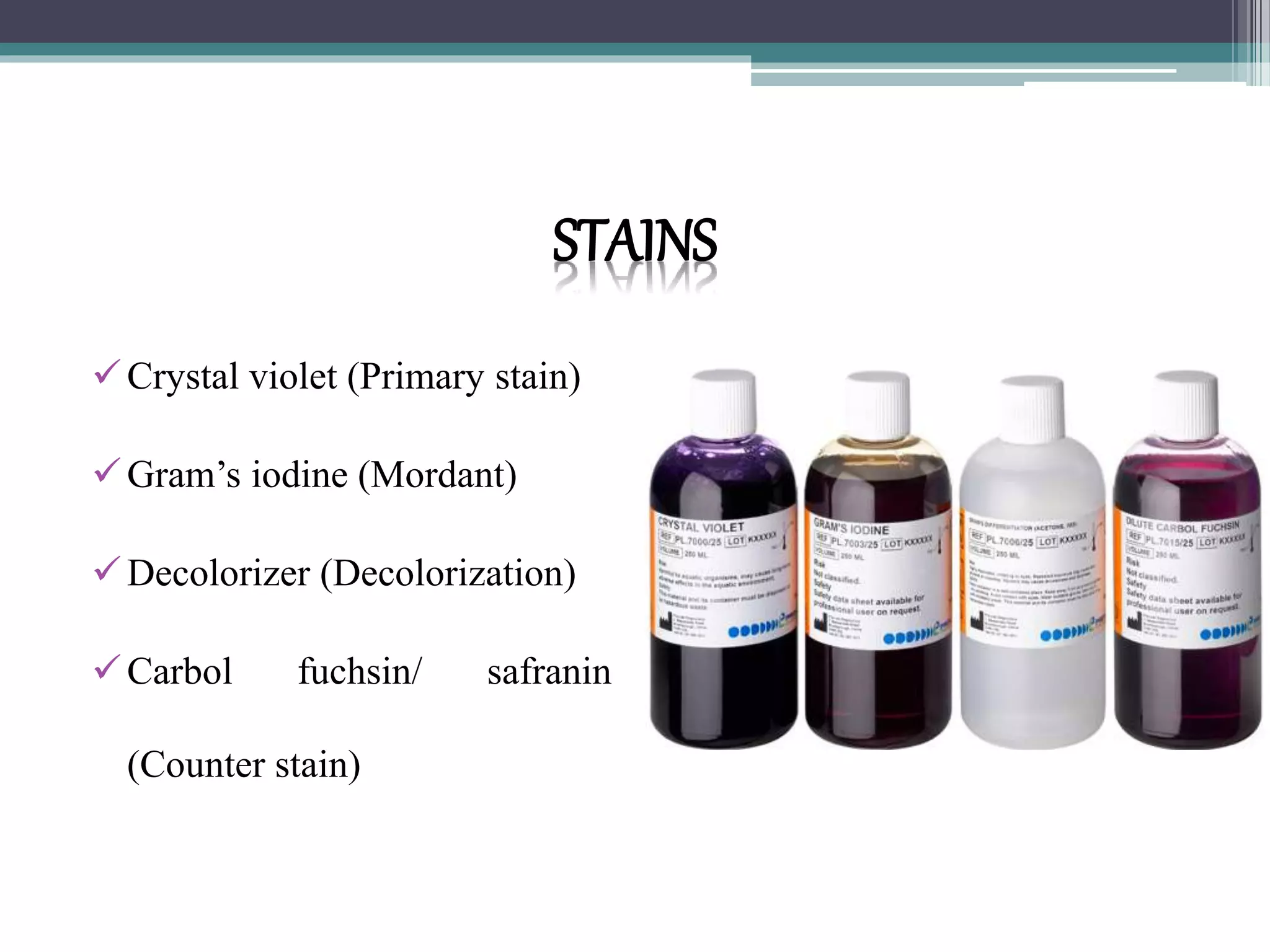
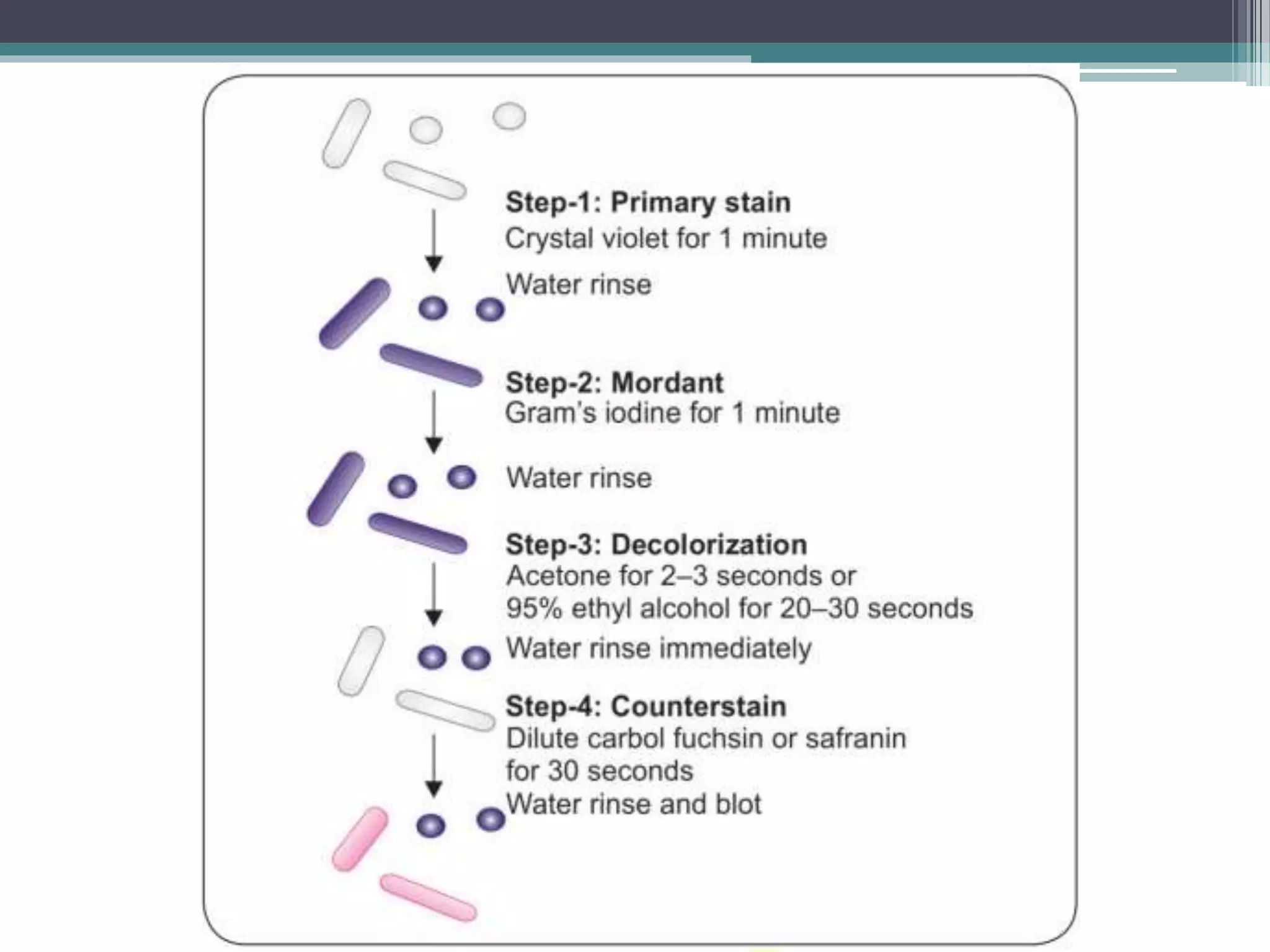
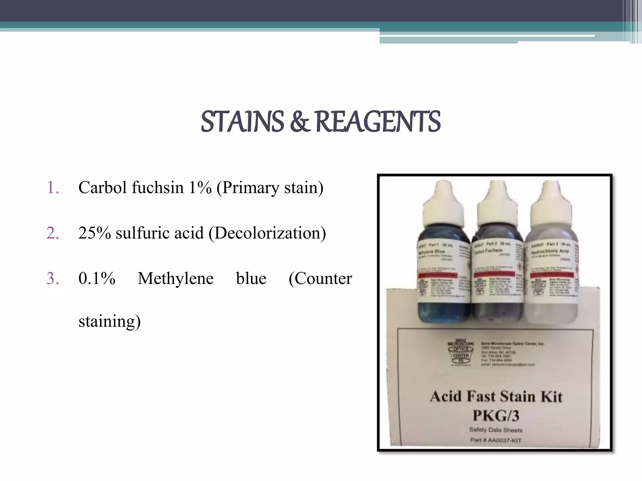
Staining Techniques
Ankur vashishtha
Staining Techniques
• Structural details of bacteria cannot be seen under light
microscope due to lack of contrast.
• Hence, it is necessary to use staining methods to produce color
contrast and thereby increase the visibility.
• Before staining, the fixation of the smear to the slide is done.
Fixation
• Fixation is the process by which the internal and external
structures of cells are preserved and fixed in position.
• It Kills and fixes the cells on to the slide.
• There are two types of fixation as follows
1. Heat Fixation
2. Chemical Fixation
Heat Fixation
• It is usually done for Bacterial
smears by gently flame heating
an air-dried film of bacteria.
• This adequately preserves
overall morphology but not
structures within the cells
Chemical Fixation
• It can be done using ethanol, acetic acid, mercuric chloride,
Formaldehyde, methanol and glutaraldehyde.
• They are used to protect the fine internal structure of the cells.
• This is useful for examination of blood smears.
Staining Techniques in Microbiology
1. Simple stain
2. Negative stain
3. Impregnation methods
4. Differential stain
Simple stain
• Use basic dyes, Such as methylene blue or basic fuchsine are
used as simple stains.
• They provide the color contrast, but impart the same color to
all the bacteria in a smear.
Negative stain
• A drop of bacteria suspension is mixed with dyes, such as
India ink or nigrosin.
• The background gets stained black where as unstained
bacterial/ yeast capsule stand out in contrast.
• This is very useful in the demonstration of bacterial/yeast
capsules which do not take up simple stains.
Impregnation methods
• Bacterial cells and structures that are too thin to be seen under
the light microscope, are thickened by impregnation of silver
salts on their surface to make them visible,
• e.g. for demonstration of bacterial flagella and spirochetes.
Differential stain
• Here, two stains are used which impart different colors to different bacteria
or bacterial structures, which help in differentiating bacteria.
• The most commonly employed differential stains are:-
 Gram stain:- It differentiates bacteria into gram-positive and gram-
negative group.
 Acid-fast stain:- It differentiates bacteria into acid-fast and non acid-
fast group.
 Albert stain:- It differentiates bacteria having metachromatic granules
from other bacteria that do not have them.
GramStain
• This staining technique was originally
developed by Hans Christian Gram
(1884).
STAINS
Crystal violet (Primary stain)
Gram’s iodine (Mordant)
Decolorizer (Decolorization)
Carbol fuchsin/ safranin
(Counter stain)
procedure
• Fixation:- The smear
made on a slide from
bacterial culture or
specimen, is air dried and
then heat fixed.
Interpretation of gram stain
• Smear is examined under oil immersion objective:-
▫ Gram-positive bacteria resist decolorization and retain the color of
primary stain i.e. violet
▫ Gram-negative bacteria are decolorized and, therefore, take counter
stain and appear pink.
Principle of gram staining
• The following theories have been put forward.
▫ pH theory
▫ Cell wall theory
Cell wall theory
• This is believed to be the most important postulate to describe
the mechanism of Gram stain.
▫ Gram-positive cell wall has a thick peptidoglycan layer. (50-100
layers thick).
pH theory
▫ Cytoplasm of gram-positive bacteria is more acidic, hence ,
can retain the basic dye (e.g. crystal violet) for longer time.
▫ Iodine serves as mordant, i.e. it combines with the primary
stain to from a dye-iodine complex which gets retained
inside the cell.
ACID-FAST STAIn
• The acid-fast stain was discovered by Paul Ehrlich and subsequently
modified by Ziehl and Neelsen.
• This staining is done to identify acid-fast organisms, such as
Mycobacterium tuberculosis and others.
• Acid-fastness is due to presence of mycolic acid in the cell wall.
Smear Preparation
• Smear measuring 2x3 cm in size is prepared in a new clean
grease free scratch free slide from the yellow purulent portion
of the sputum .
• The smear should neither be too thick nor too thin.
• Smear preparation should be done near a flame,
STAINS & REAGENTS
1. Carbol fuchsin 1% (Primary stain)
2. 25% sulfuric acid (Decolorization)
3. 0.1% Methylene blue (Counter
staining)
Procedure
5 Minutes 2-4 minutes 30 Second
Interpretation
• Mycobacterium tuberculosis
appears as long slender straight
or slightly curved & beaded,
red colored acid-fast bacillus.
ALBERT STAIN
• Albert stain is used to demonstrate the metachromatic granules of
corynebacterium diphtheriae.
Procedure
• Fixation:- the smear is heat fixed
• Smear is covered with Albert-I (Albert’s stain) for 5 Minutes
• Then washed in water, and blotted dry
• Albert – II (Iodine solution) is added for 1 minute.
• Slide is washed in water, blotted dry and examined under oil
immersion field.
Interpretation
• corynebacterium diphtheriae
appears as green colored bacilli
arranged in chines latter or
cuniform pattern, with bluish
black metachromatic granules at
polar ends.
Thank You

Staining techniques

  • 1.
  • 2.
    Staining Techniques • Structuraldetails of bacteria cannot be seen under light microscope due to lack of contrast. • Hence, it is necessary to use staining methods to produce color contrast and thereby increase the visibility. • Before staining, the fixation of the smear to the slide is done.
  • 3.
    Fixation • Fixation isthe process by which the internal and external structures of cells are preserved and fixed in position. • It Kills and fixes the cells on to the slide. • There are two types of fixation as follows 1. Heat Fixation 2. Chemical Fixation
  • 4.
    Heat Fixation • Itis usually done for Bacterial smears by gently flame heating an air-dried film of bacteria. • This adequately preserves overall morphology but not structures within the cells
  • 5.
    Chemical Fixation • Itcan be done using ethanol, acetic acid, mercuric chloride, Formaldehyde, methanol and glutaraldehyde. • They are used to protect the fine internal structure of the cells. • This is useful for examination of blood smears.
  • 6.
    Staining Techniques inMicrobiology 1. Simple stain 2. Negative stain 3. Impregnation methods 4. Differential stain
  • 8.
    Simple stain • Usebasic dyes, Such as methylene blue or basic fuchsine are used as simple stains. • They provide the color contrast, but impart the same color to all the bacteria in a smear.
  • 9.
    Negative stain • Adrop of bacteria suspension is mixed with dyes, such as India ink or nigrosin. • The background gets stained black where as unstained bacterial/ yeast capsule stand out in contrast. • This is very useful in the demonstration of bacterial/yeast capsules which do not take up simple stains.
  • 10.
    Impregnation methods • Bacterialcells and structures that are too thin to be seen under the light microscope, are thickened by impregnation of silver salts on their surface to make them visible, • e.g. for demonstration of bacterial flagella and spirochetes.
  • 11.
    Differential stain • Here,two stains are used which impart different colors to different bacteria or bacterial structures, which help in differentiating bacteria. • The most commonly employed differential stains are:-  Gram stain:- It differentiates bacteria into gram-positive and gram- negative group.  Acid-fast stain:- It differentiates bacteria into acid-fast and non acid- fast group.  Albert stain:- It differentiates bacteria having metachromatic granules from other bacteria that do not have them.
  • 12.
    GramStain • This stainingtechnique was originally developed by Hans Christian Gram (1884).
  • 13.
    STAINS Crystal violet (Primarystain) Gram’s iodine (Mordant) Decolorizer (Decolorization) Carbol fuchsin/ safranin (Counter stain)
  • 14.
    procedure • Fixation:- Thesmear made on a slide from bacterial culture or specimen, is air dried and then heat fixed.
  • 17.
    Interpretation of gramstain • Smear is examined under oil immersion objective:- ▫ Gram-positive bacteria resist decolorization and retain the color of primary stain i.e. violet ▫ Gram-negative bacteria are decolorized and, therefore, take counter stain and appear pink.
  • 19.
    Principle of gramstaining • The following theories have been put forward. ▫ pH theory ▫ Cell wall theory
  • 21.
    Cell wall theory •This is believed to be the most important postulate to describe the mechanism of Gram stain. ▫ Gram-positive cell wall has a thick peptidoglycan layer. (50-100 layers thick).
  • 22.
    pH theory ▫ Cytoplasmof gram-positive bacteria is more acidic, hence , can retain the basic dye (e.g. crystal violet) for longer time. ▫ Iodine serves as mordant, i.e. it combines with the primary stain to from a dye-iodine complex which gets retained inside the cell.
  • 23.
    ACID-FAST STAIn • Theacid-fast stain was discovered by Paul Ehrlich and subsequently modified by Ziehl and Neelsen. • This staining is done to identify acid-fast organisms, such as Mycobacterium tuberculosis and others. • Acid-fastness is due to presence of mycolic acid in the cell wall.
  • 25.
    Smear Preparation • Smearmeasuring 2x3 cm in size is prepared in a new clean grease free scratch free slide from the yellow purulent portion of the sputum . • The smear should neither be too thick nor too thin. • Smear preparation should be done near a flame,
  • 26.
    STAINS & REAGENTS 1.Carbol fuchsin 1% (Primary stain) 2. 25% sulfuric acid (Decolorization) 3. 0.1% Methylene blue (Counter staining)
  • 27.
    Procedure 5 Minutes 2-4minutes 30 Second
  • 28.
    Interpretation • Mycobacterium tuberculosis appearsas long slender straight or slightly curved & beaded, red colored acid-fast bacillus.
  • 29.
    ALBERT STAIN • Albertstain is used to demonstrate the metachromatic granules of corynebacterium diphtheriae.
  • 30.
    Procedure • Fixation:- thesmear is heat fixed • Smear is covered with Albert-I (Albert’s stain) for 5 Minutes • Then washed in water, and blotted dry • Albert – II (Iodine solution) is added for 1 minute. • Slide is washed in water, blotted dry and examined under oil immersion field.
  • 32.
    Interpretation • corynebacterium diphtheriae appearsas green colored bacilli arranged in chines latter or cuniform pattern, with bluish black metachromatic granules at polar ends.
  • 33.