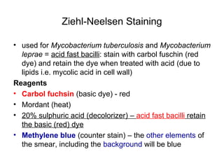
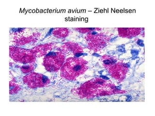
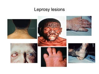
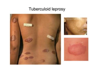
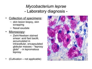
Laboratory diagnosis in infections produced by
germs of the Genus Mycobacterium
Genus Mycobacterium
General characters:
• filamentous, non-motile, non-sporulated bacilli;
• obligate aerobes
• Bacterial wall – high lipid content: mycolic acid (gives
the name of the genus) - 2 types of effects:
• 1. resistance to antiseptics
• 2. staining only possible with heating BUT after that,
discoloration with acids & alcohols is not possible →
“acid-alcohol resistant” / “acid fast” bacilli
– Gram staining not possible
– Other staining techniques required e.g. Ziehl-Neelsen
Ziehl-Neelsen Staining
• Mycobacteria – impermeable to dyes due to high
lipid and wax content of cell wall – usual staining
techniques (e.g. Gram) cannot be used
• heat and phenol (carbolic acid) help penetration
of dye inside mycobacterial cells
• gold standard for diagnosis of tuberculosis and
leprosy
• + Nocardia, Cryptosporidium
Ziehl-Neelsen Staining
• used for Mycobacterium tuberculosis and Mycobacterium
leprae = acid fast bacilli: stain with carbol fuschin (red
dye) and retain the dye when treated with acid (due to
lipids i.e. mycolic acid in cell wall)
Reagents
• Carbol fuchsin (basic dye) - red
• Mordant (heat)
• 20% sulphuric acid (decolorizer) – acid fast bacilli retain
the basic (red) dye
• Methylene blue (counter stain) – the other elements of
the smear, including the background will be blue
Mycobacterium avium – Ziehl Neelsen
staining
Genus Mycobacterium
- Clinical significance -
• 41 species of which over 20 isolated in human infections
• Tuberculosis (pulomonary & extrapulmonary) -
M.tuberculsis, M.bovis
• Other (similar) lung infections - M.avium
• Skin infections (possible dissemination) - M.ulcerans
• Leprosy - M.leprae
Robert Koch (1843-1910)
• German physician,
pioneer microbiologist
• “founder of modern
bacteriology”
• Identification of causative
agent of tuberculosis
(Mycobacterium
tuberculosis a.k.a. Koch´s
bacillus)
Mycobacterium tuberculosis
- Collection of specimens -
• Sputum:
– Challenging! – avoid contamination with saliva and
secretions from upper air ways
– Optimal moment: in the morning (higher amount of
sputum secreted during the night and stagnant in
lower air ways)
– Indirect method:
• Patient energically rinses mouth with saline solution
• Coughs and expectorates in sterile container (Petri dish)
– Direct method:
• Bronchoscopy / tracheal punctioning
Mycobacterium tuberculosis
- Collection of specimens - continued
• Pleural fluid: pleural puncture
(thoracentesis/ pleural tap)
Mycobacterium tuberculosis
- Collection of specimens - continued
Urine:
- first morning miction
- clean uro-genital area
- eliminate first flow
- collect middle flow in sterile
container
- Send to lab immediately or
store at 2-8°C
- Repeat in 2-3 days
(intermittent elimination of
bacilli)
Mycobacterium tuberculosis
- Collection of specimens - continued
• CSF: lumbar puncture (spinal
tap)
Mycobacterium tuberculosis
- Collection of specimens - continued
• Bone marrow aspiration:
suspected bone tuberculosis
• Ascites (peritoneal fluid):
paracentesis (peritoneal
puncture)
Mycobacterium tuberculosis
- Microscopy -
• Preliminary treatment of specimen:
– Homogenization with 4% NaOH (also destroys associated flora)
– 30 min at 37°C
– Centrifugation: 10-15 min, 3000 rpm → sediment used for
further examination
• Ziehl-Neelsen stained smear:
– examined for at least 10 min (minimum 100 microscopic fields)
– thin red, encurved bacilli, arranged in groups or in angulated
pairs or isolated;
– positive result expressed as “presence of acid fast bacilli”
Mycobacterium tuberculosis - Ziehl-Neelsen
Staining
Mycobacterium tuberculosis
- Isolation & Identification-
• The previously homogenized (NaOH – destruction of
associated flora) and concentrated (centrifuged)
specimen – inoculated on solid media
• Lowenstein-Jensen (egg, glycerin, asparagin) – slant in
tube
• Incubation at 37°C for 2 - 4 weeks (slow growing germs)
• Identification:
• Colonial characters: prominent, rough, irregular colonies,
slightly yellowish
• Further identification – Ziehl-Neelsen stained smear from
colonies
Mycobacterium tuberculosis – Ziehl Neelsen
staining from culture
Mycobacterium tuberculosis
- Isolation & Identification- continued
• Niacin test:
– production of niacin by M.tuberculosis in egg-containing medium →
canary yellow colour (test is negative for other mycobacteria)
– Paper strips impregnated with reagents for detection of niacin
produced by mycobacteria
• Peroxidase test: M.tuberculosis, M. bovis – positive test (other,
atypical mycobacteria – negative test)
• Catalase test: M.tuberculosis and M.bovis - weakly positive test
(atypical mycobacteria - strongly positive test)
• Tuberculostatic susceptibility tests
• Oxygen requirement:
– M.tuberculosis – aerobic
– M.bovis is microaerophilic
Mycobacterium tuberculosis
- Antimicrobial susceptibility -
• Multidrug resistant (MDR) strains – major public health
problem
• Treatment of tuberculosis is standardized – association
of antiTB drugs adjusted according to response to
treatment regimen (1st
→2nd
→ 3rd
line regimens)
(follow link below)
• http://www.cdc.gov/tb/publications/guidelines/Treatment.htm
Mycobacterium tuberculosis
- Immunization-
Bacille de Calmette et Guérin (BCG): attenuated strain
of M.bovis (lower virulence; no virulence in humans)
Vaccination policies adjusted to morbidity in various regions
(see link below)
http://www.bcgatlas.org/
In Romania BCG (TB vaccine) given to all newborns
(during the 1st
week of life)
Guidelines for TB vaccination:
• http://www.cdc.gov/tb/publications/guidelines/vaccines.htm
Mycobacterium leprae
• Clinical significance: Leprosy –
chronic infection (Hansen´s
disease, named after Gerhard
Hansen, 1841-1912 –
Norwegian physician)
Leprosy
• slowly progressing bacterial infection affecting:
– skin
– peripheral sensitive nerves in the hands and feet
– mucous membranes (nose, throat, eyes)
– Internal organs, bones
• Transmission via infectious droplets (infectivity is low):
– acquisition of disease requires close, longtime contact with
patient with active infection, not treated; after 2-3 days of
treatment – patient is not contagious any more
• 2 clinical forms:
– lepromatous
– tuberculoid
Leprosy - continued
• Lepromatous leprosy (multibacillary):
– low immune response
– spongy tumor like swellings on the face and body
– degenerative lesions of internal organs and bones
– deformity & sensitivity loss in extremities (loss of fingers, toes,
nose tip, lips)
– long evolution (20-30 years)
• Tuberculoid leprosy (paucibacillary):
– isolated, asymetrical skin lesions
– not contagious
Leprosy lesions
Tuberculoid leprosy
Mycobacterium leprae
- Laboratory diagnosis -
• Collection of specimens:
– skin lesion biopsy, skin
scrapping
– Nasal exudate
• Microscopy:
– Ziehl-Neelsen stained
smear: acid fast bacilli,
accumulated in
intracellular, encapsulated
globular masses - “leprosy
globi” - in lepromatous
leprosy
• (Cultivation – not applicable)
Prevention and treatment of leprosy
• Hygiene (washing hands), disinfection, monitoring close
contacts of leprosy diagnosed cases
• Early diagnosis – very important in order to initiate
treatment and control further transmission (infectivity of
leprosy patient tends to zero after first 2-3 days of
treatment)
• Multidrug therapy (MDT): combination of 2-3
antimicrobials: Dapsone + Rifampicin + Clofazimin – 6
months – 2 years
• + Corrective surgery

Mycobacterium

  • 1.
    Laboratory diagnosis ininfections produced by germs of the Genus Mycobacterium
  • 2.
    Genus Mycobacterium General characters: •filamentous, non-motile, non-sporulated bacilli; • obligate aerobes • Bacterial wall – high lipid content: mycolic acid (gives the name of the genus) - 2 types of effects: • 1. resistance to antiseptics • 2. staining only possible with heating BUT after that, discoloration with acids & alcohols is not possible → “acid-alcohol resistant” / “acid fast” bacilli – Gram staining not possible – Other staining techniques required e.g. Ziehl-Neelsen
  • 3.
    Ziehl-Neelsen Staining • Mycobacteria– impermeable to dyes due to high lipid and wax content of cell wall – usual staining techniques (e.g. Gram) cannot be used • heat and phenol (carbolic acid) help penetration of dye inside mycobacterial cells • gold standard for diagnosis of tuberculosis and leprosy • + Nocardia, Cryptosporidium
  • 4.
    Ziehl-Neelsen Staining • usedfor Mycobacterium tuberculosis and Mycobacterium leprae = acid fast bacilli: stain with carbol fuschin (red dye) and retain the dye when treated with acid (due to lipids i.e. mycolic acid in cell wall) Reagents • Carbol fuchsin (basic dye) - red • Mordant (heat) • 20% sulphuric acid (decolorizer) – acid fast bacilli retain the basic (red) dye • Methylene blue (counter stain) – the other elements of the smear, including the background will be blue
  • 5.
    Mycobacterium avium –Ziehl Neelsen staining
  • 6.
    Genus Mycobacterium - Clinicalsignificance - • 41 species of which over 20 isolated in human infections • Tuberculosis (pulomonary & extrapulmonary) - M.tuberculsis, M.bovis • Other (similar) lung infections - M.avium • Skin infections (possible dissemination) - M.ulcerans • Leprosy - M.leprae
  • 7.
    Robert Koch (1843-1910) •German physician, pioneer microbiologist • “founder of modern bacteriology” • Identification of causative agent of tuberculosis (Mycobacterium tuberculosis a.k.a. Koch´s bacillus)
  • 8.
    Mycobacterium tuberculosis - Collectionof specimens - • Sputum: – Challenging! – avoid contamination with saliva and secretions from upper air ways – Optimal moment: in the morning (higher amount of sputum secreted during the night and stagnant in lower air ways) – Indirect method: • Patient energically rinses mouth with saline solution • Coughs and expectorates in sterile container (Petri dish) – Direct method: • Bronchoscopy / tracheal punctioning
  • 9.
    Mycobacterium tuberculosis - Collectionof specimens - continued • Pleural fluid: pleural puncture (thoracentesis/ pleural tap)
  • 10.
    Mycobacterium tuberculosis - Collectionof specimens - continued Urine: - first morning miction - clean uro-genital area - eliminate first flow - collect middle flow in sterile container - Send to lab immediately or store at 2-8°C - Repeat in 2-3 days (intermittent elimination of bacilli)
  • 11.
    Mycobacterium tuberculosis - Collectionof specimens - continued • CSF: lumbar puncture (spinal tap)
  • 12.
    Mycobacterium tuberculosis - Collectionof specimens - continued • Bone marrow aspiration: suspected bone tuberculosis • Ascites (peritoneal fluid): paracentesis (peritoneal puncture)
  • 13.
    Mycobacterium tuberculosis - Microscopy- • Preliminary treatment of specimen: – Homogenization with 4% NaOH (also destroys associated flora) – 30 min at 37°C – Centrifugation: 10-15 min, 3000 rpm → sediment used for further examination • Ziehl-Neelsen stained smear: – examined for at least 10 min (minimum 100 microscopic fields) – thin red, encurved bacilli, arranged in groups or in angulated pairs or isolated; – positive result expressed as “presence of acid fast bacilli”
  • 14.
    Mycobacterium tuberculosis -Ziehl-Neelsen Staining
  • 15.
    Mycobacterium tuberculosis - Isolation& Identification- • The previously homogenized (NaOH – destruction of associated flora) and concentrated (centrifuged) specimen – inoculated on solid media • Lowenstein-Jensen (egg, glycerin, asparagin) – slant in tube • Incubation at 37°C for 2 - 4 weeks (slow growing germs) • Identification: • Colonial characters: prominent, rough, irregular colonies, slightly yellowish • Further identification – Ziehl-Neelsen stained smear from colonies
  • 16.
    Mycobacterium tuberculosis –Ziehl Neelsen staining from culture
  • 17.
    Mycobacterium tuberculosis - Isolation& Identification- continued • Niacin test: – production of niacin by M.tuberculosis in egg-containing medium → canary yellow colour (test is negative for other mycobacteria) – Paper strips impregnated with reagents for detection of niacin produced by mycobacteria • Peroxidase test: M.tuberculosis, M. bovis – positive test (other, atypical mycobacteria – negative test) • Catalase test: M.tuberculosis and M.bovis - weakly positive test (atypical mycobacteria - strongly positive test) • Tuberculostatic susceptibility tests • Oxygen requirement: – M.tuberculosis – aerobic – M.bovis is microaerophilic
  • 18.
    Mycobacterium tuberculosis - Antimicrobialsusceptibility - • Multidrug resistant (MDR) strains – major public health problem • Treatment of tuberculosis is standardized – association of antiTB drugs adjusted according to response to treatment regimen (1st →2nd → 3rd line regimens) (follow link below) • http://www.cdc.gov/tb/publications/guidelines/Treatment.htm
  • 19.
    Mycobacterium tuberculosis - Immunization- Bacillede Calmette et Guérin (BCG): attenuated strain of M.bovis (lower virulence; no virulence in humans) Vaccination policies adjusted to morbidity in various regions (see link below) http://www.bcgatlas.org/ In Romania BCG (TB vaccine) given to all newborns (during the 1st week of life) Guidelines for TB vaccination: • http://www.cdc.gov/tb/publications/guidelines/vaccines.htm
  • 20.
    Mycobacterium leprae • Clinicalsignificance: Leprosy – chronic infection (Hansen´s disease, named after Gerhard Hansen, 1841-1912 – Norwegian physician)
  • 21.
    Leprosy • slowly progressingbacterial infection affecting: – skin – peripheral sensitive nerves in the hands and feet – mucous membranes (nose, throat, eyes) – Internal organs, bones • Transmission via infectious droplets (infectivity is low): – acquisition of disease requires close, longtime contact with patient with active infection, not treated; after 2-3 days of treatment – patient is not contagious any more • 2 clinical forms: – lepromatous – tuberculoid
  • 22.
    Leprosy - continued •Lepromatous leprosy (multibacillary): – low immune response – spongy tumor like swellings on the face and body – degenerative lesions of internal organs and bones – deformity & sensitivity loss in extremities (loss of fingers, toes, nose tip, lips) – long evolution (20-30 years) • Tuberculoid leprosy (paucibacillary): – isolated, asymetrical skin lesions – not contagious
  • 23.
  • 24.
  • 25.
    Mycobacterium leprae - Laboratorydiagnosis - • Collection of specimens: – skin lesion biopsy, skin scrapping – Nasal exudate • Microscopy: – Ziehl-Neelsen stained smear: acid fast bacilli, accumulated in intracellular, encapsulated globular masses - “leprosy globi” - in lepromatous leprosy • (Cultivation – not applicable)
  • 26.
    Prevention and treatmentof leprosy • Hygiene (washing hands), disinfection, monitoring close contacts of leprosy diagnosed cases • Early diagnosis – very important in order to initiate treatment and control further transmission (infectivity of leprosy patient tends to zero after first 2-3 days of treatment) • Multidrug therapy (MDT): combination of 2-3 antimicrobials: Dapsone + Rifampicin + Clofazimin – 6 months – 2 years • + Corrective surgery