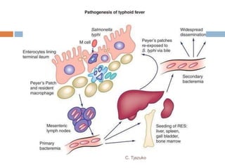
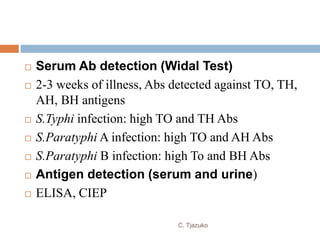
SHIGELLA AND SALMONELLA
Ms. Clemencia Tjazuko
MSc. Medical Microbiology
 Morphology
 Short gram-negative rods
 0.5 by 1-3 µm in size
 Non-motile
 Non-sporing
 Non-capsulated
 Fimbriae may be present
C. Tjazuko
Shigella
 Agent of bacillary dysentery
 Dysentery- infection of the intestines which causes
the passage of bloody stools mixed with mucus.
 Named after Japanese microbiologist Kiyoshi
Shiga, who isolated the first member 
S.dysenteriae in 1886 from epidemic dysentery
C. Tjazuko
 Antigenic Structure
 Simple
 ‘Major’ antigens and ‘Minor’ somatic antigens
 Some have K antigens
 Fimbrial antigens
C. Tjazuko
Classification
 Classified into four species based on combination
of biochemical and serological characteristics
 Serotypes distinguished within the species
C. Tjazuko
1. S.dysenteriae (Group A)
 Does not ferment mannitol
 15 serotypes
 Serotype 1- S.shigae
 Only shigella producing shiga toxin (exotoxin)
 Only member of enterobacteriaceae that is catalase
negative
 Indole negative
C. Tjazuko
 Serotype 2- S.schmitzi
 Forms indole
 Ferments sorbitol and rhamnose
 Serotype 3-7
 Formerly Large-Sachs group
C. Tjazuko
2. S.flexneri (Group B)
 Based on specific type antigen
 6 serotypes
 Complex antigenically
 Serotype 6
 Indole negative and has three biotypes
 Manchester, Newcastle and Boyd 88
C. Tjazuko
3. S. Boydii (Group C)
 19 serotypes
 Isolated least frequently
 Resemble S.flexneri biochemically but not
antigenically
 Described by Boyd (1931) from India
C. Tjazuko
4. S. Sonnei (Group C)
 Sonne (1915), Denmark
 Ferments lactose and sucrose late
 Indole negative
 Antigenically homogenous
 2 forms: Phase I and Phase II
 Phase II colonies are larger, flatter and irregular
 Mildest form of bacillary dysentry
C. Tjazuko
 Toxins
1. Endotoxin
 May be due to the LPS of Gram-negative cell wall
 Has irritating effect on intestinal wall which causes
diarrhoea and subsequently intestinal ulcers
C. Tjazuko
2. Exotoxin
 Produced by S.dysenteriae type 1
 Also called Shiga toxin or verotoxin
 Acts as enterotoxin and neurotoxin
 Inhibit protein synthesis
C. Tjazuko
3. Verocytotoxin (VT)
 Cytotoxin produced by S.dysenteriae type 1
 Acts on vero cells
 VT1 and VT2
 VT1 two subunits A and B.
A binds cytotoxin to cells
B inhibit protein synthesis
C. Tjazuko
Pathogenesis of Shigella
 Mode of transmission:
infection through contaminated fingers
contaminated food/water
flies (rare)
transmitted sexually (homosexuals), gay bowel
syndrome
 Minimum Infective Dose:
10-100 bacilli
C. Tjazuko
 Entry via M cell:
Bacilli enter mucosa via M cell
Cross the basolateral side of M cells to reach submucosa where
they are engulfed by macrophages
Bacilli released by macrophages
Cytokines released which attract inflammatory cells to infected
site inflammation acute collitis
C. Tjazuko
 Invasion:
Inside submucosa, shigellae induce own uptake
into adjacent epithelial
Determined by large virulence plasmid, codes
for ipa proteins and Type III secretion system
 Direct cell-to-cell spread:
from one host cell to the other by inducing actin
polymerisation cells of host
C. Tjazuko
Bacillary dysentery
 Short incubation period (1-7 days)
 Clinical features:
 Frequent passage of loose, scanty feces containing
blood and mucus, with abdominal cramps and
tenesmus
 Complications:
 Arthritis, toxic neuritis, conjunctivitis, parotitis,
intussusception in children
 Hemolytic uremic syndrome in severe cases
C. Tjazuko
C. Tjazuko
Laboratory Diagnosis
 Specimen
 Isolating bacillus from feces
 Fresh feces inoculated or transported in Sach’s
buffered glycerol saline or Gram-negative broth @
pH7.0-7.4
 Rectal swabs not satisfactory
 Gram-negative broth sodium deoxycholate to
inhibit gram-positive bacteria, subcultured 6-8hrs
after incubation
 Selenite F Broth/Salmonella Shigella (SS) broth
C. Tjazuko
 Microscopy- plenty of pus cells, erythrocytes and
macrophages
 Culture- best to use mucus flakes
 MacConkey, DCA/ XLD
 Incubation @ 37 ºC, plates inspected for pale or pink-
coloured colonies, identified by biochemical reactions
 Non-motile bacillus that is : urease, citrate, H2S and
KCN negative further investigated by biochemical tests
 Confirmed by slide agglutination with sera
C. Tjazuko
 Salmonella Shigella Agar - colonies colourless
 Hektoen enteric (HE) Agar – contains bile salts as
inhibitory agents and some dyes, colonies appear
green with fading colour to the periphery
C. Tjazuko
 Treatment
 Acute cases infants and young children,
dehydration corrected propmptly
 Antibiotics reserved for severe toxic cases
 Many strains still sensitive to Nalidixic acid and
Norfloxacin
C. Tjazuko
Salmonella
 Morphology
 Gram-negative rods,1-3 by 0.5 µm in size
 Fimbriae – present
 Motile with petrichate flagella
 But S.Gallinarum and S.Pullorum are non-motile
C. Tjazuko
Clinical Classification
1. Typhoidal Salmonella
 Serotypes S.Typhi and S.Paratyphi restricted to
human hosts causing enteric fever
(typhoid/paratyphoid fever)
2. Non-typhoidal Salmonella
 Remaining serotypes are the food poisoning
group, infect human beings causing
gastroenteritis, septicemia or localised infections
C. Tjazuko
Antigenic Classification
 Kauffmann-White scheme
 Based on the presence of different somatic (O) and
flagellar (H) which is detected by agglutination with
respective antisera
 Based on O antigen:
 Serogroup 2
 Serogroup 4
 Serogroup 9
 Within each group further diffrentiation by Phase 1
and Phase 2 flagellar antigens
C. Tjazuko
C. Tjazuko
Antigenic Structure
Flagellar antigen H
Somatic antigen O
Surface antigen Vi
H antigen
Present on flagella, heat labile
Destroyed by boiling or alcohol
H suspensions agglutinate rapidly to form large, loose,
fluffy clumps
C. Tjazuko
 Strongly immunogenic, induce antibody formation
 Occurs in two phases
O antigen
 Phosholipid-protein-polysaccharide complex
 Identical to endotoxin
 Heat and alcohol stable
 67 O antigens
 Less immunogenic
 O suspensions form compact, chalky, granular clumps
with antisera
C. Tjazuko
Vi antigen
 Surface polysachharide envelope or capsular antigen
covering the O antigen
 Inhibits phagocytosis, resists complement
activation, bacterial lysis
 Heat labile, Poorly immunogenic
 Present bacilli is inagglutinable with O antisera.
Becomes agglutinable after boiling or heating at 100
ºC for 1hr
 S.Paratyphi C
C. Tjazuko
C. Tjazuko
Antigenic Variation
 H-O Variation
 Associated with the loss of flagella. Induced by:
Phenol agar: inhibits flagella
Mutation: Non-motile mutant of S.Typhi 901-O
strain subcultured in Craigie’s tube
 Phase variation:
Phase 1: serotype specific (a, b,c etc)
Phase 2: nospecific/group antigens (1,2, etc)
C. Tjazuko
 V-W Variation
 V forms of S.Typhi isolates with Vi antigens are
agglutinated by Vi antisera but not O antisera
 After serial subcultures, Vi antigen is lost W
forms agglutinable by O antisera but not Vi
antisera
 Also VW forms agglutinable with both
C. Tjazuko
 S-R Variations
 Smooth to rough variation, due to loss of of O
antigen side chain from LPS leading to exposure of
core polysaccharide portion
 Smooth colonies produced by virulent strains
 Rough strains form large, round and irregular
colonies, avirulent due to loss of O antigen
C. Tjazuko
Pathogenesis of Salmonella
 S.Typhi and S.Paratyphi A, B, C  Typhoidal
salmonellae
 Cause Enteric fever also known as Typhoid fever
 Infective Dose:103 – 106 bacilli
 Risk factors: stomach acidity, intestinal integrity
 Entry through epithelial cells (M cells): lines the
mucosa. Salmonellae cause formation of
membrane ruffles. Ruffles enclose adherent
bacteria bacteria mediated endocytosis (BME)
C. Tjazuko
 Mechanism of BME: Type III secretion system
delivery of bacterial proteins into cytoplasm of
epithelial cells alters actin cytoskeleton required for
bacterial uptake
 Entry into macrophages: Salmonellae containing
vacuoles cross epithelial layersubmucosa
phagocytosed
 Survival inside macrophages:
 S.Typhi alters its surface so that it is no longer
suspectible to lysosomal enzymes of macrrophages
 Encode Type III secretion system that delivers bacterial
proteins into macrophage cytoplasm
C. Tjazuko
 Primary bacteremia: Salmonellae inside
macrophages spread via lymphatics to enter blood
stream
 Spread: disseminate throughout liver, spleen,
lymph nodes and bone marrow; and gallbladder,
kidneys and lungs further multiplication
 Secondary bacteremia: from clinical organs
C. Tjazuko
C. Tjazuko
Clinical Manifestations
 Gradual onset with headac he,
malaise,anorexia, coated tongue and abdominal
discomfort with constipation or diarrhea
 Soft, palpable spleen. Hepatomegally
 Rashes/Rose spots that fade on pressure
 Complications: gastrointestinal bleeding and
intestinal perforation in third or fourth week of
illness
C. Tjazuko
 Carriage
 10% of untreated patients become carriers and excrete
S.Typhi in urine or feces
 Two types of carriers:
 Fecal carriers: bacilli multiply in gall bladder and
excreted in feces. Common
 Urinary carriers: multiplication in kidneys and bacilli
excreted in urine
 Tyhoid Mary-chronic carrier, a New York cook who
caused 1300 cases outbreaks
C. Tjazuko
Laboratory Diagnosis
 Specimen and Culture Isolation
 Blood and Bone marrow culture:
 In first week of illness in blood culture bottle
(BHI broth, automated BACTEC)
 Stool Culture (3-4 weeks of illness)
 Enrichment broth such as Selenite F Broth,
tetrathionate broth and gram-negative broth
 MacConkey Agar NLF
 DCA, XLD agar, SS agar and Wilson Blair’s
C. Tjazuko
C. Tjazuko
C. Tjazuko
 Urine Culture
 In 3-4 weeks of illness
 On MacConkey agar
 Culture Smear and Motility
 Motile, gram-neg bacilli
 Biochemical Identification
 Catalase positive and oxidase negative
 Nitrate reduced to nitrite
 Indole and Urease negative
C. Tjazuko
 Slide Agglutination
 Confirm the serotype
C. Tjazuko
 Serum Ab detection (Widal Test)
 2-3 weeks of illness, Abs detected against TO, TH,
AH, BH antigens
 S.Typhi infection: high TO and TH Abs
 S.Paratyphi A infection: high TO and AH Abs
 S.Paratyphi B infection: high To and BH Abs
 Antigen detection (serum and urine)
 ELISA, CIEP
C. Tjazuko
 Molecular Methods
 PCR detecting flagellin gene, iroB, and fliC
gene
 Antimicrobial testing
 Done on Mueller-Hinton Agar
C. Tjazuko
Assignment due 22 Feb 2017
1. To which family does Shigella and Salmonella
belong to? What are the general properties of
that family?
2. Name the Non-Typhoidal Salmonellae
species? Write short notes about what
diseases they cause
C. Tjazuko

Shigella and Salmonella Lecture

  • 1.
    SHIGELLA AND SALMONELLA Ms.Clemencia Tjazuko MSc. Medical Microbiology
  • 2.
     Morphology  Shortgram-negative rods  0.5 by 1-3 µm in size  Non-motile  Non-sporing  Non-capsulated  Fimbriae may be present C. Tjazuko
  • 3.
    Shigella  Agent ofbacillary dysentery  Dysentery- infection of the intestines which causes the passage of bloody stools mixed with mucus.  Named after Japanese microbiologist Kiyoshi Shiga, who isolated the first member  S.dysenteriae in 1886 from epidemic dysentery C. Tjazuko
  • 4.
     Antigenic Structure Simple  ‘Major’ antigens and ‘Minor’ somatic antigens  Some have K antigens  Fimbrial antigens C. Tjazuko
  • 5.
    Classification  Classified intofour species based on combination of biochemical and serological characteristics  Serotypes distinguished within the species C. Tjazuko
  • 6.
    1. S.dysenteriae (GroupA)  Does not ferment mannitol  15 serotypes  Serotype 1- S.shigae  Only shigella producing shiga toxin (exotoxin)  Only member of enterobacteriaceae that is catalase negative  Indole negative C. Tjazuko
  • 7.
     Serotype 2-S.schmitzi  Forms indole  Ferments sorbitol and rhamnose  Serotype 3-7  Formerly Large-Sachs group C. Tjazuko
  • 8.
    2. S.flexneri (GroupB)  Based on specific type antigen  6 serotypes  Complex antigenically  Serotype 6  Indole negative and has three biotypes  Manchester, Newcastle and Boyd 88 C. Tjazuko
  • 9.
    3. S. Boydii(Group C)  19 serotypes  Isolated least frequently  Resemble S.flexneri biochemically but not antigenically  Described by Boyd (1931) from India C. Tjazuko
  • 10.
    4. S. Sonnei(Group C)  Sonne (1915), Denmark  Ferments lactose and sucrose late  Indole negative  Antigenically homogenous  2 forms: Phase I and Phase II  Phase II colonies are larger, flatter and irregular  Mildest form of bacillary dysentry C. Tjazuko
  • 11.
     Toxins 1. Endotoxin May be due to the LPS of Gram-negative cell wall  Has irritating effect on intestinal wall which causes diarrhoea and subsequently intestinal ulcers C. Tjazuko
  • 12.
    2. Exotoxin  Producedby S.dysenteriae type 1  Also called Shiga toxin or verotoxin  Acts as enterotoxin and neurotoxin  Inhibit protein synthesis C. Tjazuko
  • 13.
    3. Verocytotoxin (VT) Cytotoxin produced by S.dysenteriae type 1  Acts on vero cells  VT1 and VT2  VT1 two subunits A and B. A binds cytotoxin to cells B inhibit protein synthesis C. Tjazuko
  • 14.
    Pathogenesis of Shigella Mode of transmission: infection through contaminated fingers contaminated food/water flies (rare) transmitted sexually (homosexuals), gay bowel syndrome  Minimum Infective Dose: 10-100 bacilli C. Tjazuko
  • 15.
     Entry viaM cell: Bacilli enter mucosa via M cell Cross the basolateral side of M cells to reach submucosa where they are engulfed by macrophages Bacilli released by macrophages Cytokines released which attract inflammatory cells to infected site inflammation acute collitis C. Tjazuko
  • 16.
     Invasion: Inside submucosa,shigellae induce own uptake into adjacent epithelial Determined by large virulence plasmid, codes for ipa proteins and Type III secretion system  Direct cell-to-cell spread: from one host cell to the other by inducing actin polymerisation cells of host C. Tjazuko
  • 17.
    Bacillary dysentery  Shortincubation period (1-7 days)  Clinical features:  Frequent passage of loose, scanty feces containing blood and mucus, with abdominal cramps and tenesmus  Complications:  Arthritis, toxic neuritis, conjunctivitis, parotitis, intussusception in children  Hemolytic uremic syndrome in severe cases C. Tjazuko
  • 18.
  • 19.
    Laboratory Diagnosis  Specimen Isolating bacillus from feces  Fresh feces inoculated or transported in Sach’s buffered glycerol saline or Gram-negative broth @ pH7.0-7.4  Rectal swabs not satisfactory  Gram-negative broth sodium deoxycholate to inhibit gram-positive bacteria, subcultured 6-8hrs after incubation  Selenite F Broth/Salmonella Shigella (SS) broth C. Tjazuko
  • 20.
     Microscopy- plentyof pus cells, erythrocytes and macrophages  Culture- best to use mucus flakes  MacConkey, DCA/ XLD  Incubation @ 37 ºC, plates inspected for pale or pink- coloured colonies, identified by biochemical reactions  Non-motile bacillus that is : urease, citrate, H2S and KCN negative further investigated by biochemical tests  Confirmed by slide agglutination with sera C. Tjazuko
  • 21.
     Salmonella ShigellaAgar - colonies colourless  Hektoen enteric (HE) Agar – contains bile salts as inhibitory agents and some dyes, colonies appear green with fading colour to the periphery C. Tjazuko
  • 22.
     Treatment  Acutecases infants and young children, dehydration corrected propmptly  Antibiotics reserved for severe toxic cases  Many strains still sensitive to Nalidixic acid and Norfloxacin C. Tjazuko
  • 23.
    Salmonella  Morphology  Gram-negativerods,1-3 by 0.5 µm in size  Fimbriae – present  Motile with petrichate flagella  But S.Gallinarum and S.Pullorum are non-motile C. Tjazuko
  • 24.
    Clinical Classification 1. TyphoidalSalmonella  Serotypes S.Typhi and S.Paratyphi restricted to human hosts causing enteric fever (typhoid/paratyphoid fever) 2. Non-typhoidal Salmonella  Remaining serotypes are the food poisoning group, infect human beings causing gastroenteritis, septicemia or localised infections C. Tjazuko
  • 25.
    Antigenic Classification  Kauffmann-Whitescheme  Based on the presence of different somatic (O) and flagellar (H) which is detected by agglutination with respective antisera  Based on O antigen:  Serogroup 2  Serogroup 4  Serogroup 9  Within each group further diffrentiation by Phase 1 and Phase 2 flagellar antigens C. Tjazuko
  • 26.
  • 27.
    Antigenic Structure Flagellar antigenH Somatic antigen O Surface antigen Vi H antigen Present on flagella, heat labile Destroyed by boiling or alcohol H suspensions agglutinate rapidly to form large, loose, fluffy clumps C. Tjazuko
  • 28.
     Strongly immunogenic,induce antibody formation  Occurs in two phases O antigen  Phosholipid-protein-polysaccharide complex  Identical to endotoxin  Heat and alcohol stable  67 O antigens  Less immunogenic  O suspensions form compact, chalky, granular clumps with antisera C. Tjazuko
  • 29.
    Vi antigen  Surfacepolysachharide envelope or capsular antigen covering the O antigen  Inhibits phagocytosis, resists complement activation, bacterial lysis  Heat labile, Poorly immunogenic  Present bacilli is inagglutinable with O antisera. Becomes agglutinable after boiling or heating at 100 ºC for 1hr  S.Paratyphi C C. Tjazuko
  • 30.
  • 31.
    Antigenic Variation  H-OVariation  Associated with the loss of flagella. Induced by: Phenol agar: inhibits flagella Mutation: Non-motile mutant of S.Typhi 901-O strain subcultured in Craigie’s tube  Phase variation: Phase 1: serotype specific (a, b,c etc) Phase 2: nospecific/group antigens (1,2, etc) C. Tjazuko
  • 32.
     V-W Variation V forms of S.Typhi isolates with Vi antigens are agglutinated by Vi antisera but not O antisera  After serial subcultures, Vi antigen is lost W forms agglutinable by O antisera but not Vi antisera  Also VW forms agglutinable with both C. Tjazuko
  • 33.
     S-R Variations Smooth to rough variation, due to loss of of O antigen side chain from LPS leading to exposure of core polysaccharide portion  Smooth colonies produced by virulent strains  Rough strains form large, round and irregular colonies, avirulent due to loss of O antigen C. Tjazuko
  • 34.
    Pathogenesis of Salmonella S.Typhi and S.Paratyphi A, B, C  Typhoidal salmonellae  Cause Enteric fever also known as Typhoid fever  Infective Dose:103 – 106 bacilli  Risk factors: stomach acidity, intestinal integrity  Entry through epithelial cells (M cells): lines the mucosa. Salmonellae cause formation of membrane ruffles. Ruffles enclose adherent bacteria bacteria mediated endocytosis (BME) C. Tjazuko
  • 35.
     Mechanism ofBME: Type III secretion system delivery of bacterial proteins into cytoplasm of epithelial cells alters actin cytoskeleton required for bacterial uptake  Entry into macrophages: Salmonellae containing vacuoles cross epithelial layersubmucosa phagocytosed  Survival inside macrophages:  S.Typhi alters its surface so that it is no longer suspectible to lysosomal enzymes of macrrophages  Encode Type III secretion system that delivers bacterial proteins into macrophage cytoplasm C. Tjazuko
  • 36.
     Primary bacteremia:Salmonellae inside macrophages spread via lymphatics to enter blood stream  Spread: disseminate throughout liver, spleen, lymph nodes and bone marrow; and gallbladder, kidneys and lungs further multiplication  Secondary bacteremia: from clinical organs C. Tjazuko
  • 37.
  • 38.
    Clinical Manifestations  Gradualonset with headac he, malaise,anorexia, coated tongue and abdominal discomfort with constipation or diarrhea  Soft, palpable spleen. Hepatomegally  Rashes/Rose spots that fade on pressure  Complications: gastrointestinal bleeding and intestinal perforation in third or fourth week of illness C. Tjazuko
  • 39.
     Carriage  10%of untreated patients become carriers and excrete S.Typhi in urine or feces  Two types of carriers:  Fecal carriers: bacilli multiply in gall bladder and excreted in feces. Common  Urinary carriers: multiplication in kidneys and bacilli excreted in urine  Tyhoid Mary-chronic carrier, a New York cook who caused 1300 cases outbreaks C. Tjazuko
  • 40.
    Laboratory Diagnosis  Specimenand Culture Isolation  Blood and Bone marrow culture:  In first week of illness in blood culture bottle (BHI broth, automated BACTEC)  Stool Culture (3-4 weeks of illness)  Enrichment broth such as Selenite F Broth, tetrathionate broth and gram-negative broth  MacConkey Agar NLF  DCA, XLD agar, SS agar and Wilson Blair’s C. Tjazuko
  • 41.
  • 42.
  • 43.
     Urine Culture In 3-4 weeks of illness  On MacConkey agar  Culture Smear and Motility  Motile, gram-neg bacilli  Biochemical Identification  Catalase positive and oxidase negative  Nitrate reduced to nitrite  Indole and Urease negative C. Tjazuko
  • 44.
     Slide Agglutination Confirm the serotype C. Tjazuko
  • 45.
     Serum Abdetection (Widal Test)  2-3 weeks of illness, Abs detected against TO, TH, AH, BH antigens  S.Typhi infection: high TO and TH Abs  S.Paratyphi A infection: high TO and AH Abs  S.Paratyphi B infection: high To and BH Abs  Antigen detection (serum and urine)  ELISA, CIEP C. Tjazuko
  • 46.
     Molecular Methods PCR detecting flagellin gene, iroB, and fliC gene  Antimicrobial testing  Done on Mueller-Hinton Agar C. Tjazuko
  • 47.
    Assignment due 22Feb 2017 1. To which family does Shigella and Salmonella belong to? What are the general properties of that family? 2. Name the Non-Typhoidal Salmonellae species? Write short notes about what diseases they cause C. Tjazuko