Download as PDF, PPTX

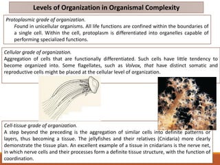

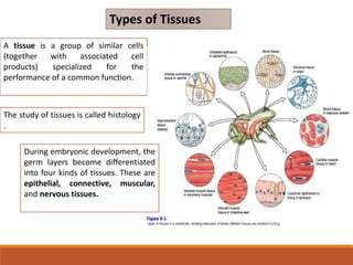
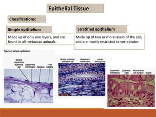
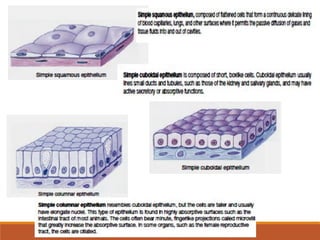
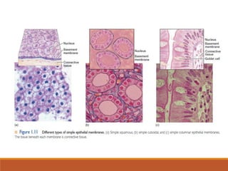
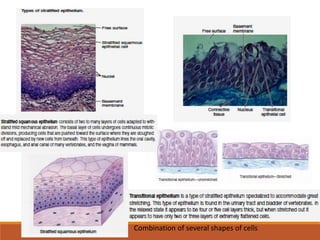
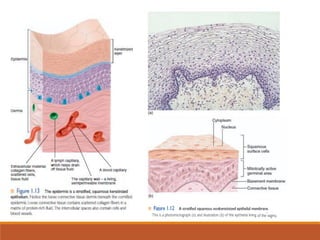
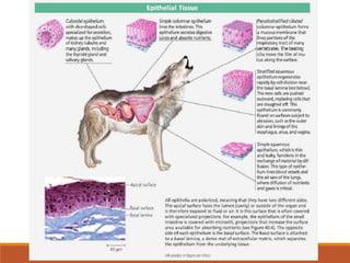
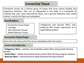
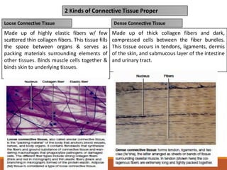
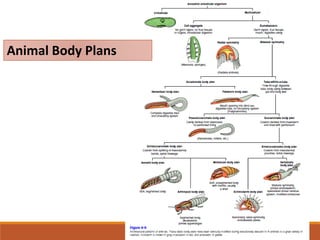


The document outlines the levels of organization in animals, detailing the protoplasmic, cellular, tissue, organ, and organ-system grades of organization, along with specialized tissues including epithelial, connective, muscular, and nervous tissues. It explains the structure and function of these tissues, their classifications, and their roles in various bodily functions. Additionally, the document discusses animal symmetry types such as spherical, radial, and bilateral symmetry, highlighting their significance in animal evolution and adaptation.


