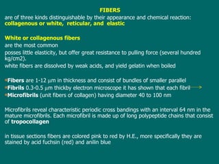
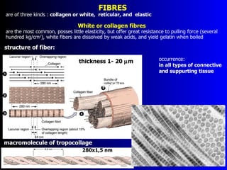
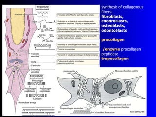
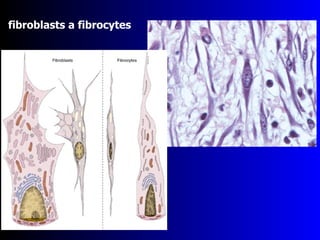
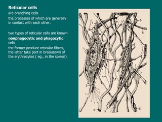
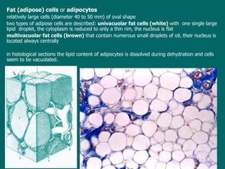
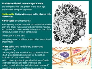
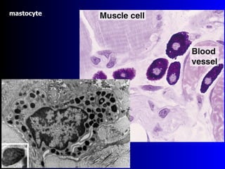
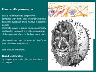
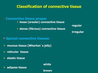
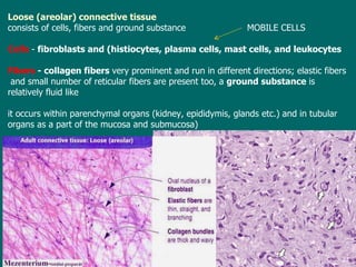
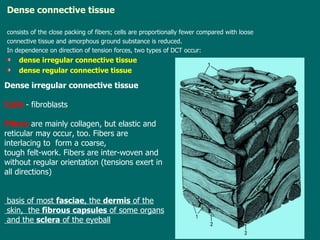
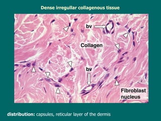
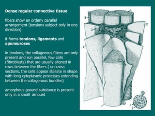
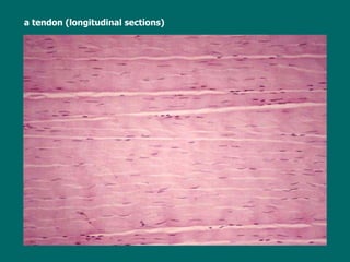
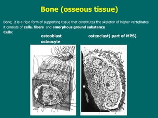
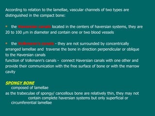
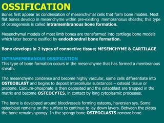
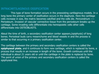
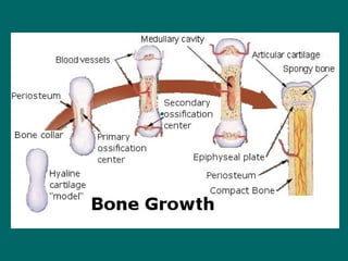
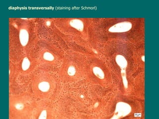
Lecture 6     GenMed_2nd semester General histology Tissues - definition, their origin and classification Connective and supporting tissue - general characteristics, components and classification of them The connective tissue proper - types, chief distribution, and function Supporting tissues: cartilage and bone - types, chief distribution, and function Histogenesis of bone tissue (ossification)
T issues of the adult and their classification tissue = a  complex of similar cells specialized in common direction and able to perform a common function  4  primary (basic, fundamental) tissues - epithelial tissue ( the  epithelium) - connective and supporting tissues - muscle (muscular) tissue - nerve tissue tissues  form elementary components of organs t he occurrence, arrangement and proportions of tissues in individual organs are different  and  are  object of study  in   the microscopic anatomy Epithelial   tissue   is composed of cells  that  are in close apposition with one another; among cells  there is present only a small amount of intercellular substance e pithelial cells are usually of regular form without extensive cytoplasmic processes a dhesion between the cells is very strong e pithelia derive from  the  all germ layers
Connective and supporting tissues unlike epithelia, contain   cells  that are separated   from one another ; th e intervening spaces are occupied by  the   intercellular  substance ( material )  produced by cells t he intercellular substance consists of two components:  fibers  and  amorphous ground substance c onnective and supporting tissues are always of  mesenchymal origin Muscle tissue   is composed of elongated  cells that  are able to  contract f or this function cells are well adapted as they contain contractile proteins: actin and myosin cells or muscle fibers tend to be aggregated in bundles  that  are conspicuously different from the surroundings tissues three  types of muscle tissue are distinguished:  smooth ,  s keletal, cardiac s mooth muscle tissue  derives  from the mesenchyme, s keletal and cardiac  from the mesoderm Nerve tissue consists of  nerve cells,  neurons , and associated supporting cells of various type called  neuroglia n eurons are highly specialized cells that have the ability to receive, generate and transmit nerve impulses e xcept the microglia, the nervous tissue derives from the neuroectoderm
Connective and supporting tissue s  - general characteristics,  their  components   and classification   posses three characteristics in common: they develop from the  mesenchyme   that  is itself derived from the  third germ layer  or  mesoderm, they are all composed of  cells  and relatively large amount  of  intercellular substance   cells lie more or less scattered, sometimes not in contact, sometimes touching only at the ends of long cytoplasmic processes, the intercellular substance consists of  fibers  and  ground amorphous substance c onnective  and supporting  tissue s  in the adult  are: Connective tissue proper  Cartilage  Bone Functions: -  mechanical function   -  bones  form skeleton of body, skeleton of organs inclusively their  stroma   etc. -  nutritional function   -   is maintained  by the intercellular  substance -  defensive function   -   some connective  cells mobilize to defend the body against bacteria and  other foreign bodies -  histiocytes, plasmocytes, leukocytes
FIBERS are of three kinds distinguishable by their appearance and chemical reaction:  collagenous or white,  reticular, and  elastic White or collagenous fibers  are the most common posses little elasticity, but offer great resistance to pulling force (several hundred kg/cm2). w hite fibers are dissolved by weak acids, and yield gelatin when boiled Fibers  are 1- 1 2   m in thickness and consist of bundles of smaller parallel  Fibrils  0.3-0.5   m thick by  electron microscope  it  ha s  shown that each fibril  Microfibrils  (unit fibers of collagen) having diameter 40 to 100 nm Microfibrils reveal characteristic periodic cross bandings with an interval 64 nm in the mature microfibrils . E ach microfibril is made up of long polypeptide chains  that consist of  tropocollagen   in tissue sections fibers are colored pink to red  by  H.E., more specifically they are stained by acid fuchsin (red) and anilin blue
macromolecule of tropocollage occurrence:  in all types of connective  and suppurting tissue 280x1,5 nm thickness 1- 20   m structure of fiber: structure of fiber: FIB RE S are of three kinds :  collagen or white,  reticular, and  elastic White or collagen fib re s  are the most common ,  posses little elasticity, but offer great resistance to pulling force (several hundred kg/cm 2 ) , w hite fibers are dissolved by weak acids, and yield gelatin when boiled
synthesis of collagenous fibers: fibroblasts, chodroblasts, osteoblasts, odontoblasts procollagen  /enzyme  procollagen peptidase tropocollagen
 
Reticular fibers  similar to white fibers  but  are usually finer in caliber do not stain appreciably with eosin, but have an affinity for silver salts  ( are termed as argyrophil ) c hemically they are composed of molecules of tropocollagen like the white fibers r eticular fibers form a nestlike supporting framework of some lymphatic organs, they occur around small blood vessels, muscle and nerve fibers, and also the fat cells Elastic fibers  characteristic of these fibers is elasticity f ibers are composed of albumoid elastin  that  shows remarkable resistance to hot water, acids and alkalis b y electron microscopy elastic fibers consist of peripheral collection of thin tubular fibrils of 10 nm diameter  that surround the  central amorphous component Elastic fibers are found in the skin, lungs,  arteries ,  veins ,  connective tissue proper ,  elastic cartilage ,  periodontal ligament , fetal tissue and other structures. they  stain  unspecifically  with eosin (pink)  and  selectively with orcein (brown) or resorcin-fuchsin (dark blue-purple)
Elastic fib re s  Characteristic of these fibers is elasticity , are branched and form networks,  d. cca  1   m,  f ib re s are composed of   glycoprotein microfibrils   and   albumoid   elastin (protein)   that  shows remarkable resistance to hot water, acids and alkalis b y electron microscopy elastic fibers consist of peripheral collection of thin tubular fibrils of 10 nm diameter  that surround the  central amorphous component e lastic fibers occur singly or in the form of sheets Electron micrographs of developing elastic fibers. A: In early stages of formation, developing fibers consist of numerous small glycoprotein microfibrils. B: With further development, amorphous aggregates of elastin are found among the microfibrils. C: The amorphous elastin accumulates, ultimately occupying the center of an elastic fiber delineated by microfibrils. Note the collagen fibrils, seen in cross section.
GROUND SUBSTANCE ground substance of connective tissue is a homogeneous semifluid material that surrounds the cells and fibers is composed of: mucopolysaccharides (newly glycosaminoglycans), proteins (proteoglycans and glycoproteins), water minerals ( Chemical  element  ) m ucopolysaccharides :  the most common are  hyaluronate , chondroitin-4-sulfate, chondroitin-6-sulfate h yaluronate  (hyaluronic acid) is very important because the viscosity of ground substance depends on the content of it i n preparations the ground substance seems to be structureless and  stains  metachromatically with the toluidine blu e T he ground substance like as fibers  is  elaborated by endoplasmic reticulum of connective tissue cells, especially of fibroblasts
CONNECTIVE TISSUE CELLS CT cells are  classified as Fixed cells   -  are responsible for production extracellular components and  Mobile wandering  cells   that  are   involved in tissue reaction to injury  and in defense reactions Fixed cells:  fibroblasts, reticular cells, adipocytes, undifferentiated cells Fibroblasts   the most  frequent  CT cells are relatively large flat  or  branching cells  that  appear  fusiform or spindle shaped  in profile contain nucleus of oval or elongated shape  with  one  or two nucleoli and small amount  of finelygranular chromatin boundaries  of fibroblasts are  often  indistinct   in histological preparations
fibroblasts a fibrocytes
Reticular cells are branching cells the processes of which are generally in contact with each othe r . t wo type s  of reticular cells are  known nonphagocytic and phagocytic  cells t he former produce reticular fibres, the latter take part in breakdown of the erythrocytes ( eg., in the spleen).
Fat (adipose) cells   or   adipocytes relatively large cells (diameter 40 to 50 mm) of oval shape t wo types of adipose cells are described:  univacuolar fat cells (white)  with  one single large lipid  droplet ,  the cytoplasm is reduced to only a thin rim, the nucleus is flat multivacuolar fat cells (brown)   that  contain numerous small droplets of oil, their nucleus is located always centrally in  histological sections the lipid content of adipocytes is dissolved during dehydration  and cells  seem to be vacuolated.
Undifferentiated mesenchymal cells are embryonic cells  that  persist to the adult  age are occurred along the capillaries Mobile cells :  h istiocytes , mast cells, plasma cells leukocytes H istiocytes  (macrophages)  are irregularly shaped cells with processes  that  usually are short and blunt ,  nucleus is ovoid, sometimes indented, and smaller and more densely staining than that of the fibroblast , n ucleoli are not conspicuous t he cytoplasm stains dark macrophages are capable of amoeboid movement and phagocytosis Mast cells  (role in defense, allergy and anyphylaxis) are irregularly oval in outline and occasionally have short  pseudopodia (locomotion for the cell), an indication of their slow mobility cells contain cytoplasmic granules  that  are refractile and water-soluble and stain with basic and metachromatic dyes (as methylene blue or azure A)   contain heparin and also histamine an serotonin
mastocyte
Plasma cells , plasmocytes bear a resemblance to lymphocytes c ompared with them, they are larger and have  basophilic cytoplasm and a nucleus in eccentric  position chromatin occurs in coarse clumps peripherally  and is often  arranged in a pattern suggestive  of the spokes of wheel or the hours on a clock p lasma cells are rare, but are more plentiful in  sites of chronic inflamations cells  produce antibodies Blood leukocytes  by lymphocytes, eosinophils, neutrophils and  monocytes
M esenchyme   unspecialized connective tissue  from  where   all connective tissues  derive  composed of mesenchymal cells whose processes are in contact and of a ground substance  that  is fluid and usually  without  fibrils it  occurs during  first  weeks of embryonic life
Connective tissue proper loose (areolar) connective tissue regular dense (fibrous) connective tissue  irregular Special connective tissues:  mucous tissue (Wharton´s jelly) reticular  tissue elastic tissue   white adipose tissue   brown Classification of connective tissu e
Loose (areolar) connective tissue   consists  of  cells, fibers and ground substance  MOBILE CELLS C ells  -  f ibroblasts and (histiocytes ,  plasma cells, mast cells, and leukocytes  Fibers  -  c ollagen fibers  very prominent and  run  in different directions; elastic fibers  and  small number of reticular fibers  are  present  too, a   ground substance  is relatively fluid like  it occurs  within parenchymal organs (kidney, epididymis, glands etc.)  a n d in tubular organs  as a part of the mucosa and submucosa )
Dense connective tissue consists of  the close packing of fibers; cells are proportionally fewer  compared with  loose  connective tissue and amorphous ground substance is reduced. I n  dependence on direction of tension forces, two types of DCT  occur: d ense irregular   connective tissue d ense regular   connective tissue  D ense irregular   connective tissue C ells  - fibroblasts  F ibers  are  mainly  collagen, but elastic and reticular  may occur, too .  Fibers  are  interlacing to  form a coarse,  tough felt-work. Fibers are inter-woven and without regular   orientation (tension s  exert in all directions) basis of most  fasciae , the  dermis  of the skin,  the  fibrous capsules  of some organs and the  sclera  of the eyeball
Dense irregullar collagenous tissue distribution:  capsules, reticular layer of the dermis
D ense regular   connective tissue   fibers  show  an orderly parallel arrangement (tensions subject only in one direction) it forms   tendons, ligaments  and  aponeuroses i n tendons, the collagenous fibers are only present and run parallel , few cells (fibroblasts) that  are usually aligned in rows between the fibers  ( on  cross  sections, the cells appear stellate in shape with  long  cytoplasmic processes extending between the collagenous bundles ) a morphous ground substance is present only in a small  amount
  a tendon (longitudinal sections)
Mucous tissue cells  are stellate fibroblasts with anastomosing processes  that  lie in a mucoid jelly-like mass, the  fibers  are  of  only collagen  type   as Wharton' s jelly in the  umbilical cord  and as  dental pulp  of the deciduous teeth
Reticular tissue   composed of reticular cells  and  reticular fibers , amorphous ground substance is negligible Cells  are stellate  with   long cytoplasmic extensions ,  large  and  pale nucle us  and abundant basophilic cytoplasm Fibers  are in close appositions to cells and form more or less complicated network D istribution :  forms the framework of lymphatic organs and bone marrow
Elastic tissue cells, fibers and not great amount of the ground substance C ells  are typical fibroblasts, sparsely scattered with nuclei markedly flattened F ibers  are mostly elastic and parallel oriented; a small number of reticular fibers was also described is occurred as the ligamenta flava, the true vocal cords, the ligamentum nuchae
Adipose tissue white adipose tissue (univacuolar) brown adipose tissue   (multivacuolar) Cell;   aggregated  fat cells +  scarsely  fibroblasts, eosinophils, and mast cells Fibers;  c ell s are  surrounded by a web of fine reticular fibers t he closely packed fat cells form  lobules   in  and between the m  there is a rich network of blood capillaries
are   cartilage  and  bone l ike connective tissue proper,  the both  are composed of  cells, fibers and amorphous ground substance but  differ from the connective tissue proper in the rigidity of  amorphous ground substance (matrix) the ground substance  of  cartilage  contains   chondromucoid  -  the protein rich in chondroitinsulfate   the ground substance  of  bone chiefly  osseomucoid  that is  impregnated with inorganic salts, especially with calcium phosphate and calcium carbonate Cartilage common characteristics of cartilage: is strong and some pliable(böjlig) tissue,  that  is  avascular ,  has no   lymphatic vessels and is  not innervated - cartilage cells are  chondrocytes  - they are large cells of  rounded shape with a central spherical nucleus ,  the  cytoplasm is basophilic   and rich in rough endoplasmic reticulum, glycogen, lipid drople ts cells are housed in small spaces within the matrix known as lacunae Supporting tissues
- fibers can be solely collagenous or a combination of collagenous and elastic depending upon the cartilage type - the surface of the hyaline and elastic cartilage is  enveloped  by a connective tissue membrane -  the perichondrium , from which the growth and nutrition of cartilage is provided t he fibrocartilage has no perichondrium Classification of the cartilage: hyaline cartilage yellow or elastic cartilage   w hite (fibrous) cartilage or fibrocartilage – no perichondrium Hyaline cartilage is firm and slightly bluish i n the microscope, its  matrix appears clear (glasslike)   because  fibers and ground substance have the same staining capacity and refractive index i n the adult ,  chondrocytes are arranged in groups, composed of  2 ,  4 , or  8  cells  that   are of the same origin (cells of each group have been derived from the same parent cell)  s uch cell groups are  termed  isogenous groups  or cell nests
  f ibrous component is represented by very  fine  collagenous fibers  that form a feltwork   Semitransparent opalescent cartilage that forms most of the  fetal skeleton  and that consists of cells that synthesize a surrounding matrix of hyaluronic acid, collagen, and protein; in the adult, it is found in the  trachea, larynx, and joint surfaces . several isogeneous   groups may unite in complexes termed as  territoria between territoria the matrix stains slightly and is called as  interterritorial matrix perichondrium
Elastic cartilage is usually  yellowish   in  colour and opa que  and more flexible than hyaline c hondrocytes are numerous and scattered singly or in small isogenous groups of two or three cells  that  are surrounded by capsules of the intercellular substance , fibers: elastic and collagenous th e matrix  (intercellular substance) shows  characteristic fibrillar appearance   in H.E. preparations   this is caused by presence of   elastic fibers  that are  never  totally masked by amorphous ground substance   elastic  fibers  may be  visualized by special dyes  as  orcein or resorcin- fuchsin
Elastic  cartilage  stained with  orcein
White (fibrous) cartilage or fibrocartilage  – NO PERICHONDRIUM is a transitional t issue  between  hyaline cartilage and dense fibrous connective tissue  of tendons and ligaments the  intercellular substance is composed of thick bundles of collagenous fibers running parallel with one another ,   c hondrocytes are not numerous, rounded and are arranged singly or in pairs in rows between bundles of collagenous fibers  t he amorphous ground substance is inconspicuous  and  found only in the immediate  vicinity of the chondrocytes f ibrocartilage has  no true perichondrium
DISTRIBUTION OF CARTILAGE HYALINE    ELASTIC  FIBROCARTILAGE Fetal skeleton  External acoustic  meatus  Intervertebral discs Pubic symphysis      (symphysis pubis) Costal cartilages  Pinna of auricle   Nose    Epiglottis    Temporomandibular   joint Larynx Trachea + bronchi  Eustachian tube    Articular cartilage  (pharyngotympanic tube)
B one (osseous tissue) Bone; It is a rigid form of  supporting  tissue that constitutes the skeleton of higher vertebrates it  consists of  cells , fibers  and  amorphous ground substance   Cells : osteoblast  osteo c last( part of MPS) osteocyt e
Interce l lular substance contains collagenous fibers and amorphous ground substance (osseomucoid)  that is  impregnated by inorganic salts   -  calcium phosphate, calcium carbonate , calcium fluoride and magnesium fluoride the  inorganic salts are responsible for the rigidity and hardness of bone while  the collagenous fibers contribute to the strength and resilience of bone Sections of bone cannot be made in the ordinary way because the bone matrix shows the great hardness two method are used to prepare  bone  for study  in a routine : either bone is softened by the use of acids ( decalcification ), or pieces of bone are dried and ground very thin ( ground specimens ) the  latter method destroys the cells; the former destroys the inorganic part  of intercellular substance, but  osteocytes and collagenous  groundwork are re tained
Macroscopically, two types of bone  are  distinguished:  the  compact (dense) the spongy (cancellous)   T he  compact bone  is  always solid ,  the  spongy bone  consists of  slender, irregular trabeculae or bars which branch and unite  with one another to form a meshwork with  few exceptions, both types  occur  in  all  bone s i n typical long bones, the shaft (diaphysis) is chiefly compact bone  that  surround s  the medullary cavity  while the  epiphysis consists of spongy bone covered by a thin shell of compact  bone   i n flat bones, two plates of compact bone enclose  a  middle layer of spongy bone ( diploe ) The external and  internal surfaces are covered by a  specialized  connective tissue  coats –  the periosteum   (good developed)  and  the endosteum   (less obvious).
Microscopically, two types of osseous tissue are  distinguished : woven or irregular bone Haversian or lamellar (regular) bone T he  W oven-/ Irregular bone resembles the fibrous (dense) connective tissue when is decalcified consists of not numerous osteocytes and calcified matrix with collagenous fibers without the precise orientation (fibers usually run in all directions or form a feltwork formations) this  bone occurs chiefly at  sites of insertion of muscles  to the skeleton; similar to the woven bone is the  cementum  of tooth
Haversian-/ Lamellar  bone  It is developmentally and functionally more perfect type of osseous tissue the  bone is composed of thin layers of bone matrix 3-7  m thick  lamellae E ach lamella consists of the  collagenous fibers + calcified ground substance . Fibers in any lamella are roughly parallel to each other and take spiral or helical course (i t is of interest that the fiber direction is always different in the adjacent lamellae ; t his  alternating arrangement in fiber direction explains why lamellae appear to be so distinct,  one from  a nother) Osteocytes  are between the lamellae and are usually scattered singly they  occupy lenticular-   shaped spaces known as  lacunae  and posses long processes  that  are  housed in tiny canals   or tunnels known as  canaliculi
osteocytes with processes (HE)
Remember :  c ompact and spongy  bones described in the anatomy are always composed of the  Haversian or lamellar  type  of osseous tissue in  the compact bone  (three dimensional diagram) ,  the lamellae are arranged in three different ways: the great majority of lamellae are arranged concentrically around longitudinal vascular channels within the  bone and to form cylindrical units called  haversian systems   or osteons o steons vary greatly in size, being made up of 4 to 20 lamellae i n cross sections, the haversian systems   appear as concentric   rings around circular opening (Haversian canal) i n longitudinal sections, lamellae are seen as closely spaced  bands parallel to a long slit besides the former lamellae, there are  lamellae without any relations to blood  v essels  that  form fields of varying size  and irregular shape  –  interstitial lamellae  or interstitial systems  they are supposed to be   rests  of  old Haversian systems,  that  have  been partially removed in the process  of bone   remodeling at the external surface, immediately  beneath  the periosteum, and on the internal surface,   subjacent the  endosteum, the lamellae are  arranged parallel to the periosteum  or around the central cavity – outer circumferential and inner circumferential lamellae
diaphysis a model an osteon  Haversian and Volkmans canals
diaphysis transversally (HE)
diaphysis transversally  (staining after Schmorl)
According to  relation to  the  lamella e,  vascular channels  of two types are distinguished in the compact bone: the  Haversian canals   located in the centers of haversian systems, they are 20 to 100   m in diameter and contain one or two blood vessels the  Volkmann's canals   - they are not surrounded by concentrically  arranged lamellae and  traverse the bone in direction perpendicular or oblique to the Haversian canals function of Volkmann's canals  -  connect Haversian canals with one other and provide their communication with the free surface of bone or with the marrow cavity SPONGY BONE composed of lamellae a s the trabeculae of spongy/ cancellous bone are relatively thin, they may not  contain complete haversian systems but only superficial or circumferential lamellae
OSSIFICATION Bones first appear as condensation of mesenchymal cells that form bone models. Most flat bones develop in mesenchyme within pre-existing  membranosus sheaths; this type of osteogenesis is called  intramembranous bone formation. Mesenchymal models of most limb bones are transformed into cartilage bone models which later become ossified by  endochondral bone formation. Bone develops in 2 types of connective tissue; MESENCHYME & CARTILAGE INTRAMEMBRANOUS OSSIFICATION This type of bone formation occurs in the mesenchyme that has formed a membranous sheath.  The mesenchyme condense and become highly vascular, some cells differentiate into  OSTEOBLAST  and begins to deposit intercellular substances – osteoid tissue or prebone. Calcium-phosphate is then deposited and the osteoblast are trapped in the matrix and become  OSTEOCYTES , in contact by long cytoplasmic processes. The bone is developed around bloodvessels forming osteons, haversian sys. Some osteoblast remains on the surface to continue to lay down layers. Between the plates the bone remains spongy. In the spongy bone  OSTEOCLASTS  remove bone.
diaphysis transverally
INTRACARTILAGINOUS OSSIFICATION This type of bone formation occurs in the preexisting cartilaginous models. In a long bone the primary center of ossification occurs in the diaphysis. Here the cartilage cells increase in size, the matrix becomes calcified and the cells die. Pericondrium ---- Periosteum. Invasion of vascular connective tissue from the periosteum breaks up the cartilage. Some invading cells differentiate into hemiopoietic cells other cells differentiate into OSTEOBLASTS. About the time of birth, a secondary ossification center appears,(epiphysis) of long bones. Periosteal buds carry mesenchyme and blood vessels in and the process is similar to that occurring in a primary ossification center.  The cartilage between the primary and secondary ossification centers is called the  epiphyseal plate , and it continues to form new cartilage, which is replaced by bone, a process that results in an increase in length of the bone. Growth continues until the individual is about 21 years old or until the cartilage in the plate is replaced by bone. The point of union of the primary and secondary ossification centers is called the epiphyseal line.
 
diaphysis transversally  (staining after Schmorl)

Lecture6

  • 1.
    Lecture 6 GenMed_2nd semester General histology Tissues - definition, their origin and classification Connective and supporting tissue - general characteristics, components and classification of them The connective tissue proper - types, chief distribution, and function Supporting tissues: cartilage and bone - types, chief distribution, and function Histogenesis of bone tissue (ossification)
  • 2.
    T issues ofthe adult and their classification tissue = a complex of similar cells specialized in common direction and able to perform a common function 4 primary (basic, fundamental) tissues - epithelial tissue ( the epithelium) - connective and supporting tissues - muscle (muscular) tissue - nerve tissue tissues form elementary components of organs t he occurrence, arrangement and proportions of tissues in individual organs are different and are object of study in the microscopic anatomy Epithelial tissue is composed of cells that are in close apposition with one another; among cells there is present only a small amount of intercellular substance e pithelial cells are usually of regular form without extensive cytoplasmic processes a dhesion between the cells is very strong e pithelia derive from the all germ layers
  • 3.
    Connective and supportingtissues unlike epithelia, contain cells that are separated from one another ; th e intervening spaces are occupied by the intercellular substance ( material ) produced by cells t he intercellular substance consists of two components: fibers and amorphous ground substance c onnective and supporting tissues are always of mesenchymal origin Muscle tissue is composed of elongated cells that are able to contract f or this function cells are well adapted as they contain contractile proteins: actin and myosin cells or muscle fibers tend to be aggregated in bundles that are conspicuously different from the surroundings tissues three types of muscle tissue are distinguished: smooth , s keletal, cardiac s mooth muscle tissue derives from the mesenchyme, s keletal and cardiac from the mesoderm Nerve tissue consists of nerve cells, neurons , and associated supporting cells of various type called neuroglia n eurons are highly specialized cells that have the ability to receive, generate and transmit nerve impulses e xcept the microglia, the nervous tissue derives from the neuroectoderm
  • 4.
    Connective and supportingtissue s - general characteristics, their components and classification posses three characteristics in common: they develop from the mesenchyme that is itself derived from the third germ layer or mesoderm, they are all composed of cells and relatively large amount of intercellular substance cells lie more or less scattered, sometimes not in contact, sometimes touching only at the ends of long cytoplasmic processes, the intercellular substance consists of fibers and ground amorphous substance c onnective and supporting tissue s in the adult are: Connective tissue proper Cartilage Bone Functions: - mechanical function - bones form skeleton of body, skeleton of organs inclusively their stroma etc. - nutritional function - is maintained by the intercellular substance - defensive function - some connective cells mobilize to defend the body against bacteria and other foreign bodies - histiocytes, plasmocytes, leukocytes
  • 5.
    FIBERS are ofthree kinds distinguishable by their appearance and chemical reaction: collagenous or white, reticular, and elastic White or collagenous fibers are the most common posses little elasticity, but offer great resistance to pulling force (several hundred kg/cm2). w hite fibers are dissolved by weak acids, and yield gelatin when boiled Fibers are 1- 1 2  m in thickness and consist of bundles of smaller parallel Fibrils 0.3-0.5  m thick by electron microscope it ha s shown that each fibril Microfibrils (unit fibers of collagen) having diameter 40 to 100 nm Microfibrils reveal characteristic periodic cross bandings with an interval 64 nm in the mature microfibrils . E ach microfibril is made up of long polypeptide chains that consist of tropocollagen in tissue sections fibers are colored pink to red by H.E., more specifically they are stained by acid fuchsin (red) and anilin blue
  • 6.
    macromolecule of tropocollageoccurrence: in all types of connective and suppurting tissue 280x1,5 nm thickness 1- 20  m structure of fiber: structure of fiber: FIB RE S are of three kinds : collagen or white, reticular, and elastic White or collagen fib re s are the most common , posses little elasticity, but offer great resistance to pulling force (several hundred kg/cm 2 ) , w hite fibers are dissolved by weak acids, and yield gelatin when boiled
  • 7.
    synthesis of collagenousfibers: fibroblasts, chodroblasts, osteoblasts, odontoblasts procollagen /enzyme procollagen peptidase tropocollagen
  • 8.
  • 9.
    Reticular fibers similar to white fibers but are usually finer in caliber do not stain appreciably with eosin, but have an affinity for silver salts ( are termed as argyrophil ) c hemically they are composed of molecules of tropocollagen like the white fibers r eticular fibers form a nestlike supporting framework of some lymphatic organs, they occur around small blood vessels, muscle and nerve fibers, and also the fat cells Elastic fibers characteristic of these fibers is elasticity f ibers are composed of albumoid elastin that shows remarkable resistance to hot water, acids and alkalis b y electron microscopy elastic fibers consist of peripheral collection of thin tubular fibrils of 10 nm diameter that surround the central amorphous component Elastic fibers are found in the skin, lungs, arteries , veins , connective tissue proper , elastic cartilage , periodontal ligament , fetal tissue and other structures. they stain unspecifically with eosin (pink) and selectively with orcein (brown) or resorcin-fuchsin (dark blue-purple)
  • 10.
    Elastic fib res Characteristic of these fibers is elasticity , are branched and form networks, d. cca 1  m, f ib re s are composed of glycoprotein microfibrils and albumoid elastin (protein) that shows remarkable resistance to hot water, acids and alkalis b y electron microscopy elastic fibers consist of peripheral collection of thin tubular fibrils of 10 nm diameter that surround the central amorphous component e lastic fibers occur singly or in the form of sheets Electron micrographs of developing elastic fibers. A: In early stages of formation, developing fibers consist of numerous small glycoprotein microfibrils. B: With further development, amorphous aggregates of elastin are found among the microfibrils. C: The amorphous elastin accumulates, ultimately occupying the center of an elastic fiber delineated by microfibrils. Note the collagen fibrils, seen in cross section.
  • 11.
    GROUND SUBSTANCE groundsubstance of connective tissue is a homogeneous semifluid material that surrounds the cells and fibers is composed of: mucopolysaccharides (newly glycosaminoglycans), proteins (proteoglycans and glycoproteins), water minerals ( Chemical element ) m ucopolysaccharides : the most common are hyaluronate , chondroitin-4-sulfate, chondroitin-6-sulfate h yaluronate (hyaluronic acid) is very important because the viscosity of ground substance depends on the content of it i n preparations the ground substance seems to be structureless and stains metachromatically with the toluidine blu e T he ground substance like as fibers is elaborated by endoplasmic reticulum of connective tissue cells, especially of fibroblasts
  • 12.
    CONNECTIVE TISSUE CELLSCT cells are classified as Fixed cells - are responsible for production extracellular components and Mobile wandering cells that are involved in tissue reaction to injury and in defense reactions Fixed cells: fibroblasts, reticular cells, adipocytes, undifferentiated cells Fibroblasts the most frequent CT cells are relatively large flat or branching cells that appear fusiform or spindle shaped in profile contain nucleus of oval or elongated shape with one or two nucleoli and small amount of finelygranular chromatin boundaries of fibroblasts are often indistinct in histological preparations
  • 13.
  • 14.
    Reticular cells arebranching cells the processes of which are generally in contact with each othe r . t wo type s of reticular cells are known nonphagocytic and phagocytic cells t he former produce reticular fibres, the latter take part in breakdown of the erythrocytes ( eg., in the spleen).
  • 15.
    Fat (adipose) cells or adipocytes relatively large cells (diameter 40 to 50 mm) of oval shape t wo types of adipose cells are described: univacuolar fat cells (white) with one single large lipid droplet , the cytoplasm is reduced to only a thin rim, the nucleus is flat multivacuolar fat cells (brown) that contain numerous small droplets of oil, their nucleus is located always centrally in histological sections the lipid content of adipocytes is dissolved during dehydration and cells seem to be vacuolated.
  • 16.
    Undifferentiated mesenchymal cellsare embryonic cells that persist to the adult age are occurred along the capillaries Mobile cells : h istiocytes , mast cells, plasma cells leukocytes H istiocytes (macrophages) are irregularly shaped cells with processes that usually are short and blunt , nucleus is ovoid, sometimes indented, and smaller and more densely staining than that of the fibroblast , n ucleoli are not conspicuous t he cytoplasm stains dark macrophages are capable of amoeboid movement and phagocytosis Mast cells (role in defense, allergy and anyphylaxis) are irregularly oval in outline and occasionally have short pseudopodia (locomotion for the cell), an indication of their slow mobility cells contain cytoplasmic granules that are refractile and water-soluble and stain with basic and metachromatic dyes (as methylene blue or azure A) contain heparin and also histamine an serotonin
  • 17.
  • 18.
    Plasma cells ,plasmocytes bear a resemblance to lymphocytes c ompared with them, they are larger and have basophilic cytoplasm and a nucleus in eccentric position chromatin occurs in coarse clumps peripherally and is often arranged in a pattern suggestive of the spokes of wheel or the hours on a clock p lasma cells are rare, but are more plentiful in sites of chronic inflamations cells produce antibodies Blood leukocytes by lymphocytes, eosinophils, neutrophils and monocytes
  • 19.
    M esenchyme unspecialized connective tissue from where all connective tissues derive composed of mesenchymal cells whose processes are in contact and of a ground substance that is fluid and usually without fibrils it occurs during first weeks of embryonic life
  • 20.
    Connective tissue properloose (areolar) connective tissue regular dense (fibrous) connective tissue irregular Special connective tissues: mucous tissue (Wharton´s jelly) reticular tissue elastic tissue white adipose tissue brown Classification of connective tissu e
  • 21.
    Loose (areolar) connectivetissue consists of cells, fibers and ground substance MOBILE CELLS C ells - f ibroblasts and (histiocytes , plasma cells, mast cells, and leukocytes Fibers - c ollagen fibers very prominent and run in different directions; elastic fibers and small number of reticular fibers are present too, a ground substance is relatively fluid like it occurs within parenchymal organs (kidney, epididymis, glands etc.) a n d in tubular organs as a part of the mucosa and submucosa )
  • 22.
    Dense connective tissueconsists of the close packing of fibers; cells are proportionally fewer compared with loose connective tissue and amorphous ground substance is reduced. I n dependence on direction of tension forces, two types of DCT occur: d ense irregular connective tissue d ense regular connective tissue D ense irregular connective tissue C ells - fibroblasts F ibers are mainly collagen, but elastic and reticular may occur, too . Fibers are interlacing to form a coarse, tough felt-work. Fibers are inter-woven and without regular orientation (tension s exert in all directions) basis of most fasciae , the dermis of the skin, the fibrous capsules of some organs and the sclera of the eyeball
  • 23.
    Dense irregullar collagenoustissue distribution: capsules, reticular layer of the dermis
  • 24.
    D ense regular connective tissue fibers show an orderly parallel arrangement (tensions subject only in one direction) it forms tendons, ligaments and aponeuroses i n tendons, the collagenous fibers are only present and run parallel , few cells (fibroblasts) that are usually aligned in rows between the fibers ( on cross sections, the cells appear stellate in shape with long cytoplasmic processes extending between the collagenous bundles ) a morphous ground substance is present only in a small amount
  • 25.
    atendon (longitudinal sections)
  • 26.
    Mucous tissue cells are stellate fibroblasts with anastomosing processes that lie in a mucoid jelly-like mass, the fibers are of only collagen type as Wharton' s jelly in the umbilical cord and as dental pulp of the deciduous teeth
  • 27.
    Reticular tissue composed of reticular cells and reticular fibers , amorphous ground substance is negligible Cells are stellate with long cytoplasmic extensions , large and pale nucle us and abundant basophilic cytoplasm Fibers are in close appositions to cells and form more or less complicated network D istribution : forms the framework of lymphatic organs and bone marrow
  • 28.
    Elastic tissue cells,fibers and not great amount of the ground substance C ells are typical fibroblasts, sparsely scattered with nuclei markedly flattened F ibers are mostly elastic and parallel oriented; a small number of reticular fibers was also described is occurred as the ligamenta flava, the true vocal cords, the ligamentum nuchae
  • 29.
    Adipose tissue whiteadipose tissue (univacuolar) brown adipose tissue (multivacuolar) Cell; aggregated fat cells + scarsely fibroblasts, eosinophils, and mast cells Fibers; c ell s are surrounded by a web of fine reticular fibers t he closely packed fat cells form lobules in and between the m there is a rich network of blood capillaries
  • 30.
    are cartilage and bone l ike connective tissue proper, the both are composed of cells, fibers and amorphous ground substance but differ from the connective tissue proper in the rigidity of amorphous ground substance (matrix) the ground substance of cartilage contains chondromucoid - the protein rich in chondroitinsulfate the ground substance of bone chiefly osseomucoid that is impregnated with inorganic salts, especially with calcium phosphate and calcium carbonate Cartilage common characteristics of cartilage: is strong and some pliable(böjlig) tissue, that is avascular , has no lymphatic vessels and is not innervated - cartilage cells are chondrocytes - they are large cells of rounded shape with a central spherical nucleus , the cytoplasm is basophilic and rich in rough endoplasmic reticulum, glycogen, lipid drople ts cells are housed in small spaces within the matrix known as lacunae Supporting tissues
  • 31.
    - fibers canbe solely collagenous or a combination of collagenous and elastic depending upon the cartilage type - the surface of the hyaline and elastic cartilage is enveloped by a connective tissue membrane - the perichondrium , from which the growth and nutrition of cartilage is provided t he fibrocartilage has no perichondrium Classification of the cartilage: hyaline cartilage yellow or elastic cartilage w hite (fibrous) cartilage or fibrocartilage – no perichondrium Hyaline cartilage is firm and slightly bluish i n the microscope, its matrix appears clear (glasslike) because fibers and ground substance have the same staining capacity and refractive index i n the adult , chondrocytes are arranged in groups, composed of 2 , 4 , or 8 cells that are of the same origin (cells of each group have been derived from the same parent cell) s uch cell groups are termed isogenous groups or cell nests
  • 32.
    fibrous component is represented by very fine collagenous fibers that form a feltwork Semitransparent opalescent cartilage that forms most of the fetal skeleton and that consists of cells that synthesize a surrounding matrix of hyaluronic acid, collagen, and protein; in the adult, it is found in the trachea, larynx, and joint surfaces . several isogeneous groups may unite in complexes termed as territoria between territoria the matrix stains slightly and is called as interterritorial matrix perichondrium
  • 33.
    Elastic cartilage isusually yellowish in colour and opa que and more flexible than hyaline c hondrocytes are numerous and scattered singly or in small isogenous groups of two or three cells that are surrounded by capsules of the intercellular substance , fibers: elastic and collagenous th e matrix (intercellular substance) shows characteristic fibrillar appearance in H.E. preparations this is caused by presence of elastic fibers that are never totally masked by amorphous ground substance elastic fibers may be visualized by special dyes as orcein or resorcin- fuchsin
  • 34.
    Elastic cartilage stained with orcein
  • 35.
    White (fibrous) cartilageor fibrocartilage – NO PERICHONDRIUM is a transitional t issue between hyaline cartilage and dense fibrous connective tissue of tendons and ligaments the intercellular substance is composed of thick bundles of collagenous fibers running parallel with one another , c hondrocytes are not numerous, rounded and are arranged singly or in pairs in rows between bundles of collagenous fibers t he amorphous ground substance is inconspicuous and found only in the immediate vicinity of the chondrocytes f ibrocartilage has no true perichondrium
  • 36.
    DISTRIBUTION OF CARTILAGEHYALINE ELASTIC FIBROCARTILAGE Fetal skeleton External acoustic meatus Intervertebral discs Pubic symphysis (symphysis pubis) Costal cartilages Pinna of auricle Nose Epiglottis Temporomandibular joint Larynx Trachea + bronchi Eustachian tube Articular cartilage (pharyngotympanic tube)
  • 37.
    B one (osseoustissue) Bone; It is a rigid form of supporting tissue that constitutes the skeleton of higher vertebrates it consists of cells , fibers and amorphous ground substance Cells : osteoblast osteo c last( part of MPS) osteocyt e
  • 38.
    Interce l lularsubstance contains collagenous fibers and amorphous ground substance (osseomucoid) that is impregnated by inorganic salts - calcium phosphate, calcium carbonate , calcium fluoride and magnesium fluoride the inorganic salts are responsible for the rigidity and hardness of bone while the collagenous fibers contribute to the strength and resilience of bone Sections of bone cannot be made in the ordinary way because the bone matrix shows the great hardness two method are used to prepare bone for study in a routine : either bone is softened by the use of acids ( decalcification ), or pieces of bone are dried and ground very thin ( ground specimens ) the latter method destroys the cells; the former destroys the inorganic part of intercellular substance, but osteocytes and collagenous groundwork are re tained
  • 39.
    Macroscopically, two typesof bone are distinguished: the compact (dense) the spongy (cancellous) T he compact bone is always solid , the spongy bone consists of slender, irregular trabeculae or bars which branch and unite with one another to form a meshwork with few exceptions, both types occur in all bone s i n typical long bones, the shaft (diaphysis) is chiefly compact bone that surround s the medullary cavity while the epiphysis consists of spongy bone covered by a thin shell of compact bone i n flat bones, two plates of compact bone enclose a middle layer of spongy bone ( diploe ) The external and internal surfaces are covered by a specialized connective tissue coats – the periosteum (good developed) and the endosteum (less obvious).
  • 40.
    Microscopically, two typesof osseous tissue are distinguished : woven or irregular bone Haversian or lamellar (regular) bone T he W oven-/ Irregular bone resembles the fibrous (dense) connective tissue when is decalcified consists of not numerous osteocytes and calcified matrix with collagenous fibers without the precise orientation (fibers usually run in all directions or form a feltwork formations) this bone occurs chiefly at sites of insertion of muscles to the skeleton; similar to the woven bone is the cementum of tooth
  • 41.
    Haversian-/ Lamellar bone It is developmentally and functionally more perfect type of osseous tissue the bone is composed of thin layers of bone matrix 3-7  m thick lamellae E ach lamella consists of the collagenous fibers + calcified ground substance . Fibers in any lamella are roughly parallel to each other and take spiral or helical course (i t is of interest that the fiber direction is always different in the adjacent lamellae ; t his alternating arrangement in fiber direction explains why lamellae appear to be so distinct, one from a nother) Osteocytes are between the lamellae and are usually scattered singly they occupy lenticular- shaped spaces known as lacunae and posses long processes that are housed in tiny canals or tunnels known as canaliculi
  • 42.
  • 43.
    Remember : c ompact and spongy bones described in the anatomy are always composed of the Haversian or lamellar type of osseous tissue in the compact bone (three dimensional diagram) , the lamellae are arranged in three different ways: the great majority of lamellae are arranged concentrically around longitudinal vascular channels within the bone and to form cylindrical units called haversian systems or osteons o steons vary greatly in size, being made up of 4 to 20 lamellae i n cross sections, the haversian systems appear as concentric rings around circular opening (Haversian canal) i n longitudinal sections, lamellae are seen as closely spaced bands parallel to a long slit besides the former lamellae, there are lamellae without any relations to blood v essels that form fields of varying size and irregular shape – interstitial lamellae or interstitial systems they are supposed to be rests of old Haversian systems, that have been partially removed in the process of bone remodeling at the external surface, immediately beneath the periosteum, and on the internal surface, subjacent the endosteum, the lamellae are arranged parallel to the periosteum or around the central cavity – outer circumferential and inner circumferential lamellae
  • 44.
    diaphysis a modelan osteon Haversian and Volkmans canals
  • 45.
  • 46.
  • 47.
    diaphysis transversally (staining after Schmorl)
  • 48.
    diaphysis transversally (staining after Schmorl)
  • 49.
    According to relation to the lamella e, vascular channels of two types are distinguished in the compact bone: the Haversian canals located in the centers of haversian systems, they are 20 to 100  m in diameter and contain one or two blood vessels the Volkmann's canals - they are not surrounded by concentrically arranged lamellae and traverse the bone in direction perpendicular or oblique to the Haversian canals function of Volkmann's canals - connect Haversian canals with one other and provide their communication with the free surface of bone or with the marrow cavity SPONGY BONE composed of lamellae a s the trabeculae of spongy/ cancellous bone are relatively thin, they may not contain complete haversian systems but only superficial or circumferential lamellae
  • 50.
    OSSIFICATION Bones firstappear as condensation of mesenchymal cells that form bone models. Most flat bones develop in mesenchyme within pre-existing membranosus sheaths; this type of osteogenesis is called intramembranous bone formation. Mesenchymal models of most limb bones are transformed into cartilage bone models which later become ossified by endochondral bone formation. Bone develops in 2 types of connective tissue; MESENCHYME & CARTILAGE INTRAMEMBRANOUS OSSIFICATION This type of bone formation occurs in the mesenchyme that has formed a membranous sheath. The mesenchyme condense and become highly vascular, some cells differentiate into OSTEOBLAST and begins to deposit intercellular substances – osteoid tissue or prebone. Calcium-phosphate is then deposited and the osteoblast are trapped in the matrix and become OSTEOCYTES , in contact by long cytoplasmic processes. The bone is developed around bloodvessels forming osteons, haversian sys. Some osteoblast remains on the surface to continue to lay down layers. Between the plates the bone remains spongy. In the spongy bone OSTEOCLASTS remove bone.
  • 51.
  • 52.
    INTRACARTILAGINOUS OSSIFICATION Thistype of bone formation occurs in the preexisting cartilaginous models. In a long bone the primary center of ossification occurs in the diaphysis. Here the cartilage cells increase in size, the matrix becomes calcified and the cells die. Pericondrium ---- Periosteum. Invasion of vascular connective tissue from the periosteum breaks up the cartilage. Some invading cells differentiate into hemiopoietic cells other cells differentiate into OSTEOBLASTS. About the time of birth, a secondary ossification center appears,(epiphysis) of long bones. Periosteal buds carry mesenchyme and blood vessels in and the process is similar to that occurring in a primary ossification center. The cartilage between the primary and secondary ossification centers is called the epiphyseal plate , and it continues to form new cartilage, which is replaced by bone, a process that results in an increase in length of the bone. Growth continues until the individual is about 21 years old or until the cartilage in the plate is replaced by bone. The point of union of the primary and secondary ossification centers is called the epiphyseal line.
  • 53.
  • 54.
    diaphysis transversally (staining after Schmorl)

Editor's Notes

  • #30 FUNCTION: Its main role is to store energy in the form of fat , although it also cushions and insulates the body + Adipose tissue also serves as an important endocrine organ [1] by producing hormones such as leptin , resistin , and the cytokine TNFα
  • #39 Resiliens betyder ordagrant förmågan att återhämta sig eller motstå olika störningar.