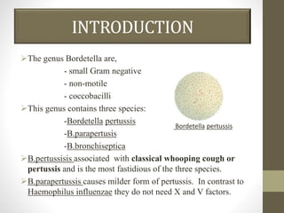
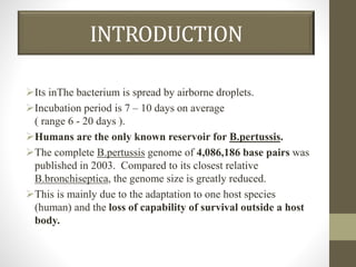
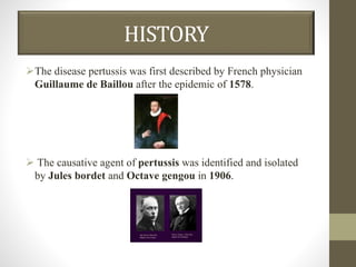
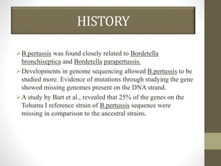
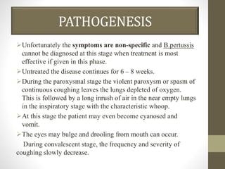
VIVEKANANDHA ARTSANDSCIENCECOLLEGEFORWOMEN,
veerachipalayam,sankari
BORDETELLA
DEPARTMENT OF MICROBIOLOGY
SUBJECT : MEDICAL BACTERIOLOGY AND MYCOLOGY
PRESENTED BY,
M.KEJAPRIYA,
I – M.Sc., MICROBIOLOGY,
VIVEKANANDHA ARTS AND SCIENCE COLLEGE FORWOMEN,
VEERACHIPALAYAM, SANKAGIRI.
GUIDED BY,
Dr.R. MYTHILI RAVICHANDHIRAN,
HEAD OF THE DEPARTMENT,
DEPARTMENT OF MICROBIOLOGY,
VIAAS,
VEERACHIPALAYAM, SANKAGIRI.
INTRODUCTION
The genus Bordetella are,
- small Gram negative
- non-motile
- coccobacilli
This genus contains three species:
-Bordetella pertussis
-B.parapertusis
-B.bronchiseptica
B.pertussisis associated with classical whooping cough or
pertussis and is the most fastidious of the three species.
B.parapertussis causes milder form of pertussis. In contrast to
Haemophilus influenzae they do not need X and V factors.
Bordetella pertussis
INTRODUCTION
Its inThe bacterium is spread by airborne droplets.
Incubation period is 7 – 10 days on average
( range 6 - 20 days ).
Humans are the only known reservoir for B.pertussis.
The complete B.pertussis genome of 4,086,186 base pairs was
published in 2003. Compared to its closest relative
B.bronchiseptica, the genome size is greatly reduced.
This is mainly due to the adaptation to one host species
(human) and the loss of capability of survival outside a host
body.
HISTORY
The disease pertussis was first described by French physician
Guillaume de Baillou after the epidemic of 1578.
 The causative agent of pertussis was identified and isolated
by Jules bordet and Octave gengou in 1906.
HISTORY
B.pertussis was found closely related to Bordetella
bronchiseptica and Bordetella parapertussis.
Developments in genome sequencing allowed B.pertussis to be
studied more. Evidence of mutations through studying the gene
showed missing genomes present on the DNA strand.
A study by Bart et al., revealed that 25% of the genes on the
Tohama I reference strain of B.pertussis sequence were
missing in comparison to the ancestral strains.
TAXONOMY
DOMAIN : Bacteria
PHYLUM : Pseudomonadota
CLASS : Betaproteobacteria
ORDER : Burkholderiales
FAMILY : Alcaligenaceae
GENUS : Bordetella
MORPHOLOGY
Earlier B.pertussis is a small ovoid Gram negative coccobacilli
1 – 1.5 µm by 0.3 µm.
It is non-motile and non-sporing.
When isolated from a clinical sample it it capsulated but
rapidly loses it on subculture.
In smears from culture, the bacilli are arranged in loose clumps
with clear spaces in between giving a thumb print like pattern.
Morphology Of Bordetella pertussis
CULTURE CHARACTERISTICS
Bordetella are aerobes and facultative anaerobes.
They grow best at 35 - 36ºC.
Bordetella are sensitive to various inhibitory substances.
Bordet and Gengou developed a special media for growing
Bordetella which is called Bordet Gengou medium. It
consists of glycerine potato blood agar.
A higher concentration of blood is added not to provide
additional nutritive factors but to neutralize inhibitory
substances.
The plates are incubated for 48 – 72 hours as the growth is
slow.
The colonies are small, dome shaped, opaque, greyish,
refractile and glistening resembling the appearance of bisected
pearls or mercury drops.
CULTURE CHARACTERISTICS
They are surrounded with hazy haemolysis.
Regan and Lowe is a selective medium comprising of charcoal,
cephalexin, amphotericin along with horse blood.
B. Pertussis on
BG medium
B.Pertussis
Alluminium paint appearance
BIOCHEMICAL REACTIONS
B.pertussis is oxidase, catalase, indole positive , reduce
nitrates, utilize citrates, splits urea and do not ferment sugars.
RESISTANCE
Killed by heat at 55ºC for 30 minutes.
Drying and disinfectants kill the organism
Survive outside for 5 days.
3 days on cloths.
Few hours on paper.
TRANSPORT MEDIUM
Transferred into Casamio acid solution att pH 7.2 in modified
Stuarts medium, Glycerin potato blood agar of Bordet Gengou.
Adding Pencillin becomes more selective.
Transport medium of
Bordetella
ANTIGENIC STRUCTURE
Bordetella is a multitoxin disease.
It produces eight different antigenically defined virulence
factors.
-Agglutinogens
-Lipopolysaccharide
-Heat labile toxin
-Tracheal cytotoxin
-Haemolysin
-Pertactin
-Pertussis toxin
-Adenylatecyclase
-Filamentous haemagglutinin
They are major
virulence factors
which also exhibit
excellent
immunogenicity
VIRULENCE FACTOR
PATHOGENESIS
Pertussis is a childhood disease.
B.pertussis causes whooping cough in 95%, B.parapertussis in
5% and B.bronchiseptica in 0.1% cases.
The route of infection is respiratory. It is amongst the most
contagious diseases.
Incubation period is 1- 2 weeks.
The disease can be divided into three stages,
-catarrhal
-paroxysmal
-convalescent
The infectivity is maximal during the incubation period and the
catarrhal stage.
PATHOGENESIS
Unfortunately the symptoms are non-specific and B.pertussis
cannot be diagnosed at this stage when treatment is most
effective if given in this phase.
Untreated the disease continues for 6 – 8 weeks.
During the paroxysmal stage the violent paroxysm or spasm of
continuous coughing leaves the lungs depleted of oxygen.
This is followed by a long inrush of air in the near empty lungs
in the inspiratory stage with the characteristic whoop.
At this stage the patient may even become cyanosed and
vomit.
The eyes may bulge and drooling from mouth can occur.
During convalescent stage, the frequency and severity of
coughing slowly decrease.
PATHOGENESIS
LABORATORY DIAGNOSIS
SPECIMEN COLLECTION:
Cough plate method, nasopharyngeal aspirate, pernasal
swab, West’s post-pharyngeal swab are the methods used for
collection of specimen.
The swab should not be of cotton. It should be either of
calcium alginate or dacron on a nichrome wire.
Cough plate method:
Culture plate is held 10 – 15 cm in front of the patient’s
mouth while coughing.
Pernasal swab:
Here a flexible swab is passed gently along the floor of the
nasal cavity till resistance is felt. It is left there for 30 seconds
LABORATORY DIAGNOSIS
and then withdrawn. This method collects specimen from the roof of
the pharyngeal wall.
MICROSCOPY:
Smears may be made from respiratory specimen and stained by
Gram’s stain or preferably by fluorescent antibody technique.
CULTURE:
The samples are plated on Bordet Gengou medium or Regan
and Lowe’s medium and incubated for 48 – 72 hours.
Colonies are identified by biochemical tests and slide agglutination.
SEROLOGICAL INVESTIGATIONS:
Four fold rise in titre of antibodies is diagnostic. It can be
demonstrated by agglutination, complement fixation and
immunofluorescent test.
TREATMENT
Whooping cough is treated by macrolides, for example
erythromycin.
Other drugs which can be used are tetracycline,
chloramphenicol and ampicillin.
The therapy is most effective when started during the
incubation period or the catarrhal period.
When applied during the paroxysmal cough phases, the time of
reconvalescence is not affected, only further transmission is
reduced to 5 – 10 days after infection.
PROPHYLAXIS
Vaccination by whole cell killed vaccine in the triple vaccine
combination (DPT) is very effective.
Three intramuscular doses at 4 – 6 month intervals starting at 6
weeks of age is the recommended schedule.
The booster does should be given at 15 – 18 months of age.
B.pertussis also acts as an adjuvant for the tetanus and
diphtheria toxoids enhancing the immunogenicity of the
vaccines.
Now an acellular vaccine comprising of pertussis toxin,
pertacting and filamentous haemagglutinin is also being used.
It has fewer side effects but is costlier.
BORDETELLA PARAPERTUSSIS
It is non fastidious and grows on nutrient agar.
Colonies are pigmented.
Urease is positive.
Whooping cough in 5% cases is attributed to it.
Bordetella paraperpertussis
BORDETELLA BRONCHISEPTICA
In contrast to other two species, it is motile with peritrichous
flagella.
Grow on nutrient agar.
It reduces nitrate to nitrite, hydrolyses urea and is oxidase and
catalase positive.
It is known to cause pertussis in 0.1% cases.
Bordetella bronchiseptica

bordetella pertussis.................................ppt

  • 1.
    VIVEKANANDHA ARTSANDSCIENCECOLLEGEFORWOMEN, veerachipalayam,sankari BORDETELLA DEPARTMENT OFMICROBIOLOGY SUBJECT : MEDICAL BACTERIOLOGY AND MYCOLOGY PRESENTED BY, M.KEJAPRIYA, I – M.Sc., MICROBIOLOGY, VIVEKANANDHA ARTS AND SCIENCE COLLEGE FORWOMEN, VEERACHIPALAYAM, SANKAGIRI. GUIDED BY, Dr.R. MYTHILI RAVICHANDHIRAN, HEAD OF THE DEPARTMENT, DEPARTMENT OF MICROBIOLOGY, VIAAS, VEERACHIPALAYAM, SANKAGIRI.
  • 2.
    INTRODUCTION The genus Bordetellaare, - small Gram negative - non-motile - coccobacilli This genus contains three species: -Bordetella pertussis -B.parapertusis -B.bronchiseptica B.pertussisis associated with classical whooping cough or pertussis and is the most fastidious of the three species. B.parapertussis causes milder form of pertussis. In contrast to Haemophilus influenzae they do not need X and V factors. Bordetella pertussis
  • 3.
    INTRODUCTION Its inThe bacteriumis spread by airborne droplets. Incubation period is 7 – 10 days on average ( range 6 - 20 days ). Humans are the only known reservoir for B.pertussis. The complete B.pertussis genome of 4,086,186 base pairs was published in 2003. Compared to its closest relative B.bronchiseptica, the genome size is greatly reduced. This is mainly due to the adaptation to one host species (human) and the loss of capability of survival outside a host body.
  • 4.
    HISTORY The disease pertussiswas first described by French physician Guillaume de Baillou after the epidemic of 1578.  The causative agent of pertussis was identified and isolated by Jules bordet and Octave gengou in 1906.
  • 5.
    HISTORY B.pertussis was foundclosely related to Bordetella bronchiseptica and Bordetella parapertussis. Developments in genome sequencing allowed B.pertussis to be studied more. Evidence of mutations through studying the gene showed missing genomes present on the DNA strand. A study by Bart et al., revealed that 25% of the genes on the Tohama I reference strain of B.pertussis sequence were missing in comparison to the ancestral strains.
  • 6.
    TAXONOMY DOMAIN : Bacteria PHYLUM: Pseudomonadota CLASS : Betaproteobacteria ORDER : Burkholderiales FAMILY : Alcaligenaceae GENUS : Bordetella
  • 7.
    MORPHOLOGY Earlier B.pertussis isa small ovoid Gram negative coccobacilli 1 – 1.5 µm by 0.3 µm. It is non-motile and non-sporing. When isolated from a clinical sample it it capsulated but rapidly loses it on subculture. In smears from culture, the bacilli are arranged in loose clumps with clear spaces in between giving a thumb print like pattern. Morphology Of Bordetella pertussis
  • 8.
    CULTURE CHARACTERISTICS Bordetella areaerobes and facultative anaerobes. They grow best at 35 - 36ºC. Bordetella are sensitive to various inhibitory substances. Bordet and Gengou developed a special media for growing Bordetella which is called Bordet Gengou medium. It consists of glycerine potato blood agar. A higher concentration of blood is added not to provide additional nutritive factors but to neutralize inhibitory substances. The plates are incubated for 48 – 72 hours as the growth is slow. The colonies are small, dome shaped, opaque, greyish, refractile and glistening resembling the appearance of bisected pearls or mercury drops.
  • 9.
    CULTURE CHARACTERISTICS They aresurrounded with hazy haemolysis. Regan and Lowe is a selective medium comprising of charcoal, cephalexin, amphotericin along with horse blood. B. Pertussis on BG medium B.Pertussis Alluminium paint appearance
  • 10.
    BIOCHEMICAL REACTIONS B.pertussis isoxidase, catalase, indole positive , reduce nitrates, utilize citrates, splits urea and do not ferment sugars.
  • 11.
    RESISTANCE Killed by heatat 55ºC for 30 minutes. Drying and disinfectants kill the organism Survive outside for 5 days. 3 days on cloths. Few hours on paper.
  • 12.
    TRANSPORT MEDIUM Transferred intoCasamio acid solution att pH 7.2 in modified Stuarts medium, Glycerin potato blood agar of Bordet Gengou. Adding Pencillin becomes more selective. Transport medium of Bordetella
  • 13.
    ANTIGENIC STRUCTURE Bordetella isa multitoxin disease. It produces eight different antigenically defined virulence factors. -Agglutinogens -Lipopolysaccharide -Heat labile toxin -Tracheal cytotoxin -Haemolysin -Pertactin -Pertussis toxin -Adenylatecyclase -Filamentous haemagglutinin They are major virulence factors which also exhibit excellent immunogenicity
  • 14.
  • 15.
    PATHOGENESIS Pertussis is achildhood disease. B.pertussis causes whooping cough in 95%, B.parapertussis in 5% and B.bronchiseptica in 0.1% cases. The route of infection is respiratory. It is amongst the most contagious diseases. Incubation period is 1- 2 weeks. The disease can be divided into three stages, -catarrhal -paroxysmal -convalescent The infectivity is maximal during the incubation period and the catarrhal stage.
  • 16.
    PATHOGENESIS Unfortunately the symptomsare non-specific and B.pertussis cannot be diagnosed at this stage when treatment is most effective if given in this phase. Untreated the disease continues for 6 – 8 weeks. During the paroxysmal stage the violent paroxysm or spasm of continuous coughing leaves the lungs depleted of oxygen. This is followed by a long inrush of air in the near empty lungs in the inspiratory stage with the characteristic whoop. At this stage the patient may even become cyanosed and vomit. The eyes may bulge and drooling from mouth can occur. During convalescent stage, the frequency and severity of coughing slowly decrease.
  • 17.
  • 18.
    LABORATORY DIAGNOSIS SPECIMEN COLLECTION: Coughplate method, nasopharyngeal aspirate, pernasal swab, West’s post-pharyngeal swab are the methods used for collection of specimen. The swab should not be of cotton. It should be either of calcium alginate or dacron on a nichrome wire. Cough plate method: Culture plate is held 10 – 15 cm in front of the patient’s mouth while coughing. Pernasal swab: Here a flexible swab is passed gently along the floor of the nasal cavity till resistance is felt. It is left there for 30 seconds
  • 19.
    LABORATORY DIAGNOSIS and thenwithdrawn. This method collects specimen from the roof of the pharyngeal wall. MICROSCOPY: Smears may be made from respiratory specimen and stained by Gram’s stain or preferably by fluorescent antibody technique. CULTURE: The samples are plated on Bordet Gengou medium or Regan and Lowe’s medium and incubated for 48 – 72 hours. Colonies are identified by biochemical tests and slide agglutination. SEROLOGICAL INVESTIGATIONS: Four fold rise in titre of antibodies is diagnostic. It can be demonstrated by agglutination, complement fixation and immunofluorescent test.
  • 20.
    TREATMENT Whooping cough istreated by macrolides, for example erythromycin. Other drugs which can be used are tetracycline, chloramphenicol and ampicillin. The therapy is most effective when started during the incubation period or the catarrhal period. When applied during the paroxysmal cough phases, the time of reconvalescence is not affected, only further transmission is reduced to 5 – 10 days after infection.
  • 21.
    PROPHYLAXIS Vaccination by wholecell killed vaccine in the triple vaccine combination (DPT) is very effective. Three intramuscular doses at 4 – 6 month intervals starting at 6 weeks of age is the recommended schedule. The booster does should be given at 15 – 18 months of age. B.pertussis also acts as an adjuvant for the tetanus and diphtheria toxoids enhancing the immunogenicity of the vaccines. Now an acellular vaccine comprising of pertussis toxin, pertacting and filamentous haemagglutinin is also being used. It has fewer side effects but is costlier.
  • 22.
    BORDETELLA PARAPERTUSSIS It isnon fastidious and grows on nutrient agar. Colonies are pigmented. Urease is positive. Whooping cough in 5% cases is attributed to it. Bordetella paraperpertussis
  • 23.
    BORDETELLA BRONCHISEPTICA In contrastto other two species, it is motile with peritrichous flagella. Grow on nutrient agar. It reduces nitrate to nitrite, hydrolyses urea and is oxidase and catalase positive. It is known to cause pertussis in 0.1% cases. Bordetella bronchiseptica