Introduction
Connective tissueprovides a matrix that
connects and binds the cells and organs
and gives support to the body.
Structurally, connective tissue is formed
by three classes of components:
Cells.
Fibers.
Ground substance.
3.
The majorconstituent of connective
tissue is the extracellular matrix.
Unlike the other tissues (epithelium,
muscle, and nerve), which are
formed mainly by cells.
Extracellular matrices consist of
different combinations of protein
fibers (collagen, reticular, and
elastic) and ground substance.
4.
The connectivetissue matrix is the
medium through which nutrients
and metabolic wastes are
exchanged between cells and their
blood supply.
5.
The widevariety of connective
tissue types in the body reflects
variations in the composition and
amount of the three components
(cells, fibers, and ground
substance) that are responsible for
the remarkable structural,
functional, and pathological variety
of connective tissue.
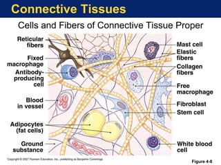
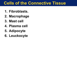
Cells of theConnective Tissue
Fibroblasts
Fibroblasts synthesize the fibers and
components of extracellular matrices.
Fibroblasts are the most common cells in
connective tissue.
Two stages of activity—active and
quiescent—are observed in these cells.
Fibroblasts are active cells and fibrocytes are
quiescent cells.
10.
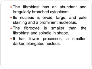
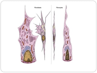
The fibroblasthas an abundant and
irregularly branched cytoplasm.
Its nucleus is ovoid, large, and pale
staining and a prominent nucleolus.
The fibrocyte is smaller than the
fibroblast and spindle in shape.
It has fewer processes; a smaller,
darker, elongated nucleus.
12.
Macrophages & theMononuclear
Phagocyte System
Macrophages characterized by
phagocytic ability.
Macrophages have a wide spectrum of
morphological features that
corresponds to their state of functional
activity and to the tissue they inhabit.
They are characterized by an irregular
surface.
They generally have many lysosomes
and an oval or kidney-shaped nucleus
located eccentrically.
13.
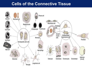
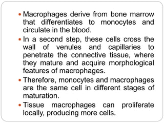
Macrophages derivefrom bone marrow
that differentiates to monocytes and
circulate in the blood.
In a second step, these cells cross the
wall of venules and capillaries to
penetrate the connective tissue, where
they mature and acquire morphological
features of macrophages.
Therefore, monocytes and macrophages
are the same cell in different stages of
maturation.
Tissue macrophages can proliferate
locally, producing more cells.
14.
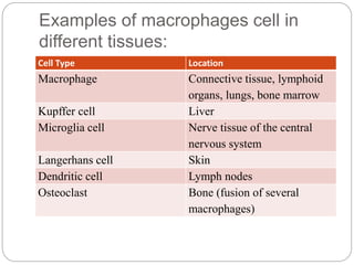
Examples of macrophagescell in
different tissues:
Cell Type Location
Macrophage Connective tissue, lymphoid
organs, lungs, bone marrow
Kupffer cell Liver
Microglia cell Nerve tissue of the central
nervous system
Langerhans cell Skin
Dendritic cell Lymph nodes
Osteoclast Bone (fusion of several
macrophages)
16.
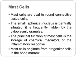
Mast Cells
Mastcells are oval to round connective
tissue cells.
The small, spherical nucleus is centrally
situated; it is frequently hidden by the
cytoplasmic granules.
The principal function of mast cells is the
storage of chemical mediators of the
inflammatory response.
Mast cells originate from progenitor cells
in the bone marrow.
18.
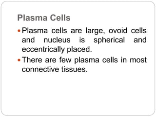
Plasma Cells
Plasmacells are large, ovoid cells
and nucleus is spherical and
eccentrically placed.
There are few plasma cells in most
connective tissues.
20.
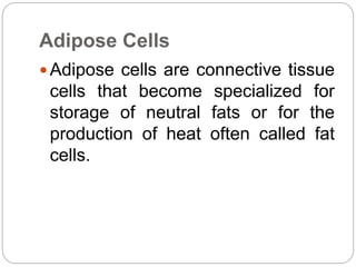
Adipose Cells
Adiposecells are connective tissue
cells that become specialized for
storage of neutral fats or for the
production of heat often called fat
cells.
21.
Leukocytes
The normalconnective tissue
contains leukocytes that migrate
from the blood vessels by
diapedesis.
Leukocytes or white blood
corpuscles are the wandering cells
of the connective tissue.
22.
They migratethrough the walls of
capillaries and postcapillary venules
from the blood to connective tissues
by a process called diapedesis.
This process increases greatly
during inflammation.
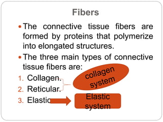
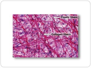
Fibers
The connectivetissue fibers are
formed by proteins that polymerize
into elongated structures.
The three main types of connective
tissue fibers are:
1. Collagen.
2. Reticular.
3. Elastic. Elastic
system
25.
Collagen andreticular fibers are formed by
the protein collagen
Elastic fibers are composed mainly of the
protein elastin.
These fibers are distributed unequally
among the types of connective tissue.
26.
Collagen Fibers
Thistype is present in the skin, bone,
cartilage, smooth muscle, and basal
lamina.
Collagen is the most abundant protein in
the human body, representing 30% of its
dry weight.
28.
Based ontheir structure and functions, they can
be classified into the following groups:
1. Collagens That Form Long Fibrils .
2. Collagens associated fibrils
3. Collagens That Form Networks.
4. Collagens That Form Anchoring Fibrils.
Collagen fibers are colourless strands, when they
are present in great numbers the tissues (e.g.,
tendons) the tissue will be white.
29.
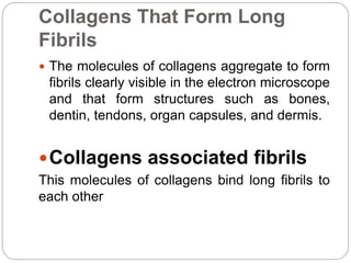
Collagens That FormLong
Fibrils
The molecules of collagens aggregate to form
fibrils clearly visible in the electron microscope
and that form structures such as bones,
dentin, tendons, organ capsules, and dermis.
Collagens associated fibrils
This molecules of collagens bind long fibrils to
each other
30.
Collagens That FormNetworks
The molecules of network-forming collagen
assemble in a meshwork that constitutes the
structural component of the basal lamina.
31.
Collagens That FormAnchoring
Fibrils
Anchoring collagen is present in the anchoring
fibrils that bind collagen fibers to the basal
lamina.
32.
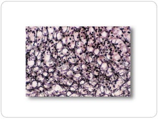
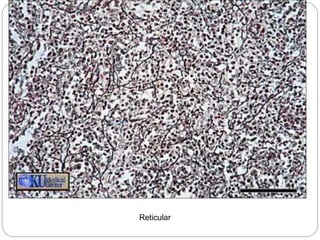
Reticular Fibers
Reticularfibers are extremely thin and
they form an extensive network in
certain organs.
They are not visible in hematoxylin and
eosin (H&E) preparations but can be
easily stained black by impregnation
with silver salts.
33.
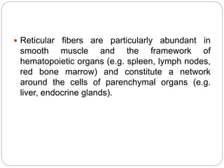
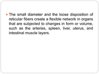
Reticular fibersare particularly abundant in
smooth muscle and the framework of
hematopoietic organs (e.g. spleen, lymph nodes,
red bone marrow) and constitute a network
around the cells of parenchymal organs (e.g.
liver, endocrine glands).
34.
The smalldiameter and the loose disposition of
reticular fibers create a flexible network in organs
that are subjected to changes in form or volume,
such as the arteries, spleen, liver, uterus, and
intestinal muscle layers.
36.
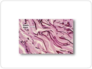
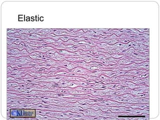
The Elastic FibersSystem
The elastic fiber system, constitutes a family of fibers
whose variable functional characteristics are
adapted to local tissue requirements.
May produced by fibroblasts in connective tissue and
by smooth muscle cells in blood vessels.
Elastin (the elastic fiber) is resistant to boiling, acid
and alkali extraction, and digestion by the usual
proteases.
Also these fibers can be found the dermis connects
the elastic system to the basal lamina.
38.
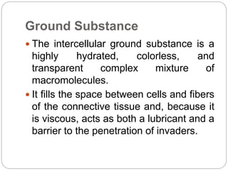
Ground Substance
Theintercellular ground substance is a
highly hydrated, colorless, and
transparent complex mixture of
macromolecules.
It fills the space between cells and fibers
of the connective tissue and, because it
is viscous, acts as both a lubricant and a
barrier to the penetration of invaders.
39.
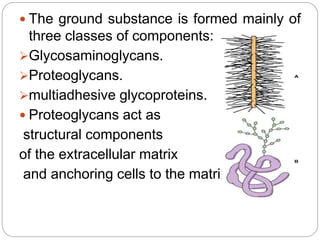
The groundsubstance is formed mainly of
three classes of components:
Glycosaminoglycans.
Proteoglycans.
multiadhesive glycoproteins.
Proteoglycans act as
structural components
of the extracellular matrix
and anchoring cells to the matrix.
40.
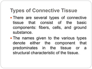
Types of ConnectiveTissue
There are several types of connective
tissue that consist of the basic
components: fibers, cells, and ground
substance.
The names given to the various types
denote either the component that
predominates in the tissue or a
structural characteristic of the tissue.
41.
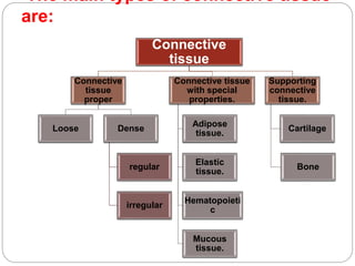
The main typesof connective tissue
are:
Connective
tissue
Connective
tissue
proper
Loose Dense
regular
irregular
Connective tissue
with special
properties.
Adipose
tissue.
Elastic
tissue.
Hematopoieti
c
Mucous
tissue.
Supporting
connective
tissue.
Cartilage
Bone
42.
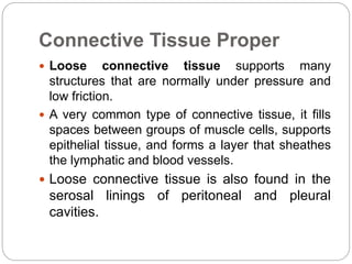
Connective Tissue Proper
Loose connective tissue supports many
structures that are normally under pressure and
low friction.
A very common type of connective tissue, it fills
spaces between groups of muscle cells, supports
epithelial tissue, and forms a layer that sheathes
the lymphatic and blood vessels.
Loose connective tissue is also found in the
serosal linings of peritoneal and pleural
cavities.
43.
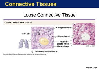
Loose connectivetissue comprises all the main
components of connective tissue proper.
There is no predominant element in this tissue.
The most numerous cells are fibroblasts and
macrophages, but all the other types of
connective tissue cells are also present.
A moderate amount of collagen, elastic, and
reticular fibers appears in this tissue.
Loose connective tissue is flexible, well
vascularized, and not very resistant to stress.
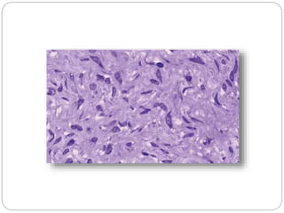
Dense connectivetissue is adapted to offer
resistance and protection.
It consists of the same components found in
loose connective tissue, but there are fewer cells
and a clear predominance of collagen fibers.
Dense connective tissue is less flexible and far
more resistant to stress than is loose connective
tissue.
47.
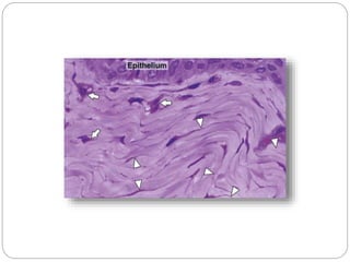
Dense irregularconnective tissue when the
collagen fibers are arranged in bundles without a
definite orientation.
The collagen fibers form a three-dimensional
network in dense irregular tissue and provide
resistance to stress from all directions.
This type of tissue is encountered in areas such as
the dermis.
49.
Dense regularconnective tissue it collagen
bundles are arranged according to a definite
pattern.
The collagen fibers of this tissue are aligned with
the linear orientation of fibroblasts in response to
prolonged stresses exerted in the same
direction; consequently they offer great
resistance to traction forces.
50.
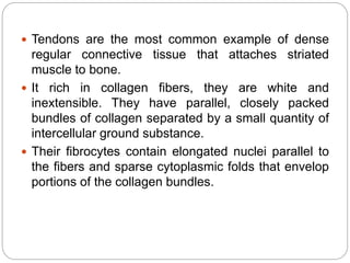
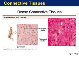
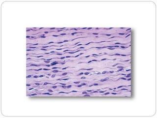
Tendons arethe most common example of dense
regular connective tissue that attaches striated
muscle to bone.
It rich in collagen fibers, they are white and
inextensible. They have parallel, closely packed
bundles of collagen separated by a small quantity of
intercellular ground substance.
Their fibrocytes contain elongated nuclei parallel to
the fibers and sparse cytoplasmic folds that envelop
portions of the collagen bundles.
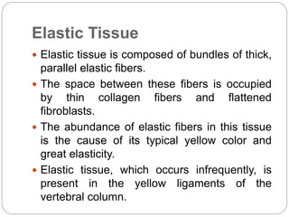
Elastic Tissue
Elastictissue is composed of bundles of thick,
parallel elastic fibers.
The space between these fibers is occupied
by thin collagen fibers and flattened
fibroblasts.
The abundance of elastic fibers in this tissue
is the cause of its typical yellow color and
great elasticity.
Elastic tissue, which occurs infrequently, is
present in the yellow ligaments of the
vertebral column.
54.
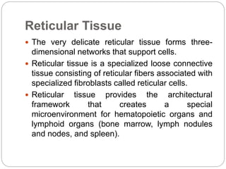
Reticular Tissue
Thevery delicate reticular tissue forms three-
dimensional networks that support cells.
Reticular tissue is a specialized loose connective
tissue consisting of reticular fibers associated with
specialized fibroblasts called reticular cells.
Reticular tissue provides the architectural
framework that creates a special
microenvironment for hematopoietic organs and
lymphoid organs (bone marrow, lymph nodules
and nodes, and spleen).
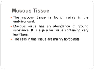
Mucous Tissue
Themucous tissue is found mainly in the
umbilical cord.
Mucous tissue has an abundance of ground
substance. It is a jellylike tissue containing very
few fibers.
The cells in this tissue are mainly fibroblasts.
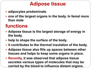
Adipose tissue
adipocytespredominate.
one of the largest organs in the body. In femal more
than male
functions
Adipose tissue is the largest storage of energy in
the body.
help to shape the surface of the body.
it contributes to the thermal insulation of the body.
Adipose tissue also fills up spaces between other
tissues and helps to keep some organs in place.
Recently, it was observed that adipose tissue
secretes various types of molecules that may be
carried by the blood to influence distant organs.
62.
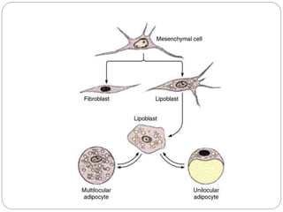
The two knowntypes of adipose
tissue
Unilocular (common, or yellow) adipose
tissue.
Multilocular (or brown) adipose tissue.
63.
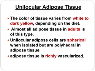
Unilocular Adipose Tissue
The color of tissue varies from white to
dark yellow, depending on the diet.
Almost all adipose tissue in adults is
of this type.
Unilocular adipose cells are spherical
when isolated but are polyhedral in
adipose tissue.
adipose tissue is richly vascularized.
64.
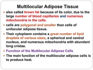
Multilocular Adipose Tissue
also called brown fat because of its color, due to the
large number of blood capillaries and numerous
mitochondria in the cells.
cells are polygonal and smaller than cells of
unilocular adipose tissue.
Their cytoplasm contains a great number of lipid
droplets of various sizes, a spherical and central
nucleus, and numerous mitochondria with abundant
long cristae.
Function of the Multilocular Adipose Cells
The main function of the multilocular adipose cells is
to produce heat.
Quiz
1. The componentsof connective tissue are ______,
______, and _______.
2. The active form of fibroblast is called _________.
3. The fibers of connective tissue found in hematopoietic
organs are __________.
4. The umbilical cord is a type of connective tissue called
______.
5. The best example of a dense regular connective tissue is
______.