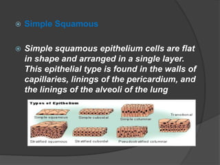
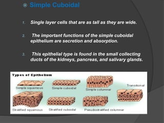
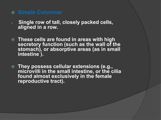
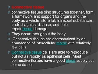
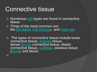
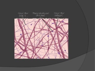
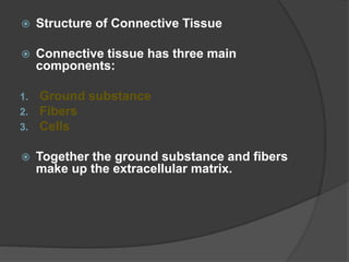
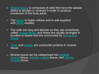
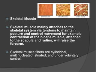
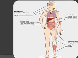
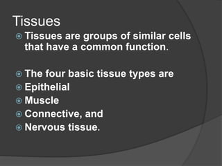
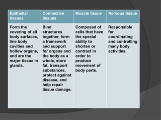
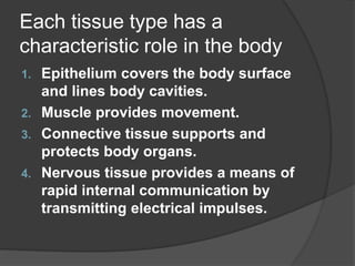
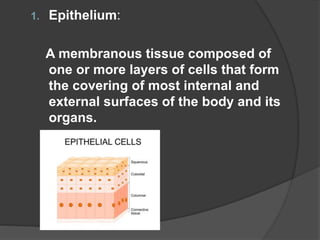
This document provides an overview of the four main types of tissues in the body: epithelial, connective, muscular, and nervous tissue. It describes the characteristics and functions of each tissue type. For epithelial tissue, it outlines the different categories (simple, stratified, transitional etc.) and provides examples. For connective tissue, it discusses the ground substance, fibers and cells. It also describes the different types of connective tissues (areolar, adipose, cartilage etc.). The document concludes with brief descriptions of muscular tissue (skeletal, cardiac and smooth muscle) and membranes that cover the body.
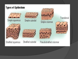
![ I .Simple Epithelia
Simple epithelium consists of a single layer of cells.
They are typically where absorption, secretion and
filtration occur.
Simple epithelial tissues are generally classified by the
shape of their cells.
The four major classes of simple epithelium are:
1]Simple squamous;
2) Simple cuboidal;
3) Simple columnar; and
4) Pseudostratified.](https://image.slidesharecdn.com/tissue-210517105754/85/Tissue-11-320.jpg)