
Recommended
More Related Content
Similar to STRUCTURAL ORGANISATION IN ANIMALS TISSUE NOTES.pdf
Similar to STRUCTURAL ORGANISATION IN ANIMALS TISSUE NOTES.pdf (20)
More from DrUpadhyay
More from DrUpadhyay (20)
Recently uploaded
Recently uploaded (20)
STRUCTURAL ORGANISATION IN ANIMALS TISSUE NOTES.pdf
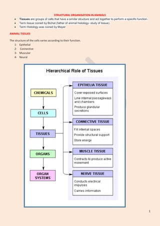
- 1. 1 STRUCTURAL ORGANISATION IN ANIMALS Tissues are groups of cells that have a similar structure and act together to perform a specific function. Term tissue coined by Bichat (father of animal histology- study of tissue). Term Histology was coined by Mayer ANIMAL TISSUES The structure of the cells varies according to their function. 1- Epithelial 2- Connective 3- Muscular 4- Neural
- 2. 2 Epithelial Tissue Covers both external and internal surfaces of the animal body. The cells are compactly packed with little intercellular matrix. Blood vessels are not present in the epithelial tissue. Types of epithelial tissue 1- simple epithelium 2- compound epithelium. TISSUE CELLS FUNCTION simple epithelium composed of a single layer of cells It functions as a lining for body cavities, ducts, and tubes compound epithelium consists of two or more cell layers protective function against chemical and mechanical stresses. They cover the dry surface of the skin, the moist surface of buccal cavity, pharynx, inner lining of ducts of salivary glands and of pancreatic ducts TYPES OF SIMPLE EPITHELIUM a- Squamous b- Cuboidal c- Columnar
- 3. 3 SIMPLE EPITHELIUM CELSS LOCATION FUNCTION Squamous epithelium ( pavement epithelium) Single layer, Thin, polygonal, flattened cells with irregular boundaries. - walls of blood vessels - air sacs (alveoli) of lungs - kidney tubules Forming a diffusion boundary. Protection, excretion, gas exchange and secretion of coelomic fluid. Cuboidal epithelium Single layer of cube-like cells. May be smooth or bear Microvilli - ducts of glands and tubular parts of nephrons. - PCT of nephron has microvilli - ovaries seminiferous tubules Secretion and absorption. columnar epithelium single layer of tall and slender cells Their nuclei are located at the base. Free surface may have microvilli lining of stomach and intestine Secretion and absorption. Some of its cells produce mucus, called goblet cells. Ciliated epithelium If the columnar or cuboidal cells bear cilia on their free surface Inner surface of hollow organs like bronchioles and fallopian tubes. Their function is to move particles or mucus in a specific direction over the epithelium. Glandular epithelium columnar or cuboidal cells get specialised for secretion isolated glandular cells (goblet cells of the alimentary canal), and multicellular, consisting of cluster of cells (salivary gland). Endocrine gland Exocrine glands SPECIALISED JUNCTIONS All cells in epithelium are held together with little intercellular material. In nearly all animal tissues, specialised junctions provide both structural and functional links between its individual cells. TYPES- Tight junctions help to stop substances from leaking across a tissue Adhering junctions Adhering junctions/ cementing Gap junctions facilitate the cells to communicate with each other by connecting the cytoplasm of adjoining cells, for rapid transfer of ions, small molecules and sometimes big molecules
- 4. 4 Connective Tissue Most abundant Linking and supporting other tissues/organs of the body. They range from soft connective tissues to specialised types, which include cartilage, bone, adipose, and blood. In all connective tissues except blood, the cells secrete fibres of structural proteins called collagen or elastin. The fibres provide strength, elasticity and flexibility to the tissue. These cells also secrete modified polysaccharides, which accumulate between cells and fibres and act as matrix (ground substance). Types of Connective tissues (i) Loose connective tissue (ii) Dense connective tissue (iii) Specialised connective tissue. Loose connective tissue has cells and fibres loosely arranged in a semi-fluid ground substance Areolar tissue present beneath the skin it serves as a support framework for epithelium. It contains fibroblasts (cells that produce and secrete fibres), macrophages and mast cells. Adipose tissue- located mainly beneath the skin. store fats Dense connective tissue Dense Regular connective tissue - collagen fibres are present in rows between many parallel bundles of fibres. Tendons - attach skeletal muscles to bones Ligaments - attach one bone to another Dense irregular connective tissue- has fibroblasts and many fibres (mostly collagen) that are oriented differently This tissue is present in the skin. Specialised connective tissue 1- Cartilage 2- Bones 3- blood CARTILAGE - soft. Cells of this tissue (chondrocytes) are enclosed in small cavities within the matrix secreted by them. Most of the cartilages in vertebrate embryos are replaced by bones in adults. Present in the tip of nose, outer ear joints, between adjacent bones of the vertebral column, limbs and hands in adults BONES - have a hard and non-pliable ground substance rich in calcium salts
- 5. 5 and collagen fibres which give bone its strength. It is the main tissue that provides structural frame to the body. Bones support and protect softer tissues and organs. The bone cells (osteocytes) are present in the spaces called lacunae. They also interact with skeletal muscles attached to them to bring about movements. The bone marrow in some bones is the site of production of blood cells. BLOOD – it is a fluid connective tissue. Contains plasma, red blood cells (RBC), white blood cells (WBC) and platelets It is the main circulating fluid that helps in the transport of various substances.
- 6. 6 Muscle Tissue Each muscle is made of many long, cylindrical fibres arranged in parallel arrays. These fibres are composed of numerous fine fibrils, called myofibrils. Show contraction Types of muscle tissue 1- Skeletal 2- Smooth 3- Cardiac. SKELETAL Tissue is closely attached to skeletal bones. In a typical muscle such as the biceps, striated (striped) skeletal muscle fibres are bundled together in a parallel fashion Voluntary SMOOTH Fibers taper at both ends (fusiform) and do not show striations. Cell junctions hold them together and they are bundled together in a connective tissue sheath. The wall of internal organs such as the blood vessels, stomach and intestine contains it. Smooth muscles are ‘involuntary’. CARDIAC Contractile tissue present only in the heart. Characters of both unstriped and striped muscle fibres. These fibres never get fatigue.
- 7. 7 Neural Tissue Neurons The Neuroglial cells which constitute the rest of the neural system protect and support neurons. Neuroglia makes up more than one half the volume of neural tissue in our body. Dr R. K. UPADHYAY KV 39 GTC, VARANASI