Bio 160 Parasitology - Blood protozoans
•Download as DOC, PDF•
3 likes•832 views

BLOOD PROTOZOA OF MAJOR CLINICAL SIGNIFICANCE

Recommended
More Related Content
What's hot
What's hot (20)
Viewers also liked
Viewers also liked (16)
Similar to Bio 160 Parasitology - Blood protozoans
Similar to Bio 160 Parasitology - Blood protozoans (20)
More from Shaina Mavreen Villaroza
More from Shaina Mavreen Villaroza (20)
Recently uploaded
Recently uploaded (20)
Bio 160 Parasitology - Blood protozoans
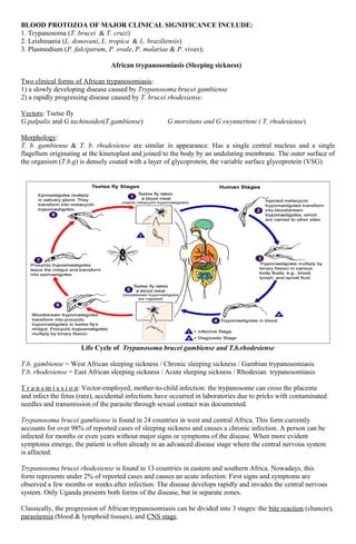
- 1. BLOOD PROTOZOA OF MAJOR CLINICAL SIGNIFICANCE INCLUDE: 1. Trypanosoma (T. brucei & T. cruzi) 2. Leishmania (L. donovani, L. tropica & L. braziliensis) 3. Plasmodium (P. falciparum, P. ovale, P. malariae & P. vivax); African trypanosomiasis (Sleeping sickness) Two clinical forms of African trypanosomiasis: 1) a slowly developing disease caused by Trypanosoma brucei gambiense 2) a rapidly progressing disease caused by T. brucei rhodesiense. Vectors: Tsetse fly G.palpalis and G.tachinoides(T.gambiense) G.morsitans and G.swynnertoni ( T. rhodesiense) Morphology: T. b. gambiense & T. b. rhodesiense are similar in appearance. Has a single central nucleus and a single flagellum originating at the kinetoplast and joined to the body by an undulating membrane. The outer surface of the organism (T.b.g) is densely coated with a layer of glycoprotein, the variable surface glycoprotein (VSG). Life Cycle of Trypanosoma brucei gambiense and T.b.rhodesiense T.b. gambiense = West African sleeping sickness / Chronic sleeping sickness / Gambian trypanosomiasis T.b. rhodesiense = East African sleeping sickness / Acute sleeping sickness / Rhodesian trypanosomiasis T r a n s m i s s i o n: Vector-employed, mother-to-child infection: the trypanosome can cross the placenta and infect the fetus (rare), accidental infections have occurred in laboratories due to pricks with contaminated needles and transmission of the parasite through sexual contact was documented. Trypanosoma brucei gambiense is found in 24 countries in west and central Africa. This form currently accounts for over 98% of reported cases of sleeping sickness and causes a chronic infection. A person can be infected for months or even years without major signs or symptoms of the disease. When more evident symptoms emerge, the patient is often already in an advanced disease stage where the central nervous system is affected. Trypanosoma brucei rhodesiense is found in 13 countries in eastern and southern Africa. Nowadays, this form represents under 2% of reported cases and causes an acute infection. First signs and symptoms are observed a few months or weeks after infection. The disease develops rapidly and invades the central nervous system. Only Uganda presents both forms of the disease, but in separate zones. Classically, the progression of African trypanosomiasis can be divided into 3 stages: the bite reaction (chancre), parasitemia (blood & lymphoid tissues), and CNS stage.
- 2. Painful sensitivity of palms & ulnar region to pressure (Kerandel's sign) may develop in some Caucasians. Very characteristic of Gambian disease is visible enlargement of the glands of the posterior cervical region (Winterbottom's sign). The clinical features of Rhodesian disease are similar but briefer & more acute. The acuteness & severity of disease do not allow typical sleeping sickness. Death is due to cardiac failure within 6-9 months. American trypanosomiasis (Chagas’ Disease) Chagas' disease is caused by the protozoan hemoflagellate, Trypanosoma cruzi. Morphology: Depending on its host environment, the org. occurs in 3 different forms: 1) metacyclic trypomastigotes - in the terminal part of the digestive and urinary tracts of vectors nuclei near the posterior of their bodies free flagellum 2) epimastigote (crithidial) - replicative form of the parasite in the insect vector and in the acellular culture medium more adaptable to survive the insect's intestines flagellum attached near The center of the body 3) epimastigote - replicative form of the parasite in the insect vector and in the acellular culture medium more adaptable to survive the insect's intestines flagellum attached near the center of the body Life Cycle of Trypanosoma cruzi Vectors: many species of kissing/ cone-nosed (riduvid/triatomine) bug, most prominently by Triatoma infestans, T. sordida, Panstrongylus megistus & Rhodnius prolixus. T r a n s m i s s i o n: Mainly transmitted by contact with feces/urine of infected blood-sucking triatomine Bugs (vector-employed), transfusion of infected blood (containing trypomastigotes), vertical or congenital transmission of the parasite occurs in 2-10% of infected women who are pregnant (passage from an infected mother to her newborn during pregnancy or childbirth, oral transmission (consumption of food contaminated with T. cruzi through, for example, contact with infected triatomine bug feces), organ transplants using organs from infected donors, and laboratory accidents. Chagas' disease can be divided into 3 stages: the primary lesion (chagoma-Ramana’s sign= orbital edema), the acute stage, & the chronic stage. Unlike T. brucei, T. cruzi does not alter its antigenic coat.
- 3. Leismaniasis Several sps. of Leishmania are pathogenic for man: L. donovani causes visceral leishmaniasis (Kala-azar, black disease, dumdum fever); L. tropica (L. t. major, L. t. minor & L. ethiopica) cause cutaneous leishmaniasis (oriental sore, Delhi ulcer, Aleppo, Delhi or Baghdad boil); & L. braziliensis (also, L. mexicana & L. peruviana) are etiologic agents of mucocutaneous leishmaniasis (espundia, Uta, chiclero ulcer). 3 main forms of leishmaniases: 1) visceral (often known as kala-azar and the most serious form of the disease) 2) cutaneous (the most common) 3) mucocutaneous. Visceral leishmaniasis (VL also known as kala-azar) is fatal if left untreated. - It is characterized by irregular bouts of fever, weight loss, enlargement of the spleen and liver, and anemia. It is highly endemic in the Indian subcontinent and in East Africa. An estimated 200 000 to 400 000 new cases of VL occur worldwide each year. Over 90% of new cases occur in 6 countries: Bangladesh, Brazil, Ethiopia, India, South Sudan and Sudan. Cutaneous leishmaniasis (CL) is the most common form of leishmaniasis and causes skin lesions, mainly ulcers, on exposed parts of the body, leaving life-long scars and serious disability. About 95% of CL cases occur in the Americas, the Mediterranean basin, the Middle East and Central Asia. Over two thirds of new CL cases occur in 6 countries: Afghanistan, Algeria, Brazil, Colombia, Iran (Islamic Republic of) and the Syrian Arab Republic. An estimated 0.7 million to 1.3 million new cases occur worldwide annually. Mucocutaneous leishmaniasis leads to partial or total destruction of mucous membranes of the nose, mouth and throat. Almost 90% of mucocutaneous leishmaniasis cases occurs in the Plurinational State of Bolivia, Brazil and Peru. Morphology: Amastigote (leishmanial form) is oval. Leptomonad measures 14 - 20 microns by 1.5 - 4 microns, a similar size to trypanosomes. Vectors: bites of infected female phlebotomine sandflies (Phlebotomus) * Some 70 animal species, including humans, have been found as natural reservoir hosts of Leishmania parasites. Life Cycle of Leishmania spp.
- 4. Malaria Life Cycle of Plasmodium species Differences between the species include: 1) Blood-stage morphology 2) Minor life cycle variations • P. vivax and P. ovale exhibit the hypnozoite stage and can cause true relapses. • trophozoite- and schizont-infected RBCs of P. falciparum sequester in the microvasculature and are not found in the peripheral circulation. 3) host erythrocyte preference • P. vivax and P. ovale prefer reticulocytes (immature erythrocytes). • P.ovale prefer enlarged RBCs (other refs.). • P. malariae prefers senescent erythrocytes. • P. falciparum exhibits no preference (non-enlarged RBCs) . 4) Disease and clinical manifestation.
- 5. Life Cycle of Babesia microti Life Cycle of Toxoplasma gondii
- 6. Life Cycle of Babesia microti Life Cycle of Toxoplasma gondii