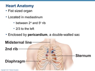
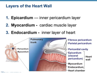
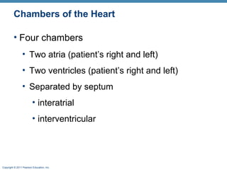
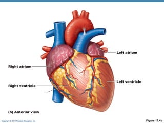
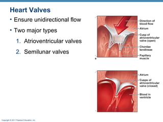
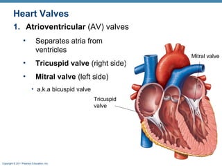
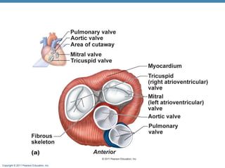
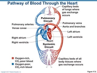
Chapter 17 Overview
Layers of heart tissue
Heart Chambers
Valves
Heart contraction

Copyright © 2011 Pearson Education, Inc.
Heart Anatomy
• Fist sized organ
• Located in mediastinum
• between 2nd and 5th rib
• 2/3 to the left

• Enclosed by pericardium, a double-walled sac

Midsternal line
2nd rib
Sternum
Diaphragm
Copyright © 2011 Pearson Education, Inc.
Pericardium
• Outer fibrous layer
• Anchors heart

• Parietal layer lines fibrous layer
• Visceral layer (epicardium) contacts heart
• Separated by pericardial fluid

Copyright © 2011 Pearson Education, Inc.
Layers of the Heart Wall
1. Epicardium — inner pericardium layer
2. Myocardium - cardiac muscle layer
3. Endocardium - inner layer of heart
Pulmonary
trunk
Pericardium
Myocardium

Copyright © 2011 Pearson Education, Inc.

Fibrous pericardium
Parietal pericardium
Pericardial cavity
Epicardium
(visceral
Heart
pericardium) wall
Myocardium
Endocardium
Heart chamber
Chambers of the Heart
• Four chambers
• Two atria (patient’s right and left)
• Two ventricles (patient’s right and left)
• Separated by septum
• interatrial
• interventricular

Copyright © 2011 Pearson Education, Inc.
Left atrium

Right atrium

Right ventricle

Left ventricle

(b) Anterior view
Copyright © 2011 Pearson Education, Inc.

Figure 17.4b
Atria: The Receiving Chambers
• Right atria
• Left atria
• Low O2 blood

• High O2 blood

• Blood from:

• Blood from:

• Sup. & inf. Vena cava
• Coronary sinus

Sup. Vena cava

Right atria
R. pulm. vein

Inf. Vena cava
Copyright © 2011 Pearson Education, Inc.

• R & L pulmonary veins
• From lungs
L. Pulmonary
vein

Left atria
Ventricles: The Discharging Chambers
Aorta

• Right ventricle
• pumps blood to the lungs
• Via pulmonary trunk

Pulmonary
trunk

• Left ventricle
• Pumps blood to the body
• Via the aorta

Copyright © 2011 Pearson Education, Inc.

Right
ventricle

Left
ventricle
Heart Valves
• Ensure unidirectional flow
• Two major types
1. Atrioventricular valves
2. Semilunar valves

Copyright © 2011 Pearson Education, Inc.
Heart Valves
1. Atrioventricular (AV) valves
•

Separates atria from
ventricles

•

Tricuspid valve (right side)

•

Mitral valve (left side)
• a.k.a bicuspid valve
Tricuspid
valve

Copyright © 2011 Pearson Education, Inc.

Mitral valve
Heart Valves
• Semilunar (SL) valves
• Separate ventricles from circulation
• Aortic semilunar valve
• Left ventricle / aorta

• Pulmonary semilunar valve
• Right ventricle / pulmonary trunk
Pulmonary
SL

Copyright © 2011 Pearson Education, Inc.

Aortic SL
Copyright © 2011 Pearson Education, Inc.
Left atrium
Right atrium

Mitral (bicuspid)
valve
Aortic valve

Tricuspid valve
Right ventricle
Chordae tendineae

Pulmonary valve
Left ventricle
Papillary muscle

(e) Frontal section
Copyright © 2011 Pearson Education, Inc.

Figure 17.4e
PATHWAY OF BLOOD

Copyright © 2011 Pearson Education, Inc.
Pathway of Blood Through the Heart
• The heart is basically two pumps
• Right side
• Pulmonary circuit
• to the lungs

• Left side
• Systemic circuit
• to the body

Copyright © 2011 Pearson Education, Inc.
Pathway of Blood Through the Heart
• R. atrium  tricuspid
valve  R. ventricle 
pulmonary valve 
pulmonary trunk 
pulmonary arteries 
lungs
• Blood through R. atrium &
ventricle is low O2
• In lungs becomes high O2
Copyright © 2011 Pearson Education, Inc.
Pathway of Blood Through the Heart
• High O2 blood enters left
atrium
• Lungs  pulmonary veins
 L. atrium  bicuspid
valve  L. ventricle 
aortic semilunar valve 
aorta  systemic
circulation

Copyright © 2011 Pearson Education, Inc.
Pathway of Blood Through the Heart

Pulmonary
Circuit
Pulmonary arteries
Venae cavae

Capillary beds
of lungs where
gas exchange
occurs
Pulmonary veins
Aorta and branches
Left atrium
Left ventricle

Right atrium
Right ventricle

Oxygen-rich,
CO2-poor blood
Oxygen-poor,
CO2-rich blood
Copyright © 2011 Pearson Education, Inc.

Heart
Systemic
Circuit

Capillary beds of all
body tissues where
gas exchange occurs
Figure 17.5
Pathway of Blood Through the Heart
• Left ventricle pumps blood through entire
body
• Thick myocardium, to generate large force

• Right ventricle pumps blood to lungs
• Thinner myocardium

Copyright © 2011 Pearson Education, Inc.
Coronary Circulation
• Supplies the heart with blood
• The heart is a muscle that requires its own blood supply

• Arteries
• Right and left coronary arteries

• Veins
• Coronary sinus, drains blood into the right
atrium

Copyright © 2011 Pearson Education, Inc.
Coronary vessels

Copyright © 2011 Pearson Education, Inc.

Heart 1 online

  • 1.
    Chapter 17 Overview Layersof heart tissue Heart Chambers Valves Heart contraction Copyright © 2011 Pearson Education, Inc.
  • 2.
    Heart Anatomy • Fistsized organ • Located in mediastinum • between 2nd and 5th rib • 2/3 to the left • Enclosed by pericardium, a double-walled sac Midsternal line 2nd rib Sternum Diaphragm Copyright © 2011 Pearson Education, Inc.
  • 3.
    Pericardium • Outer fibrouslayer • Anchors heart • Parietal layer lines fibrous layer • Visceral layer (epicardium) contacts heart • Separated by pericardial fluid Copyright © 2011 Pearson Education, Inc.
  • 4.
    Layers of theHeart Wall 1. Epicardium — inner pericardium layer 2. Myocardium - cardiac muscle layer 3. Endocardium - inner layer of heart Pulmonary trunk Pericardium Myocardium Copyright © 2011 Pearson Education, Inc. Fibrous pericardium Parietal pericardium Pericardial cavity Epicardium (visceral Heart pericardium) wall Myocardium Endocardium Heart chamber
  • 5.
    Chambers of theHeart • Four chambers • Two atria (patient’s right and left) • Two ventricles (patient’s right and left) • Separated by septum • interatrial • interventricular Copyright © 2011 Pearson Education, Inc.
  • 6.
    Left atrium Right atrium Rightventricle Left ventricle (b) Anterior view Copyright © 2011 Pearson Education, Inc. Figure 17.4b
  • 7.
    Atria: The ReceivingChambers • Right atria • Left atria • Low O2 blood • High O2 blood • Blood from: • Blood from: • Sup. & inf. Vena cava • Coronary sinus Sup. Vena cava Right atria R. pulm. vein Inf. Vena cava Copyright © 2011 Pearson Education, Inc. • R & L pulmonary veins • From lungs L. Pulmonary vein Left atria
  • 8.
    Ventricles: The DischargingChambers Aorta • Right ventricle • pumps blood to the lungs • Via pulmonary trunk Pulmonary trunk • Left ventricle • Pumps blood to the body • Via the aorta Copyright © 2011 Pearson Education, Inc. Right ventricle Left ventricle
  • 9.
    Heart Valves • Ensureunidirectional flow • Two major types 1. Atrioventricular valves 2. Semilunar valves Copyright © 2011 Pearson Education, Inc.
  • 10.
    Heart Valves 1. Atrioventricular(AV) valves • Separates atria from ventricles • Tricuspid valve (right side) • Mitral valve (left side) • a.k.a bicuspid valve Tricuspid valve Copyright © 2011 Pearson Education, Inc. Mitral valve
  • 11.
    Heart Valves • Semilunar(SL) valves • Separate ventricles from circulation • Aortic semilunar valve • Left ventricle / aorta • Pulmonary semilunar valve • Right ventricle / pulmonary trunk Pulmonary SL Copyright © 2011 Pearson Education, Inc. Aortic SL
  • 12.
    Copyright © 2011Pearson Education, Inc.
  • 13.
    Left atrium Right atrium Mitral(bicuspid) valve Aortic valve Tricuspid valve Right ventricle Chordae tendineae Pulmonary valve Left ventricle Papillary muscle (e) Frontal section Copyright © 2011 Pearson Education, Inc. Figure 17.4e
  • 14.
    PATHWAY OF BLOOD Copyright© 2011 Pearson Education, Inc.
  • 15.
    Pathway of BloodThrough the Heart • The heart is basically two pumps • Right side • Pulmonary circuit • to the lungs • Left side • Systemic circuit • to the body Copyright © 2011 Pearson Education, Inc.
  • 16.
    Pathway of BloodThrough the Heart • R. atrium  tricuspid valve  R. ventricle  pulmonary valve  pulmonary trunk  pulmonary arteries  lungs • Blood through R. atrium & ventricle is low O2 • In lungs becomes high O2 Copyright © 2011 Pearson Education, Inc.
  • 17.
    Pathway of BloodThrough the Heart • High O2 blood enters left atrium • Lungs  pulmonary veins  L. atrium  bicuspid valve  L. ventricle  aortic semilunar valve  aorta  systemic circulation Copyright © 2011 Pearson Education, Inc.
  • 18.
    Pathway of BloodThrough the Heart Pulmonary Circuit Pulmonary arteries Venae cavae Capillary beds of lungs where gas exchange occurs Pulmonary veins Aorta and branches Left atrium Left ventricle Right atrium Right ventricle Oxygen-rich, CO2-poor blood Oxygen-poor, CO2-rich blood Copyright © 2011 Pearson Education, Inc. Heart Systemic Circuit Capillary beds of all body tissues where gas exchange occurs Figure 17.5
  • 19.
    Pathway of BloodThrough the Heart • Left ventricle pumps blood through entire body • Thick myocardium, to generate large force • Right ventricle pumps blood to lungs • Thinner myocardium Copyright © 2011 Pearson Education, Inc.
  • 20.
    Coronary Circulation • Suppliesthe heart with blood • The heart is a muscle that requires its own blood supply • Arteries • Right and left coronary arteries • Veins • Coronary sinus, drains blood into the right atrium Copyright © 2011 Pearson Education, Inc.
  • 21.
    Coronary vessels Copyright ©2011 Pearson Education, Inc.