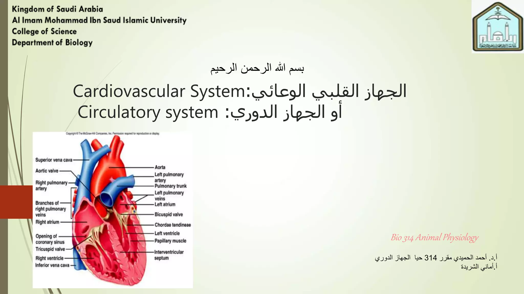
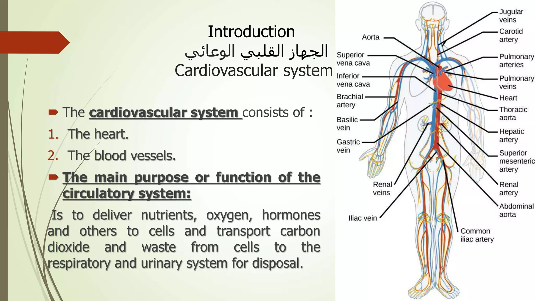
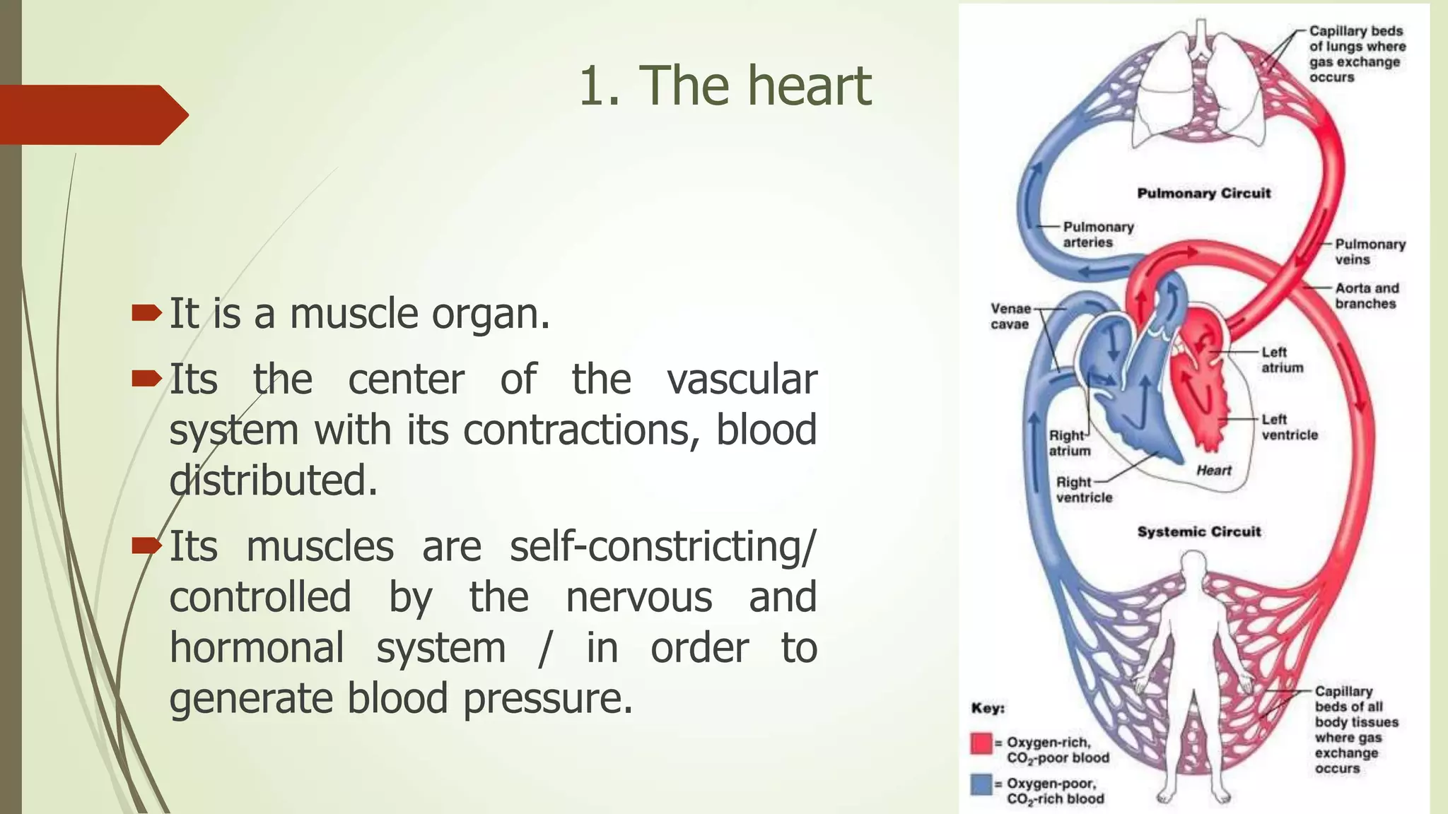
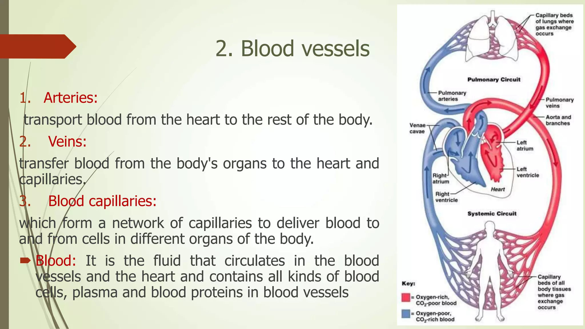
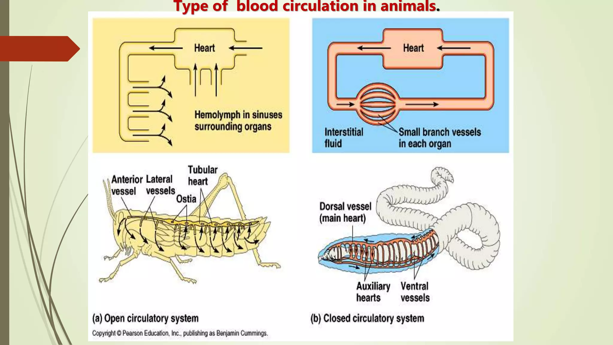
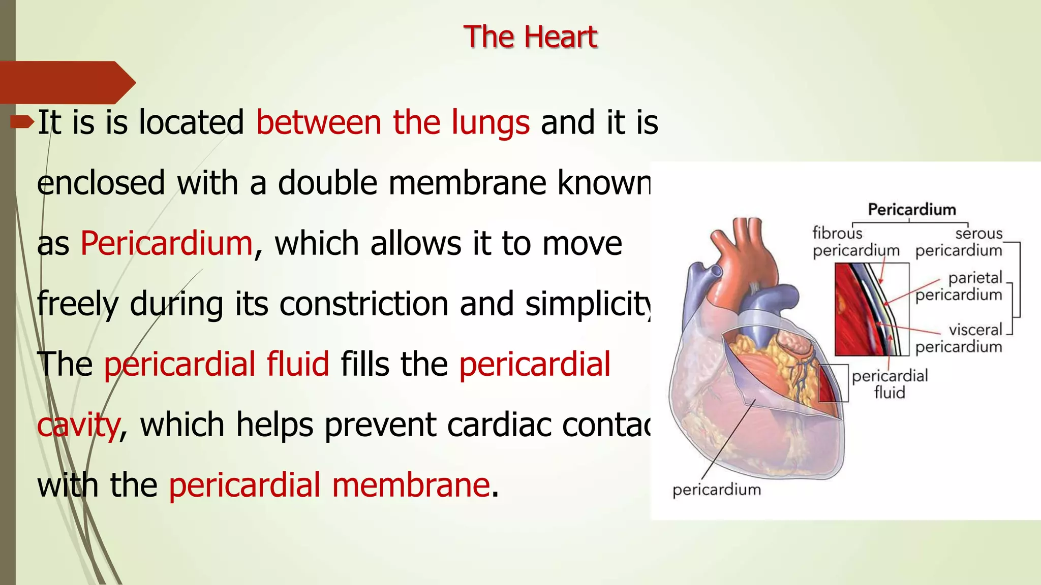
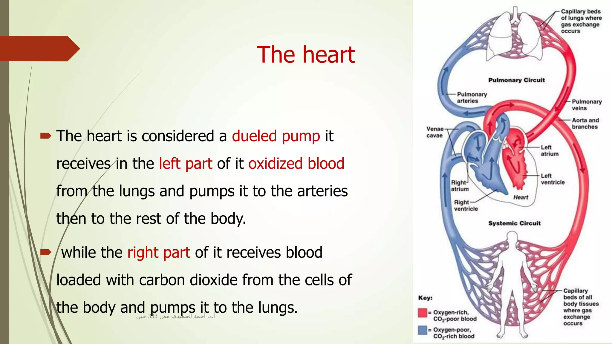
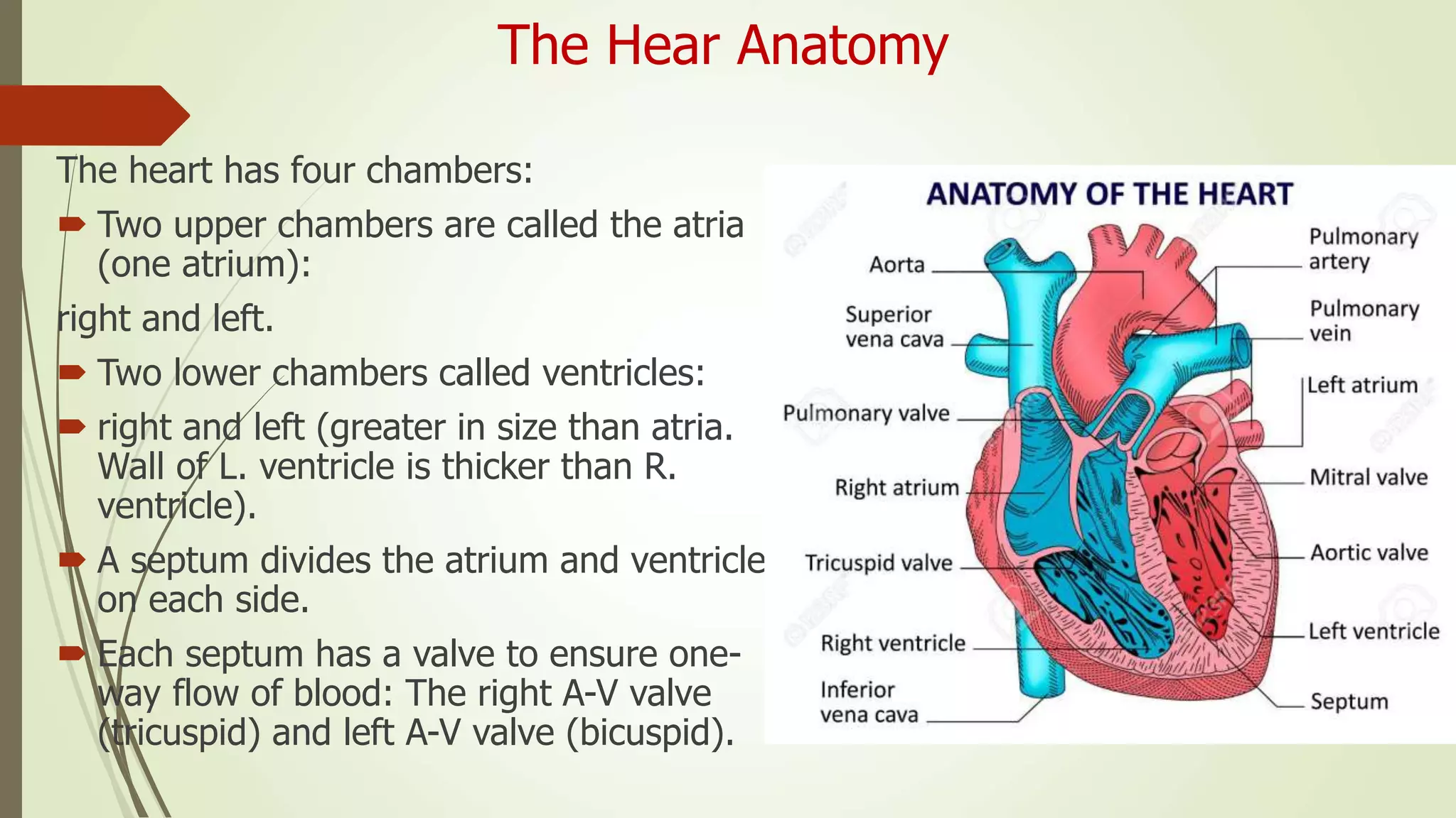
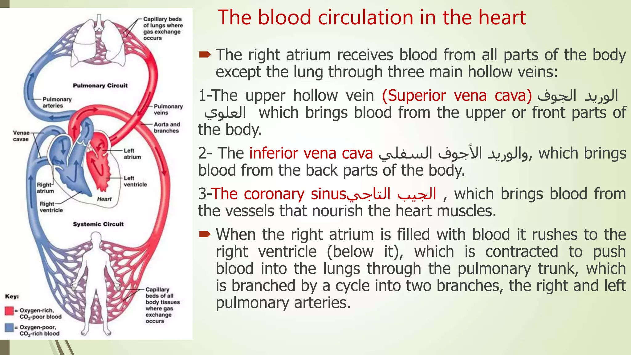
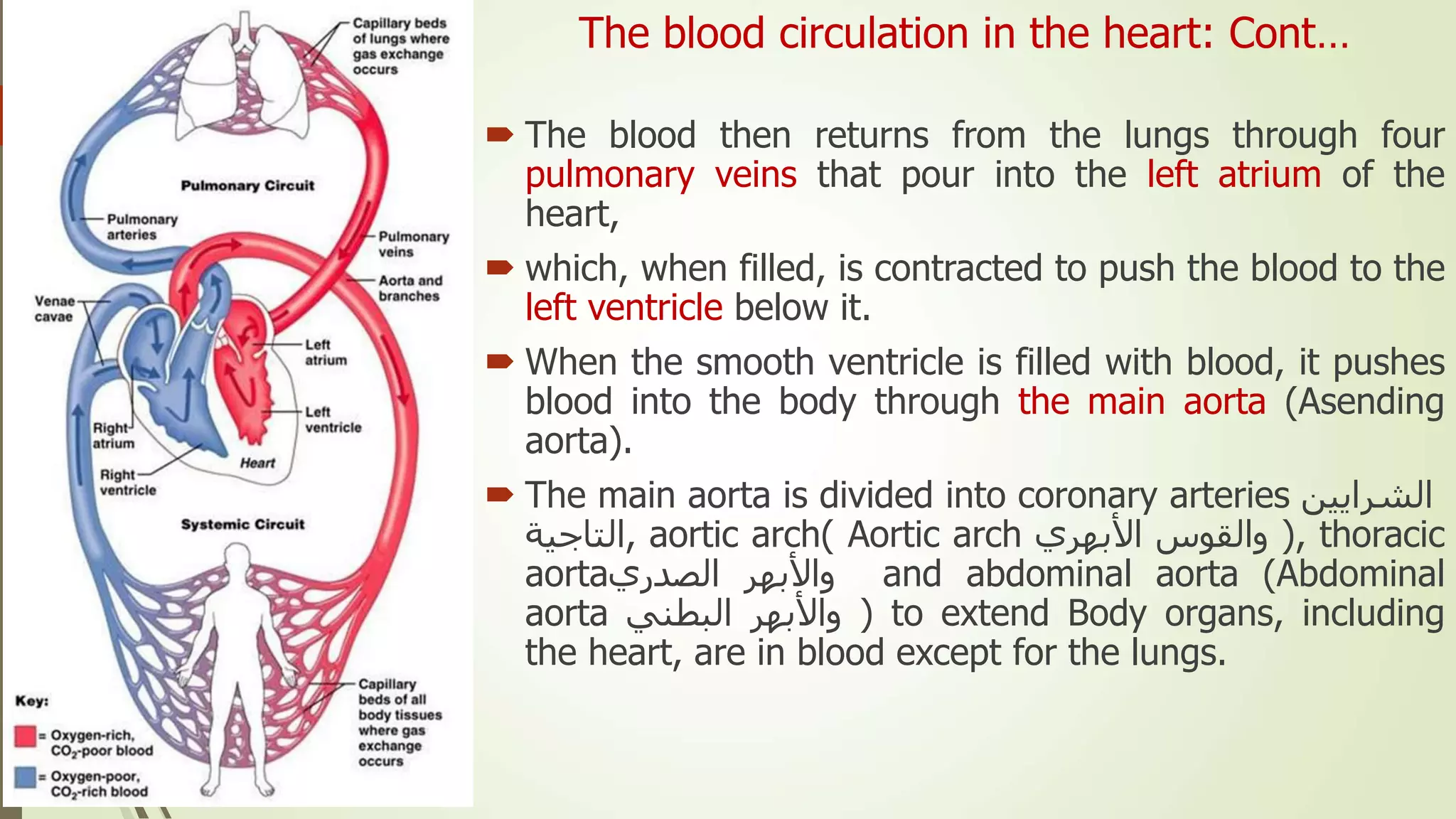
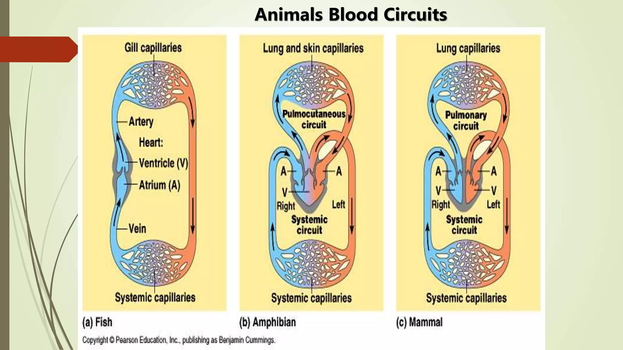
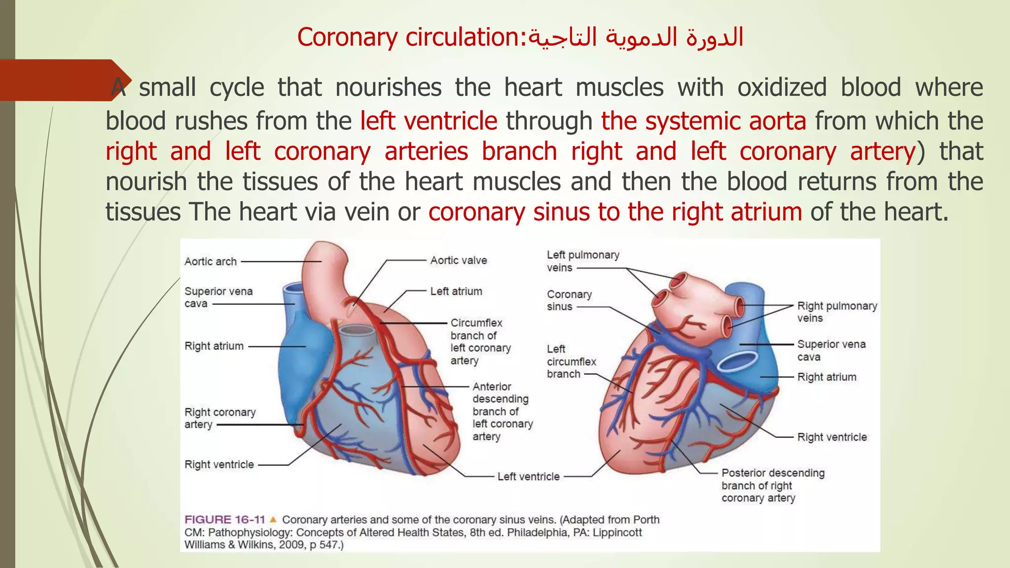
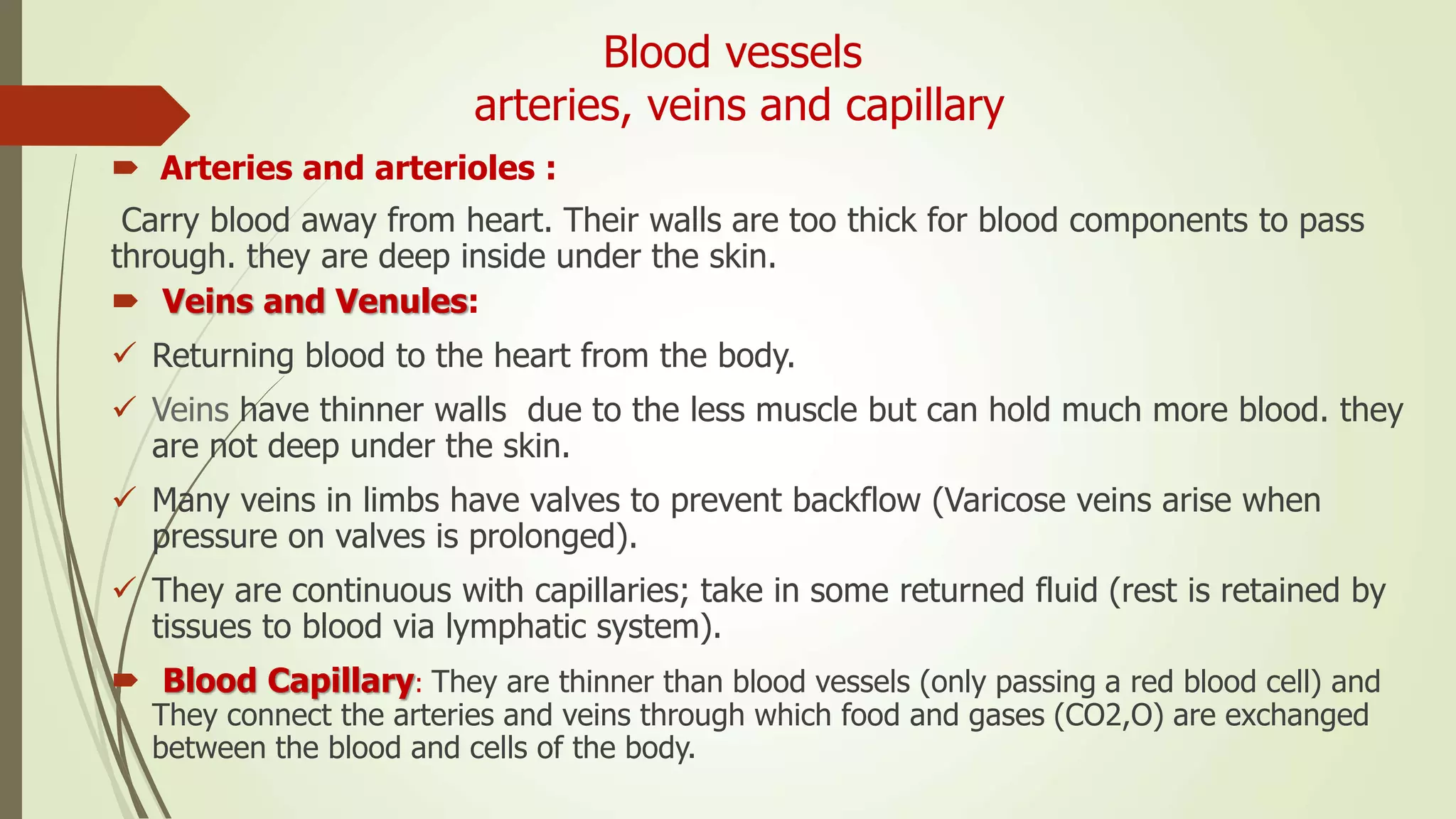
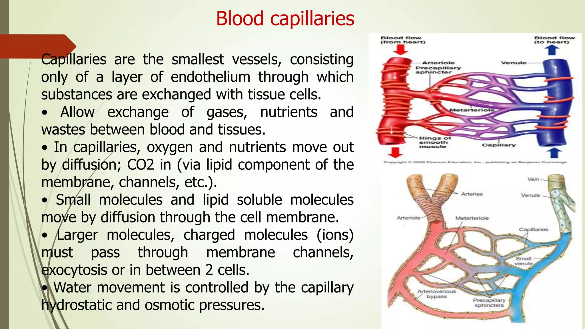
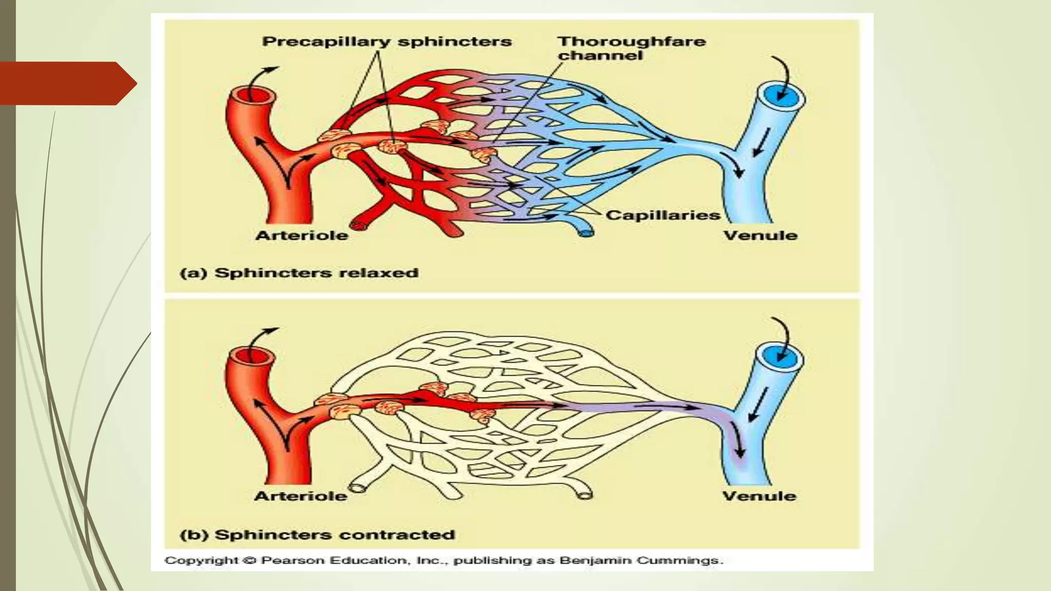
The cardiovascular system can be thought of as the transport system of the body.
This system has three main components: the heart, the blood vessel and the blood itself.
The heart is the system’s pump and the blood vessels are like the delivery routes.
The respiratory system is the network of organs and tissues that help you breathe. It includes your airways, lungs, and blood vessels. The muscles that power your lungs are also part of the respiratory system. These parts work together to move oxygen throughout the body and clean out waste gases like carbon dioxide.
This presentation provides a clear understanding of the physiology of the circulatory system. It focus lies on the division and component of the circulatory system, the three major function of the circulatory system, blood composition, structure of the heart, blood circulation; pulmonary and systemic circuit, valves of the heart, the pathway of blood flow through the heart, the cardiac cycle, pressure changes during the cardiac cycle; systole and diastole, cardiac output, heart sounds among others.
This presentation was designed by Fasama H. Kollie and presented by Benetta N. Kekulah, Cordelia Capehart and Abraham Peters.
The blood vessels are the components of the circulatory system that transport blood throughout the human body. These vessels transport blood cells, nutrients, and oxygen to the tissues of the body. They also take waste and carbon dioxide away from the tissues.
The muscular system is composed of specialized cells called muscle fibers. Their predominant function is contractibility. Muscles, attached to bones or internal organs and blood vessels, are responsible for movement. Nearly all movement in the body is the result of muscle contraction.
The Human Blood Circulatory system
Humans and other vertebrates have a closed blood circulatory system:
This system consists of
the heart (pump),
series of blood vessels
the blood that flows through them.
This means that circulating blood is pumped through a system of vessels.
Functions of Human Blood Circulatory System
1. oxygen
2. carbon dioxide
3 nutrients
4. water
5. ions
6. hormones
7. antibodies
8. metabolic wastes
The circulatory system, also called the cardiovascular system or the vascular system, is an organ system that permits blood to circulate and transport nutrients (such as amino acids and electrolytes), oxygen, carbon dioxide, hormones, and blood cells to and from the cells in the body to provide nourishment and help in fighting diseases, stabilize temperature and pH, and maintain homeostasis.
A powerpoint designed for the South African Life Sciences syllabus for grade 11. Includes information about blood and it's transportation, the human heart, the lymph system etc. Hope it helps :)
Anatomy & Physiology of Cardiovascular system,pulmonary and Systemic circuits, Heart Anatomy, blood,Layers of the heart wall, Coronary Circulation, The cardiac cycle, Electrophysiology and Contraction, Electrophysiology of Cardiac Cells, Action potentials and impulse conduction, Circulation, Differences between arteries and veins, Actin-myosin interaction, Hemodynamics, Cardiac Output
Deep Behavioral Phenotyping in Systems Neuroscience for Functional Atlasing a...Ana Luísa Pinho
Functional Magnetic Resonance Imaging (fMRI) provides means to characterize brain activations in response to behavior. However, cognitive neuroscience has been limited to group-level effects referring to the performance of specific tasks. To obtain the functional profile of elementary cognitive mechanisms, the combination of brain responses to many tasks is required. Yet, to date, both structural atlases and parcellation-based activations do not fully account for cognitive function and still present several limitations. Further, they do not adapt overall to individual characteristics. In this talk, I will give an account of deep-behavioral phenotyping strategies, namely data-driven methods in large task-fMRI datasets, to optimize functional brain-data collection and improve inference of effects-of-interest related to mental processes. Key to this approach is the employment of fast multi-functional paradigms rich on features that can be well parametrized and, consequently, facilitate the creation of psycho-physiological constructs to be modelled with imaging data. Particular emphasis will be given to music stimuli when studying high-order cognitive mechanisms, due to their ecological nature and quality to enable complex behavior compounded by discrete entities. I will also discuss how deep-behavioral phenotyping and individualized models applied to neuroimaging data can better account for the subject-specific organization of domain-general cognitive systems in the human brain. Finally, the accumulation of functional brain signatures brings the possibility to clarify relationships among tasks and create a univocal link between brain systems and mental functions through: (1) the development of ontologies proposing an organization of cognitive processes; and (2) brain-network taxonomies describing functional specialization. To this end, tools to improve commensurability in cognitive science are necessary, such as public repositories, ontology-based platforms and automated meta-analysis tools. I will thus discuss some brain-atlasing resources currently under development, and their applicability in cognitive as well as clinical neuroscience.
Earliest Galaxies in the JADES Origins Field: Luminosity Function and Cosmic ...Sérgio Sacani
We characterize the earliest galaxy population in the JADES Origins Field (JOF), the deepest
imaging field observed with JWST. We make use of the ancillary Hubble optical images (5 filters
spanning 0.4−0.9µm) and novel JWST images with 14 filters spanning 0.8−5µm, including 7 mediumband filters, and reaching total exposure times of up to 46 hours per filter. We combine all our data
at > 2.3µm to construct an ultradeep image, reaching as deep as ≈ 31.4 AB mag in the stack and
30.3-31.0 AB mag (5σ, r = 0.1” circular aperture) in individual filters. We measure photometric
redshifts and use robust selection criteria to identify a sample of eight galaxy candidates at redshifts
z = 11.5 − 15. These objects show compact half-light radii of R1/2 ∼ 50 − 200pc, stellar masses of
M⋆ ∼ 107−108M⊙, and star-formation rates of SFR ∼ 0.1−1 M⊙ yr−1
. Our search finds no candidates
at 15 < z < 20, placing upper limits at these redshifts. We develop a forward modeling approach to
infer the properties of the evolving luminosity function without binning in redshift or luminosity that
marginalizes over the photometric redshift uncertainty of our candidate galaxies and incorporates the
impact of non-detections. We find a z = 12 luminosity function in good agreement with prior results,
and that the luminosity function normalization and UV luminosity density decline by a factor of ∼ 2.5
from z = 12 to z = 14. We discuss the possible implications of our results in the context of theoretical
models for evolution of the dark matter halo mass function.
Seminar of U.V. Spectroscopy by SAMIR PANDASAMIR PANDA
Spectroscopy is a branch of science dealing the study of interaction of electromagnetic radiation with matter.
Ultraviolet-visible spectroscopy refers to absorption spectroscopy or reflect spectroscopy in the UV-VIS spectral region.
Ultraviolet-visible spectroscopy is an analytical method that can measure the amount of light received by the analyte.
Introduction:
RNA interference (RNAi) or Post-Transcriptional Gene Silencing (PTGS) is an important biological process for modulating eukaryotic gene expression.
It is highly conserved process of posttranscriptional gene silencing by which double stranded RNA (dsRNA) causes sequence-specific degradation of mRNA sequences.
dsRNA-induced gene silencing (RNAi) is reported in a wide range of eukaryotes ranging from worms, insects, mammals and plants.
This process mediates resistance to both endogenous parasitic and exogenous pathogenic nucleic acids, and regulates the expression of protein-coding genes.
What are small ncRNAs?
micro RNA (miRNA)
short interfering RNA (siRNA)
Properties of small non-coding RNA:
Involved in silencing mRNA transcripts.
Called “small” because they are usually only about 21-24 nucleotides long.
Synthesized by first cutting up longer precursor sequences (like the 61nt one that Lee discovered).
Silence an mRNA by base pairing with some sequence on the mRNA.
Discovery of siRNA?
The first small RNA:
In 1993 Rosalind Lee (Victor Ambros lab) was studying a non- coding gene in C. elegans, lin-4, that was involved in silencing of another gene, lin-14, at the appropriate time in the
development of the worm C. elegans.
Two small transcripts of lin-4 (22nt and 61nt) were found to be complementary to a sequence in the 3' UTR of lin-14.
Because lin-4 encoded no protein, she deduced that it must be these transcripts that are causing the silencing by RNA-RNA interactions.
Types of RNAi ( non coding RNA)
MiRNA
Length (23-25 nt)
Trans acting
Binds with target MRNA in mismatch
Translation inhibition
Si RNA
Length 21 nt.
Cis acting
Bind with target Mrna in perfect complementary sequence
Piwi-RNA
Length ; 25 to 36 nt.
Expressed in Germ Cells
Regulates trnasposomes activity
MECHANISM OF RNAI:
First the double-stranded RNA teams up with a protein complex named Dicer, which cuts the long RNA into short pieces.
Then another protein complex called RISC (RNA-induced silencing complex) discards one of the two RNA strands.
The RISC-docked, single-stranded RNA then pairs with the homologous mRNA and destroys it.
THE RISC COMPLEX:
RISC is large(>500kD) RNA multi- protein Binding complex which triggers MRNA degradation in response to MRNA
Unwinding of double stranded Si RNA by ATP independent Helicase
Active component of RISC is Ago proteins( ENDONUCLEASE) which cleave target MRNA.
DICER: endonuclease (RNase Family III)
Argonaute: Central Component of the RNA-Induced Silencing Complex (RISC)
One strand of the dsRNA produced by Dicer is retained in the RISC complex in association with Argonaute
ARGONAUTE PROTEIN :
1.PAZ(PIWI/Argonaute/ Zwille)- Recognition of target MRNA
2.PIWI (p-element induced wimpy Testis)- breaks Phosphodiester bond of mRNA.)RNAse H activity.
MiRNA:
The Double-stranded RNAs are naturally produced in eukaryotic cells during development, and they have a key role in regulating gene expression .
Slide 1: Title Slide
Extrachromosomal Inheritance
Slide 2: Introduction to Extrachromosomal Inheritance
Definition: Extrachromosomal inheritance refers to the transmission of genetic material that is not found within the nucleus.
Key Components: Involves genes located in mitochondria, chloroplasts, and plasmids.
Slide 3: Mitochondrial Inheritance
Mitochondria: Organelles responsible for energy production.
Mitochondrial DNA (mtDNA): Circular DNA molecule found in mitochondria.
Inheritance Pattern: Maternally inherited, meaning it is passed from mothers to all their offspring.
Diseases: Examples include Leber’s hereditary optic neuropathy (LHON) and mitochondrial myopathy.
Slide 4: Chloroplast Inheritance
Chloroplasts: Organelles responsible for photosynthesis in plants.
Chloroplast DNA (cpDNA): Circular DNA molecule found in chloroplasts.
Inheritance Pattern: Often maternally inherited in most plants, but can vary in some species.
Examples: Variegation in plants, where leaf color patterns are determined by chloroplast DNA.
Slide 5: Plasmid Inheritance
Plasmids: Small, circular DNA molecules found in bacteria and some eukaryotes.
Features: Can carry antibiotic resistance genes and can be transferred between cells through processes like conjugation.
Significance: Important in biotechnology for gene cloning and genetic engineering.
Slide 6: Mechanisms of Extrachromosomal Inheritance
Non-Mendelian Patterns: Do not follow Mendel’s laws of inheritance.
Cytoplasmic Segregation: During cell division, organelles like mitochondria and chloroplasts are randomly distributed to daughter cells.
Heteroplasmy: Presence of more than one type of organellar genome within a cell, leading to variation in expression.
Slide 7: Examples of Extrachromosomal Inheritance
Four O’clock Plant (Mirabilis jalapa): Shows variegated leaves due to different cpDNA in leaf cells.
Petite Mutants in Yeast: Result from mutations in mitochondrial DNA affecting respiration.
Slide 8: Importance of Extrachromosomal Inheritance
Evolution: Provides insight into the evolution of eukaryotic cells.
Medicine: Understanding mitochondrial inheritance helps in diagnosing and treating mitochondrial diseases.
Agriculture: Chloroplast inheritance can be used in plant breeding and genetic modification.
Slide 9: Recent Research and Advances
Gene Editing: Techniques like CRISPR-Cas9 are being used to edit mitochondrial and chloroplast DNA.
Therapies: Development of mitochondrial replacement therapy (MRT) for preventing mitochondrial diseases.
Slide 10: Conclusion
Summary: Extrachromosomal inheritance involves the transmission of genetic material outside the nucleus and plays a crucial role in genetics, medicine, and biotechnology.
Future Directions: Continued research and technological advancements hold promise for new treatments and applications.
Slide 11: Questions and Discussion
Invite Audience: Open the floor for any questions or further discussion on the topic.
Comparing Evolved Extractive Text Summary Scores of Bidirectional Encoder Rep...University of Maribor
Slides from:
11th International Conference on Electrical, Electronics and Computer Engineering (IcETRAN), Niš, 3-6 June 2024
Track: Artificial Intelligence
https://www.etran.rs/2024/en/home-english/
Multi-source connectivity as the driver of solar wind variability in the heli...Sérgio Sacani
The ambient solar wind that flls the heliosphere originates from multiple
sources in the solar corona and is highly structured. It is often described
as high-speed, relatively homogeneous, plasma streams from coronal
holes and slow-speed, highly variable, streams whose source regions are
under debate. A key goal of ESA/NASA’s Solar Orbiter mission is to identify
solar wind sources and understand what drives the complexity seen in the
heliosphere. By combining magnetic feld modelling and spectroscopic
techniques with high-resolution observations and measurements, we show
that the solar wind variability detected in situ by Solar Orbiter in March
2022 is driven by spatio-temporal changes in the magnetic connectivity to
multiple sources in the solar atmosphere. The magnetic feld footpoints
connected to the spacecraft moved from the boundaries of a coronal hole
to one active region (12961) and then across to another region (12957). This
is refected in the in situ measurements, which show the transition from fast
to highly Alfvénic then to slow solar wind that is disrupted by the arrival of
a coronal mass ejection. Our results describe solar wind variability at 0.5 au
but are applicable to near-Earth observatories.
3. Introduction
القلبي الجهازالوعائي
Cardiovascular system
The cardiovascular system consists of :
1. The heart.
2. The blood vessels.
The main purpose or function of the
circulatory system:
Is to deliver nutrients, oxygen, hormones
and others to cells and transport carbon
dioxide and waste from cells to the
respiratory and urinary system for disposal.
4. 1. The heart
It is a muscle organ.
Its the center of the vascular
system with its contractions, blood
distributed.
Its muscles are self-constricting/
controlled by the nervous and
hormonal system / in order to
generate blood pressure.
5. 2. Blood vessels
1. Arteries:
transport blood from the heart to the rest of the body.
2. Veins:
transfer blood from the body's organs to the heart and
capillaries.
3. Blood capillaries:
which form a network of capillaries to deliver blood to
and from cells in different organs of the body.
Blood: It is the fluid that circulates in the blood
vessels and the heart and contains all kinds of blood
cells, plasma and blood proteins in blood vessels
6. Cont. introduction
The Small blood circulation
It is ( microcirculation) from the lungs to the heart.
It was first described by the doctor Ibn al-Nafis in1268.
Then by the scientist (Micheal Harvey) 1553.
Then William Harvey 1628 he wrote about the work of the heart =(De Mout
Cordis) he clear that the blood circulates in the body and that the heart is
pumping to some extent, then receives it from the blood and the blood runs
through the arteries of the heart and then returns to it through the veins.
7. blood circulation
Blood circulation in animal organisms:
A large number of animal organisms lack blood circulation, they are
non-existent in the spongy, hydra, flatworms and cylindrical worms.
Ringworms have a good circulating device (but no heart).
Arthropods and molluscs they have a complete circulating system.
The highest degree of circulating system are in vertebrates.
8. Type of blood circulation
1. Closed Circulatory System:
The circulatory system in ringworms and vertebrates is closed. The
blood circulates and then returns through the blood vessels
through the capillaries.
The valves are available that make the blood go in one direction.
2. Opened Circulatory System:
The circulatory system in molluscs and arthropods is open in this type
the arteries do not connect the veins by capillary bristles, but the
arteries pour into the cavities between tissues called sinuses with
walls consisting of a single layer of cells.
10. The Heart
It is is located between the lungs and it is
enclosed with a double membrane known
as Pericardium, which allows it to move
freely during its constriction and simplicity.
The pericardial fluid fills the pericardial
cavity, which helps prevent cardiac contact
with the pericardial membrane.
11. The heart
The heart is considered a dueled pump it
receives in the left part of it oxidized blood
from the lungs and pumps it to the arteries
then to the rest of the body.
while the right part of it receives blood
loaded with carbon dioxide from the cells of
the body and pumps it to the lungs.أ.د.مقرر الحميدي أحمد333حين
12. The Hear Anatomy
The heart has four chambers:
Two upper chambers are called the atria
(one atrium):
right and left.
Two lower chambers called ventricles:
right and left (greater in size than atria.
Wall of L. ventricle is thicker than R.
ventricle).
A septum divides the atrium and ventricle
on each side.
Each septum has a valve to ensure one-
way flow of blood: The right A-V valve
(tricuspid) and left A-V valve (bicuspid).
13. The heart layer
It is enclosed in an outer covering
consisting of two layers called the
pericardium.
If take a cross section in heart, it
consist of 3 layer:
1. The lining of the inner surface of the
heart is called the endocardium.
2. Middle layer is myocardium
3. The external layer is called
epicardium.
14. The heart septum
The heart in all vertebrates (except
amphibians and fish) is divided into four
cavities (chambers) of 2 atria and 2
ventricle.
The 2 atria were separated by intra atrial
septum.
while the 2 ventricular are separated by
the inter ventricular septum.
The wall of the atria is thin than the
ventricles.
15. القلب صماماتHeart Valves
The Atrioventricular valves are:
1. Tricuspid valve:
located between both the atrium and the right ventricle has three
plates.
2. Bicuspid valve:
located between the left atrium and the left ventricle has two
plates connecting the edges of the Bicuspid cords.
These valves work to direct blood flow between the heart
chambers and prevent the return of blood from the ventricle
to the atrium.
3. There are also valves between the heart and the related
blood vessels (semilunar or semicrescent vavles) such as:
A. Aortic valves: located at the beginning of the aorta (Aorta)
located between the left ventricle and aorta.
B. Pulmonary valve or pulmonary artery valve: located between
pulmonary and the right ventricle, which prevents blood from
reversing direction.
17. The blood circulation in the heart
The right atrium receives blood from all parts of the body
except the lung through three main hollow veins:
1-The upper hollow vein (Superior vena cava) الوريدالجوف
العلوي which brings blood from the upper or front parts of
the body.
2- The inferior vena cava والوريداألجوفالسفلي , which brings
blood from the back parts of the body.
3-The coronary sinus الجيبالتاجي , which brings blood from
the vessels that nourish the heart muscles.
When the right atrium is filled with blood it rushes to the
right ventricle (below it), which is contracted to push
blood into the lungs through the pulmonary trunk, which
is branched by a cycle into two branches, the right and left
pulmonary arteries.
18. The blood circulation in the heart: Cont…
The blood then returns from the lungs through four
pulmonary veins that pour into the left atrium of the
heart,
which, when filled, is contracted to push the blood to the
left ventricle below it.
When the smooth ventricle is filled with blood, it pushes
blood into the body through the main aorta (Asending
aorta).
The main aorta is divided into coronary arteries الشرايين
,التاجية aortic arch( Aortic arch والقوساألبهري ), thoracic
aorta واألبهرالصدري and abdominal aorta (Abdominal
aorta واألبهرالبطني ) to extend Body organs, including
the heart, are in blood except for the lungs.
20. The time of the heart circulation
A human heart beat rate is about 75 beats/minute means that each pulse takes
60 seconds .75 beats = 0.8 seconds.
During the first tenth of seconds (0.1), the two atrum are contact and the
ventricles are relax, as the ventricle valves open and the
semi-crescent valves close.
Then, in the next 0.3 seconds, the two vents are flat and the ventricles are
opened.
During the remaining time of the heart cycle (0.4 seconds) known as the
period of relaxation of all the heart chambers in the diastolic role,
so the two vents are spun for 0.1 seconds and the vents are spun for 0.7
seconds. (see the fig).
22. The heart beat
The sound of the heart beat is from the close of the valves between the 2
atrium and the 2 ventricle
The sound of the heart contraction ( Systoic sound) (Lubb)
The second sound is the diastolic (Dupp) of the semilunar valves. Then a
Pause between the two.
So the doctor can be known the damage of the heart valves from these
sounds through the hearing of doctor ear speaker..
23.
24. The hear conducting system
The cardiac conduction system of the heart is a series of
specialized cardiac muscle cells that carries impulses
(electrical signals) throughout the heart musculature,
signaling the heart chambers to contract in proper
sequence.
The components of the conducting system are:
• Sinoatrial (SA) node cells (pacemaker)
• Internodal fibers
• Atrioventricular (AV)node cells.
• Atrioventricular bundle (bundle of His)
• Right and left branches
• Purkinje fibers
• The impulse that signals each heartbeat begins at the 1-
sinoatrial (SA) node: a crescent shaped mass of muscle
cells that lies in the wall of the right atrium just below the
opening of the superior vena cava.
25. Cont . The conducting system
1. The 1-sinoatrial node, the heart’s own
pacemaker, sets the basic heart rate by
generating 70-80 impulses per minute.
2. Impulses from the SA node spread in a wave
along the cardiac muscle fibers of the atria
signaling the atria to contract.
3. Some of these impulses travel along the
intranodal pathway to the 2-atrioventricular (AV)
node in the inferior part of the interatrial septum,
enters the septum as 3-aterventricular bundle and
divides into right and left 4-bundle branches.
4. the 4-Bundle fibers, (crura), become 5-bundles of
Purkinje fibers which approach the apex of the
heart, then turn superiorly into the ventricular
walls
27. Neural regulation of heartbeat
The cardiovascular (CV) center is located in the medulla oblongata
The Central nerve system (CNS) controls the heart rate through
innervating the S-A by branches of the sympathetic (also to myocardium)
and parasympathetic divisions.
The sympathetic Nervous system (SNS) impulses increase the speed of
heart rate. It is activated by stress, anxiety, excitement, or exercise.
The parasympathetic nervous system (PNS) impulses decrease the heart
rate by the parasympathetic vagus nerve (from the brain).
Almost all vessels, such as arteries, arterioles, venules and veins are
innervated, except capillaries.
29. Neural regulation
Sympathetic Fibers
Innervate SA and AV nodes and ventricles
Release noradrenaline
Increase heart rate
Increase contractility (constrictor nerves)
Increase pressure
The norepinephrine is released from sympathetic nerve terminals in the heart and
in the blood vessels.
Parasympathetic Nerve
Innervates SA node and AV node
Releases acetylcholine which may regulate sympathetic release of norepinephrine
and vice versa.
Slows heart rate
Lowers pressure
30. Hormones that increase blood pressure
(Vasoconstriction)
When blood volume falls or blood flow decreases, the renin (an enzyme) is released
by kidney.
The Renin activates the secretion of angiotensinogen by the liver which then circulates
in the blood and converted into angiotensin I (prohormone) under the effect of renin .
Angiotensin I is activated by a lung enzyme, Angiotensin-Activating Enzyme (ACE)
and becomes angiotensin II (active hormone).
Angiotensin II is a powerful vasoconstrictor of blood vessel smooth muscles. It also
triggers the secretion of aldosterone by the adrenal cortex.
The aldosterone promotes reabsorption of both H2O and salt (mainly sodium ions)
with increases the blood pressure.
Angiotensin II stimulates thirst center in hypothalamus and drinking behavior. So
blood volume is increased.
Also angiotensin II stimulates secretion of ADH from posterior pituitary (AP).
Renin-Angiotensin-Aldosterone System
32. Hormones that increase blood pressure (Vasoconstriction)
Vasopressin = Anti-diuretic hormone (ADH)
It is secreted by the posterior pituitary in response to dehydration or decreased
blood volume.
It promotes water reabsorption by the kidney tubules H2O moves back into
the blood less urine formed.
It causes vasoconstriction and increases the arterial blood pressure.
Norepinephrine and epinephrine
Are released by sympathetic nerve terminals in the blood vessels and by the medulla of adrenal
gland.
It increases the rate and force of heart contractions.
Circulating Epinephrine and Norepinefrine from the adrenal medullae have almost the same effects
on the different organs as the effects caused by direct sympathetic stimulation, except, the
effects are longer lasting.the effects are longer lasting.
Other solutes: Ca2+ ions
33. Hormones that increase blood pressure (Vasoconstriction)
Vasopressin = Anti-diuretic hormone (ADH)
Norepinephrine and epinephrine
34. Hormones that decrease blood pressure (Vasodilation)
Nitric oxide (NO) = local regulator (a gas)
Nitric oxide
Increased blood flow in arterioles causes the release of NO by the vascular endothelial cells
(endothelium relaxing factor).
It diffuses into adjacent vascular smooth muscle where it activates soluble guanylate
cyclase, produces cGMP and causes vasodilatation (This causes small arteries upstream to
relax).
Atrial Natriuretic Peptide (ANP)
It is secreted by the atria in response to the increase of atrial pressure. It causes
vasodilation through the relaxation of vascular smooth muscle.
Bradykinin
This is a vasodilator substance which is formed in tissues during inflammation or increased
tissue activity. Bradykinin is a mediator of vasodilatation in sweat glands and digestive
glands when they become activated.
Other solutes
K+ ions, Mg2+ ions, H+ ions, CO2
35. Factors that affect the hear beats
1- Body temperature: where the center of body temperature regulation located in the
brain's (Hypothalamus) which senses minor changes in blood temperature sends signals to the
(Vasomotor center) in the spinal cord.
2- Breathing: The vascular motor center is affected by the change in the amount of oxygen
and carbon dioxide in the blood. The rise of carbon dioxide increases the tension of the
arteries and therefore cause the high blood pressure
3 - Psychological state: the heart rate decreases when sadness or psychological distress
and increases when joy, fear and anger. It increases with motor or athletic activity.
4- Hormones: from the adrenal gland, The adrenaline and noradrenaline
5- Prescription drugs: atropine increases the heart beat rate. Muscarine, is the active
substance in poisonous mushrooms, lowers the pulse of heart, (Nicotine) initially causes a
decrease in the pulse rate and then followed by an increase in the heart beat rate
6- Age and sex: pulse in women faster than men, children more than adult (120 pulses/d)
7- body size: in the mouse 300-500 Pulse / in elephant 28 pulse/min, in rabbit 220
pulse/min.,In canary birds 1000 /min. in human 75/min...
36. الكهربائي القلبي التخطيطElectroncardiogram (ECG)
Preceded by the muscular contraction of the heart during the pulse a change in the
electrical voltage on both sides of the cardiac muscle fiber membranes,
So the wave of muscle contraction that runs from the sinoatrial node to the top of
the heart is preceded by a few ml. or a fraction of a second wave of voltage going in
the same direction so that the contracting tissue is more negative than tissue that
has not yet contracted and when the contracting areas are relax the it became
positive and returns.
On this basis, the pulse is electrically a wave of polarization during the contraction
followed by a wave of repolarization during the diastolic.
37. Electroncardiogram ECG
The electrical change accompanying the pulse moves to the surface of the
body, but the amount of difference in electrical voltage on both sides of the
body is minimal and can only be recorded by the electroncardiograph, which
amplifies the voltage difference.
The electrodes of the registered device of the voltage difference connect to the
right arm, left arm and left leg and measure the difference in voltage between
a couple of these electrodes and there are three ways to connect:
1. Conduct (RL) right arm (R) and left arm (L)
2. Conduction (RF) right arm (R) The left leg (F)
3. Conduct (LF) left arm (L) and left leg (F) and the device records voltage
change on a graph of electrocardiogram.
38. Blood pressure
It is the force exerted against the walls of the arteries. It is affected by heart action,
blood volume, peripheral resistance, and blood viscosity.
Arterial walls are able to expand and recoil because of the pressure of elastic fibers in the
arterial wall.
Systole = is the result of the contraction of the ventricles (normal 110-140) (pressure
when heart contracts).
Diastole = pressure when heart relaxes (70- 80).
Arterial blood pressure rises and falls following a pattern established by the cardiac cycle.
During ventricular contraction, arterial pressure is at its highest (systolic pressure). When
ventricles are relaxing, arterial pressure is at its lowest (diastolic pressure).
39. الشرايين في الدم ضغطArterial blood pressure
Blood pressure means the force that blood directs on the walls of blood
vessels as a result of ventricular contraction, and pressure is measured
in millimeters of mercury.
From a medical point of view, pressure within the systemic arteries only.
It is measured in humans usually by the brachial artery, and this is done
by an arterial blood pressure meter with the number of millimeters of
air capable of raising the mercury column in the glass tube of the
pressure device and the rate. The appropriate pressure for a healthy
human being 120/70 first (120) represents contraction pressure
(Systolic pressure) and the second (70) represents (Diastolic pressure.)
41. Hypertension
Individuals with hypertension (high blood pressure) have a sustained
elevation of pressure in the arteries.
High Blood Pressure: >140 (systolic)/90 (diastolic)
Hypertension may go undetected until complications such as heart
attack, stroke, or visual problems arise.
High blood pressure strains the heart, damages the arteries, and
increases risk of heart attack, stroke, kidney failure, and blindness.
Reduces with regular exercise, healthy diet (reduced salt, increased
potassium and fiber), and moderation of alcohol intake.
Importance: Blood pressure is a key factor for providing blood (thus
oxygen and energy) to organs.
Systolic BP must be a minimum of 70 to sustain kidney filtration and
adequate blood flow to the brain.
43. Cardiovascular diseases
Cardiovascular diseases include those that affect the heart and those that
affect the peripheral vascular system. The heart and blood vessels may be
primarily attacked by these diseases or they may be secondarily affected
as a consequence of another disease.
Atherosclerosis:
Arteries are narrowed by deposits of fat, cholesterol, and other
substances called plaques.
Once narrowed by a plaque, an artery is vulnerable to blockage by blood
clots.
Blockage in the coronary arteries (coronary heart disease) can lead to a
heart attack.
Blockage in the brain can cause a stroke.
45. The Blood Circuits
Blood vessels that carry blood to the lung form the pulmonary circuit.
الدورةالدمويةالصغرىأوالدورةالرئوية
Blood vessels carrying blood to the body form the systemic circuit. الدورة
الدمويةالكبرىاوالدورةالجهازية
Deoxygenated Blood returning from the body enters the right atrium and
passes into the right ventricle.
The right ventricle pumps the blood to the lungs via the pulmonary trunk.
Blood in the pulmonary circuit is oxygen poor and carbon dioxide rich
Once in the lungs, the blood unloads carbon dioxide and picks up oxygen.
Freshly oxygenated, the blood is carried back to the heart by the
pulmonary veins pour into L. atrium.
The left ventricle pumps the oxygenated blood into the aorta and from
there into many distributing arteries.
48. Cardiovascular Diseases (CVD)
1. Heart attack (myocardial infarction)
Damage to, or death of, part of the heart muscle, sometimes resulting in a failure
of the heart to deliver enough blood to the body; myocardial infarction.
2. Arrhythmia
An irregularity in the force or rhythm of the heartbeat.
3. Sudden cardiac death
A non-traumatic, unexpected death from sudden cardiac arrest, most often due to
arrhythmia (in association with underlying heart disease).
4. Peripheral Vascular Disease
Disease of the peripheral vessels. The lack of proper circulation (caused by
blockages in arteries that feed the legs) may cause fluids to pool in the extremities.
Symptoms include leg pain, cramping, numbness, tingling, coldness and loss of hair
to the affected limbs. The disease process may be extensive before the person is
symptomatic, as the heart and brain are more sensitive to a decreased blood flow
as compared to the extremities.
49. Risk factors that can’t be changed:
1. Heredity
Children of parents with CVD are more likely to develop it themselves
2. Being Male
CVD is the leading killer of both men and women but men face a greater risk of
heart attack than women, especially earlier in life. Estrogen production may
offer premenopausal women some protection against CVD.
3. Age
The risk of heart attack increases significantly after age 65. Over 70% of all
heart attack victims are over age 65.
4. Race/Ethnicity
Death rates from heart disease vary among the ethnic groups. African
Americans have a much higher rate of HTN, heart disease and stroke than
other groups. Asian Americans historically have had lower rates of CVD than
white Americans.
50. Risk factors that can be changed
1. Cigarette/Tobacco Use:
About 1 in 5 deaths from CVD can be contributed to smoking
Women who smoke and use contraceptives have 39 times more risk of heart attack and 22 times more
likely to have a stroke than women who do not smoke and take contraceptives.
Smoking damages the linings of the arteries and causes platelets to be stickier.
Carbon monoxide in smoke displaces oxygen in the blood, reducing the amount of oxygen available to
the heart.
2. Cholesterol:
Any body who has a liver produces cholesterol. It is also obtained through the foods we eat. It refers to the
fatty substance that circulates the blood stream and is an important component for cell membranes, sex
hormones, nerves. Cholesterol is carried in protein-lipid packages called lipoproteins (LDL and HDL).
• Low-density lipoprotein (LDL) = unhealthy (“bad”) cholesterol; (containing more cholesterol than
protein) excess amounts are deposited in artery walls and can clog arteries and increase the risk for
CVD.
• High-density lipoprotein (HDL) = healthy (“good”) cholesterol; shuttle unused cholesterol back to the
liver for recycling. (less cholesterol than LDL)
51. Risk factors that can be changed
3. Obesity
In 1998, obesity was declared a major risk factor for heart disease. Greater
than 30% are above the recommended weight.
4. Triglycerides
Greater than 190 mg/dl tends to increase blood thickness resulting in
sluggish blood flow which makes it more difficult in delivering oxygen and
nutrients to the heart.
52. Who to Reduce Your Heart Attack Risk?
1. Quit Smoking.
2. Know and manage your blood cholesterol level.
3. Maintain a healthy body weight.
4. Exercise regularly.
5. Healthy Eating Plan.
6. No alcohol consumption.
7. Know and manage your blood pressure.
8. Handle stress and anger effectively.
53. A small cycle that nourishes the heart muscles with oxidized blood where
blood rushes from the left ventricle through the systemic aorta from which the
right and left coronary arteries branch right and left coronary artery) that
nourish the tissues of the heart muscles and then the blood returns from the
tissues The heart via vein or coronary sinus to the right atrium of the heart.
Coronary circulation: الدورةالتاجية الدموية
55. Blood vessels
arteries, veins and capillary
Arteries and arterioles :
Carry blood away from heart. Their walls are too thick for blood components to pass
through. they are deep inside under the skin.
Veins and Venules:
Returning blood to the heart from the body.
Veins have thinner walls due to the less muscle but can hold much more blood. they
are not deep under the skin.
Many veins in limbs have valves to prevent backflow (Varicose veins arise when
pressure on valves is prolonged).
They are continuous with capillaries; take in some returned fluid (rest is retained by
tissues to blood via lymphatic system).
Blood Capillary: They are thinner than blood vessels (only passing a red blood cell) and
They connect the arteries and veins through which food and gases (CO2,O) are exchanged
between the blood and cells of the body.
57. Blood capillaries
Capillaries are the smallest vessels, consisting
only of a layer of endothelium through which
substances are exchanged with tissue cells.
• Allow exchange of gases, nutrients and
wastes between blood and tissues.
• In capillaries, oxygen and nutrients move out
by diffusion; CO2 in (via lipid component of the
membrane, channels, etc.).
• Small molecules and lipid soluble molecules
move by diffusion through the cell membrane.
• Larger molecules, charged molecules (ions)
must pass through membrane channels,
exocytosis or in between 2 cells.
• Water movement is controlled by the capillary
hydrostatic and osmotic pressures.
61. الدم وظائف Blood functions
1. Transfer oxygen from the lungs to tissue cells and transfer carbon dioxide
from body cells to the lungs.
2. Transfer digested nutrients from the digestive tract to tissues
3. Transporting waste (metabolic metabolites) from the body tissue cells to be
disposed of by the kidneys.
4. Transporting hormones to the target tissues that work or are affected by the
hormone.
5. Blood plasma proteins act as a regulated substance to prevent sudden pH
change.
6. Blood contributes to the preservation of the body's water balance
7. Blood contributes to maintaining the body's temperature
8. Blood work to defend the body against infection (because it contains
antibodies)
9. Blood characterized by the characteristic of clotting to prevent loss of blood
and body fluids.
62. الدم خواصBlood Characters
Blood volume:
The blood volume of each living organism varies by age and sex by
different environmental and physiological conditions and varies between
6-9% of body weight. Human body weight 70 kg have 5 litter of blood.
The blood circulation:
Contains 50% of the blood intake, the rest in the liver is 20%, the
spleen is 20%, the skin (10%).
The Blood pH:
(pH) ranges from (7.4-7.35 pH)
The blood pH variation is small or low because it contains regulated
serum solutions such as weak carbonic acid (H2CO3) which ionize in
the presence of excess acid and alkali in blood in order to maintain the
pH for blood
as flow: NaOH + H2CO3 H2O + NaHCO3.
The hemoglobin and blood plasma proteins help to save (pH).
63. Composition of the whole blood
The blood consists of:
• Plasma
Water, dissolved plasma proteins, other solutes
• Blood Cells:
- Red blood cells (RBCs) or erythrocytes: Transport oxygen
- White blood cells (WBCs) or leukocytes: Part of the immune
system
- Platelets or thrombocytes: Cell fragments involved in clotting
66. Red blood cells (RBCs) or erythrocytes
• Red blood cells (RBCs) make up 99.9% of blood’s
• Small and highly specialized discs.
• Thin in middle and thicker at edge
• Quickly absorbs and releases oxygen they don’t contain
nucleus. They contain hemoglobin protein.
Hemoglobin The red pigment that gives whole blood its color
• Binds and transports oxygen HbO2and CO2
• form from a Complex quaternary structure of four globular
protein subunits
( 2 alfa and 2 beta globulin + heme (ferrous iron)
67. Red blood cells (RBCs) or erythrocytes
Red blood cells are produced in the red bone marrow. )yellow marrow for the
production of fat cells) such as ribs, sternum, vertebrae, skull and the end of long
bones.
In the fetus it is produced in the liver spleen and lymph nodes.
Human erythrocytes live 124 days, 50 days in rabbit, 120 in dogs.
The RBC broken by retinal cells in the spleen and liver where they consume their
substance and excess hemoglobin stored in the liver and spleen in the form of a
substance called Ferritin.
Anemia:
Anaemia of malignant anemia caused by vitamin B12 deficiency (B12).
The iron deficiency necessary for the formation of hemoglobin leads to anemia
Malnutrition, especially protein leads to anemia.
Myeloid anemia is caused by the failure of the bone marrow to produce blood cells
as a result of exposure to ionizing radiation.
68. White blood cells (WBCs) or leukocytes
• Can migrate out of bloodstream.
• Have amoeboid movement.
• Attracted to chemical stimuli (+ chemotaxis).
• WBC for Protecting body against germs ,antigen defense
and immunity. Form in the bone marrow
• The amount of WBC = 6-10milione /cm3
WBCs type:
granulocytes Polymorphnuclear
1. Neutrophils =no.62% Phagocytosis
2. Basophils=0.5% produce Heparin and histamine
3. Acidophils or Eosinophils= (2.3%) phagocytes, increase in
allergy, get read of antigen,
A granulocytes :
1. Monocytes (5%) and
2. Lymphocytes 30%. Produce Antibodies
70. Platelets or thrombocytes
Three functions of Platelets:
• Release important clotting chemicals: to stop bleeding by forming a
plug
• Temporarily patch damaged vessel walls
• Actively contract tissue after clot formation
71. الدم بالزماBlood Plasma
Plasma is a blood fluid of a transparent color without blood cells,
Compose about 55% of the total blood volume
Its is composed of 90% water 10% solid materials mostly proteins and represents 6-8% of the
plasma,
While the rest of the solids are nutrients from digestion processes and substances consisting of
metabolisms such as urea, uric acid, creatinine and Lactic acid, respiratory gases, hormones
and enzymes carried by blood.
Some salts, sodium ions, potassium, calcium, magnesium and carbonate,
Blood plasma proteins and their functions:
3main proteins: Albumins, 55%, Globulins, 38%, fibrinogen, 7%
Every 100ml contain 6-8 g of protein.
Blood protein functions:
1. fiprorogen and albumin have a major role in blood clotting.
2. Glutamine in immunity
3. Proteins maintain viscosity (Viscosity of blood)
4. Osmotic pressure
5. The total blood volume.
Most blood proteins are manufactured in the liver.
72. الدم تجلط آليةBlood Clotining processe:
Blood clots are formed according to the following reactions:
1- The Pro-thrombombin is transformed into an active thromboin in the
precence of thromboplastin. Ca ++
2 - Thrombin converts dissolved fibrinogen into Fibrin as there are 15 blood
clotting factors and if one of them is lost, the person becomes bleeding.
The fipronogen is the number one factor I and prothrombin #2 II.
Thromboplastin
1- Prothrombin active Thrombin
Ca ++
-Thrombin
2-Fibrinogen Fibrin
If clotting factors are isolated from blood plasma it is called serum blood
The clotting process needs vitamin K (absorbed from the intestines to the
liver)
75. الليمف وظائف: Lymph function
1. Lymph is the medium between the blood and cells of the inter-cell fluid tissue.
2. lymph transfers proteins that cannot pass through the walls of blood vessels
and transfer them to the circulatory system through the thoracic lymph
channel.
3. Lymph absorbs and transfers fat from the areas absorbed by the intestines to
the blood.
4. Lymphatic tissues perform defensive functions in the body through T cells and
B cells, which are characterized by plasma cells for their ability to release
antibodies against antigens on the body.
77. الطحالSpleen
The spleen is the largest accumulation of lymph tissue in the body, 15 cm tall and 10cm x
10cm dark red, and is located in the upper left part of the abdomen behind the
stomach under the diaphragm and above the left kidney,
The spleen has nothing to do with the digestive system and is considered from the
lymphatic system and spleen functions:
1. plays an important role in the defense Against the organisms that pass through the
cells of the acrobat.
2. The lymphocytes and single-nucleus cells in the embryonic stage produce blood cells.
3. The spleen breaks down red blood cells and platelets through cellular swallowing.
4. The spleen plays an immune role through the resulting cells and antibodies of these
cells.
5. The spleen is a store of blood, especially when he bleeds blood or in cases of carbon
monoxide poisoning, where the spleen vacuums expand to about 350 ml of blood to
compensate for the need
6. The spleen is important in the formation of the dye Bile and the storage of iron and
some other metabolism.
78. Summary of circulatory system
The circulatory system is made up of two major pieces: the heart to
pump the blood around the body and the blood vessels to guide the
blood to different places like the brain.
Heart is a four chambered, hollow muscular organ.
Coronary circulation – the circulation of blood within the heart.
Pulmonary circulation – the flow of blood between the heart and lungs.
Systemic circulation – the flow of blood between the heart and the cells
of the body.
79. cont. Summary of circulatory system
The force of blood on the wall of the arteries is known as blood pressure.
Neural control (cardiac center) of circulation is centered in the medulla
oblongata.
The humoral regulation of circulation is controlled by substances secreted
or absorbed into the body fluids such as angiotensin II, Renin, ADH and
NO