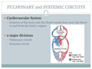
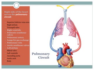
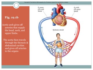
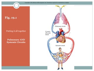
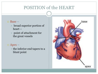
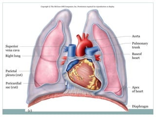
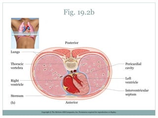
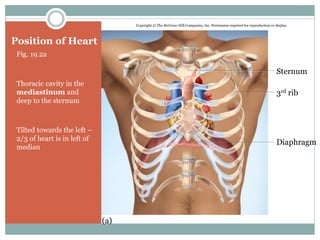
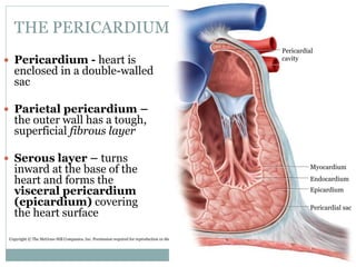
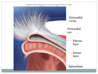
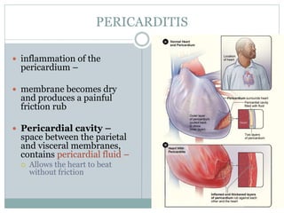
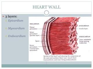
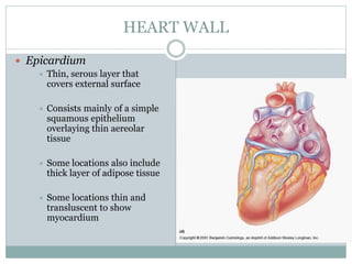
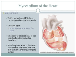
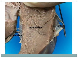
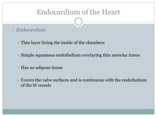
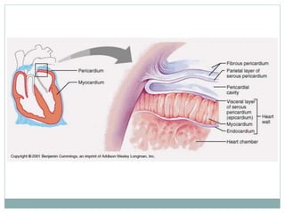
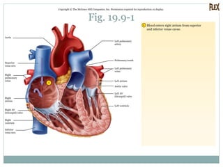
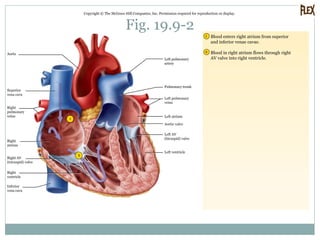
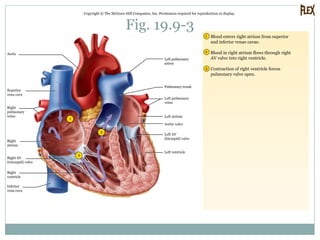
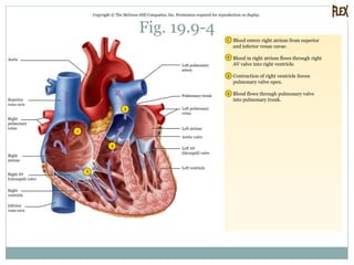
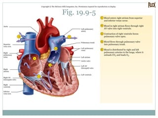
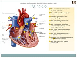
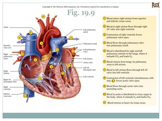
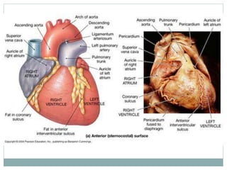
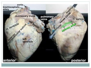
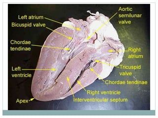
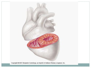
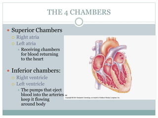
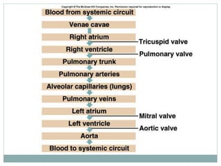
THE HEART
H E A T H E R L D R A K E
N E W Y O R K C H I R O P R A C T I C C O L L E G E , 2 0 1 3
S A L A D I N , “ U N I T Y O F F O R M A N D F U N C T I O N ”
The Circulatory System
OVERVIEW
 Objectives
 Define and distinguish between the pulmonary and systemic
circuits
 Describe the general location, size, and shape of the heart
 Describe the pericardial sac that encloses the heart
PULMONARY and SYSTEMIC CIRCUITS
 Cardiovascular System
 Consists of the heart and the blood vessels that carry the blood
to and from the body’s organs
 2 major divisions
 Pulmonary circuit
 Systemic circuit
2 DIVISIONS
 Pulmonary Circuit
 Carries blood to the lungs for gas exchange and returns it to the heart
 The right side of the heart serves the pulmonary circuit
 Receives blood that has circulated throughout the body, unloaded its oxygen
and nutrients, and picked up a load of carbon dioxide/other wastes.
 Pumps into the pulmonary trunk, which divides into right and left pulmonary
arteries, transporting into alveoli, carbon dioxide unloaded/oxygen loaded.
 Oxygen-rich blood then flows via pulmonary veins to the left side of the heart
 Systemic Circuit
 Supplies blood every organ of the body, including other parts of the
lungs and wall of the heart itself
Pulmonary
Circuit
 Right side of the heart
serves the pulmonary
circuit
 Superior/inferior vena cava
 Right atrium
 Tricuspid valve
 Right ventricle
 Pulmonic semilunar
valves
 Pulmonary artery
 Lungs for gas exchange
 Pulmonary vein
 Aortic semilunar valves
 Left atrium
 Mitral valve
 Left ventricle
 Ascending aorta
 Aortic arch
 Body
2 DIVISIONS
 Pulmonary Circuit
 Carries blood to the lungs for gas exchange and returns it to the
heart
 Systemic Circuit
 Supplies blood every organ, including wall of the heart itself
 Blood leaves the left side of the heart via the aorta, taking an
inverted U-turn, the aortic arch, and passes downward, dorsal to
the heart
Systemic Circuit
 Systemic Circuit
 Supplies blood every organ of the body, including other parts of
the lungs and wall of the heart itself
 Blood leaves the left side of the heart via the aorta, taking an
inverted U-turn, the aortic arch, and passes downward, dorsal to
the heart
Fig. 19.1b
Aortic arch gives off
arteries that supply
the head, neck, and
upper limbs.
The aorta then travels
through the thoracic &
abdominal cavities
and gives off arteries
to the organs
Copyright © The McGraw-Hill Companies, Inc. Permission required for reproduction or display.
O2-poor,
CO2-rich
blood
O2-rich,
CO2-poor
blood
Systemic circuit
O2
CO2
Systemic
Circuit
Supplies:
-- Every organ
-- Heart muscle
- Parts of the lungs
 Systemic Circuit
 After circulating through the body, the
now-deoxygenated systemic blood
returns to the right side of the heart
mainly via:
 Superior vena cava (draining the head,
neck, upper limbs and thoracic organs)
 Inferior vena cava (drains organs below
the diaphragm)
Fig. 19.1
Putting it all together
Pulmonary AND
Systemic Circuits
Copyright © The McGraw-Hill Companies, Inc. Permission required for reproduction or display.
O2
O2CO2
Pulmonary circuit
O2-poor,
CO2-rich
blood
O2-rich,
CO2-poor
blood
Systemic circuit
CO2
POSITION of the HEART
 Base –
 broad superior portion of
heart –
 point of attachment for
the great vessels
 Apex –
 the inferior end tapers to a
blunt point
Fig. 19.2c
Copyright © The McGraw-Hill Companies, Inc. Permission required for reproduction or display.
Superior
vena cava
Right lung
Parietal
pleura (cut)
Pericardial
sac (cut)
Aorta
Pulmonary
trunk
Baseof
heart
Apex
of heart
Diaphragm
(c)
Fig. 19.2b
Copyright © The McGraw-Hill Companies, Inc. Permission required for reproduction or display.
Posterior
Lungs
Pericardial
cavity
Left
ventricle
Interventricular
septum
Anterior
Sternum
Right
ventricle
Thoracic
vertebra
(b)
Position of Heart
Fig. 19.2a
Thoracic cavity in the
mediastinum and
deep to the sternum
Tilted towards the left –
2/3 of heart is in left of
median
Copyright © The McGraw-Hill Companies, Inc. Permission required for reproduction or display.
(a)
Sternum
3rd rib
Diaphragm
THE PERICARDIUM
 Pericardium - heart is
enclosed in a double-walled
sac
 Parietal pericardium –
the outer wall has a tough,
superficial fibrous layer
 Serous layer – turns
inward at the base of the
heart and forms the
visceral pericardium
(epicardium) covering
the heart surface
Fig. 19.3a
Copyright © The McGraw-Hill Companies, Inc. Permission required for reproduction or display.
Pericardial
cavity
Myocardium
Endocardium
Epicardium
Pericardial sac
Fig. 19.3b
Copyright © The McGraw-Hill Companies, Inc. Permission required for reproduction or display.
Pericardial
cavity
Pericardial
sac:
Fibrous
layer
Serous
layer
Epicardium
PERICARDITIS
 inflammation of the
pericardium –
 membrane becomes dry
and produces a painful
friction rub
 Pericardial cavity –
space between the parietal
and visceral membranes,
contains pericardial fluid –
 Allows the heart to beat
without friction
QUIZ
1. Distinguish between the pulmonary and systemic
circuits and state which part of the heart serves
each one
2. Make a two color sketch of the pericardium;
1. Use one color for the fibrous pericardium and another for the
serous pericardium and another for the serous pericardium
and show their relationship to the heart wall and pericardial
activity
GROSS ANATOMY OF THE HEART
 OBJECTIVES
 Describe the 3 layers of the heart wall
 Identify the four chambers of the heart
 Identify the surface features of the heart and correlate them with its
internal four-chambered anatomy
 Identify the four valves of the heart
 Trace the flow of blood through the four chambers of the heart and
adjacent blood vessels
 Describe the arteries that nourish the myocardium & veins that drain it
HEART WALL
 3 layers:
 Epicardium
 Myocardium
 Endocardium
HEART WALL
 Epicardium
 Thin, serous layer that
covers external surface
 Consists mainly of a simple
squamous epithelium
overlaying thin aereolar
tissue
 Some locations also include
thick layer of adipose tissue
 Some locations thin and
transluscent to show
myocardium
Myocardium of the Heart
 Myocardium
 Thick, muscular middle layer
– composed of cardiac muscle
 Thickest layer
 – performs the work of the
heart
 Thickness is proportional to the
workload on the individual
chambers
 Muscle spirals around the heart,
so when the ventricles contract,
they exhibit a twisting/wringing
motion
Endocardium of the Heart
 Endocardium
 Thin layer lining the inside of the chambers
 Simple squamous endothelium overlaying thin aereolar tissue
 Has no adipose tissue
 Covers the valve surfaces and is continuous with the endothelium
of the bl vessels
ANIMATION OF BLOOD FLOW THROUGH HEART
 http://www.wellesley.edu/Biology/Courses/111/Adu
ltHeart.gif
Fig. 19.9-1
Copyright © The McGraw-Hill Companies, Inc. Permission required for reproduction or display.
1
1 Blood enters right atrium from superior
and inferior venae cavae.
Aorta
Superior
vena cava
Right
pulmonary
veins
Right
atrium
Right AV
(tricuspid) valve
Right
ventricle
Inferior
vena cava
Left pulmonary
artery
Pulmonary trunk
Left pulmonary
veins
Left ventricle
Left AV
(bicuspid) valve
Aortic valve
Left atrium
Fig. 19.9-2
Copyright © The McGraw-Hill Companies, Inc. Permission required for reproduction or display.
2
1
2
1 Blood enters right atrium from superior
and inferior venae cavae.
Blood in right atrium flows through right
AV valve into right ventricle.
Aorta
Superior
vena cava
Right
pulmonary
veins
Right
atrium
Right AV
(tricuspid) valve
Right
ventricle
Inferior
vena cava
Left pulmonary
artery
Pulmonary trunk
Left pulmonary
veins
Left ventricle
Left AV
(bicuspid) valve
Aortic valve
Left atrium
Fig. 19.9-3
Copyright © The McGraw-Hill Companies, Inc. Permission required for reproduction or display.
3
2
1
2
3
1 Blood enters right atrium from superior
and inferior venae cavae.
Blood in right atrium flows through right
AV valve into right ventricle.
Contraction of right ventricle forces
pulmonary valve open.
Aorta
Superior
vena cava
Right
atrium
Right AV
(tricuspid) valve
Right
ventricle
Inferior
vena cava
Left pulmonary
artery
Pulmonary trunk
Left pulmonary
veins
Left ventricle
Left AV
(bicuspid) valve
Aortic valve
Left atrium
Right
pulmonary
veins
Fig. 19.9-4
Copyright © The McGraw-Hill Companies, Inc. Permission required for reproduction or display.
3
4
2
1
2
3
4
1 Blood enters right atrium from superior
and inferior venae cavae.
Blood in right atrium flows through right
AV valve into right ventricle.
Contraction of right ventricle forces
pulmonary valve open.
Blood flows through pulmonary valve
into pulmonary trunk.
Aorta
Superior
vena cava
Right
atrium
Right AV
(tricuspid) valve
Right
ventricle
Inferior
vena cava
Left pulmonary
artery
Pulmonary trunk
Left pulmonary
veins
Left ventricle
Left AV
(bicuspid) valve
Aortic valve
Left atrium
Right
pulmonary
veins
Fig. 19.9-5
Copyright © The McGraw-Hill Companies, Inc. Permission required for reproduction or display.
3
4
55
2
1
2
3
4
5
1 Blood enters right atrium from superior
and inferior venae cavae.
Blood in right atrium flows through right
AV valve into right ventricle.
Contraction of right ventricle forces
pulmonary valve open.
Blood flows through pulmonary valve
into pulmonary trunk.
Blood is distributed by right and left
pulmonary arteries to the lungs, where it
unloads CO2 and loads O2.
Aorta
Superior
vena cava
Right
atrium
Right AV
(tricuspid) valve
Right
ventricle
Inferior
vena cava
Left pulmonary
artery
Pulmonary trunk
Left pulmonary
veins
Aortic valve
Left atrium
Right
pulmonary
veins
Left ventricle
Left AV
(bicuspid) valve
Fig. 19.9-6
Copyright © The McGraw-Hill Companies, Inc. Permission required for reproduction or display.
3
6
4
55
2
1
6
2
3
4
5
6
1 Blood enters right atrium from superior
and inferior venae cavae.
Blood in right atrium flows through right
AV valve into right ventricle.
Contraction of right ventricle forces
pulmonary valve open.
Blood flows through pulmonary valve
into pulmonary trunk.
Blood is distributed by right and left
pulmonary arteries to the lungs, where it
unloads CO2 and loads O2.
Blood returns from lungs via pulmonary
veins to left atrium.
Aorta
Superior
vena cava
Right
pulmonary
veins
Right
atrium
Right AV
(tricuspid) valve
Right
ventricle
Inferior
vena cava
Left pulmonary
artery
Pulmonary trunk
Left pulmonary
veins
Left ventricle
Left AV
(bicuspid) valve
Aortic valve
Left atrium
Fig. 19.9-7
Copyright © The McGraw-Hill Companies, Inc. Permission required for reproduction or display.
3
6
4
55
2
1
7
6
2
3
4
5
6
7
1 Blood enters right atrium from superior
and inferior venae cavae.
Blood in right atrium flows through right
AV valve into right ventricle.
Contraction of right ventricle forces
pulmonary valve open.
Blood flows through pulmonary valve
into pulmonary trunk.
Blood is distributed by right and left
pulmonary arteries to the lungs, where it
unloads CO2 and loads O2.
Blood returns from lungs via pulmonary
veins to left atrium.
Blood in left atrium flows through left AV
valve into left ventricle.
Aorta
Superior
vena cava
Right
pulmonary
veins
Right
atrium
Right AV
(tricuspid) valve
Right
ventricle
Inferior
vena cava
Left pulmonary
artery
Pulmonary trunk
Left pulmonary
veins
Left ventricle
Left AV
(bicuspid) valve
Aortic valve
Left atrium
Fig. 19.9-8
Copyright © The McGraw-Hill Companies, Inc. Permission required for reproduction or display.
3
6
4
55
2
1
7
8
6
2
3
4
5
6
7
8
1 Blood enters right atrium from superior
and inferior venae cavae.
Blood in right atrium flows through right
AV valve into right ventricle.
Contraction of right ventricle forces
pulmonary valve open.
Blood flows through pulmonary valve
into pulmonary trunk.
Blood is distributed by right and left
pulmonary arteries to the lungs, where it
unloads CO2 and loads O2.
Blood returns from lungs via pulmonary
veins to left atrium.
Blood in left atrium flows through left AV
valve into left ventricle.
Aorta
Superior
vena cava
Right
pulmonary
veins
Right
atrium
Right AV
(tricuspid) valve
Right
ventricle
Left pulmonary
artery
Pulmonary trunk
Left pulmonary
veins
Left ventricle
Left AV
(bicuspid) valve
Aortic valve
Left atrium
Contraction of left ventricle (simultaneous with
step 3 ) forces aortic valve open.
Fig. 19.9-9
Copyright © The McGraw-Hill Companies, Inc. Permission required for reproduction or display.
3
6
4
55
2
1
7
8
9
6
2
3
4
5
6
7
8
9
1 Blood enters right atrium from superior
and inferior venae cavae.
Blood in right atrium flows through right
AV valve into right ventricle.
Contraction of right ventricle forces
pulmonary valve open.
Blood flows through pulmonary valve
into pulmonary trunk.
Blood is distributed by right and left
pulmonary arteries to the lungs, where it
unloads CO2 and loads O2.
Blood returns from lungs via pulmonary
veins to left atrium.
Blood in left atrium flows through left AV
valve into left ventricle.
Blood flows through aortic valve into
ascending aorta.
Aorta
Superior
vena cava
Right
pulmonary
veins
Right
atrium
Right AV
(tricuspid) valve
Right
ventricle
Left pulmonary
artery
Pulmonary trunk
Left pulmonary
veins
Left ventricle
Left AV
(bicuspid) valve
Aortic valve
Left atrium
Contraction of left ventricle (simultaneous with
step 3 ) forces aortic valve open.
Fig. 19.9-10
Copyright © The McGraw-Hill Companies, Inc. Permission required for reproduction or display.
3
6
4
55
2
1
7
8
9
6
2
3
4
5
6
7
8
9
1 0
1 Blood enters right atrium from superior
and inferior venae cavae.
Blood in right atrium flows through right
AV valve into right ventricle.
Contraction of right ventricle forces
pulmonary valve open.
Blood flows through pulmonary valve
into pulmonary trunk.
Blood is distributed by right and left
pulmonary arteries to the lungs, where it
unloads CO2 and loads O2.
Blood returns from lungs via pulmonary
veins to left atrium.
Blood in left atrium flows through left AV
valve into left ventricle.
Blood flows through aortic valve into
ascending aorta.
Blood in aorta is distributed to every organ in
the body, where it unloads O2 and loads CO2.
Aorta
Superior
vena cava
Right
pulmonary
veins
Right
atrium
Right AV
(tricuspid) valve
Right
ventricle
10
Left pulmonary
artery
Pulmonary trunk
Left pulmonary
veins
Left ventricle
Left AV
(bicuspid) valve
Aortic valve
Left atrium
Contraction of left ventricle (simultaneous with
step 3 ) forces aortic valve open.
Fig. 19.9
Copyright © The McGraw-Hill Companies, Inc. Permission required for reproduction or display.
3
6
4
55
2
1
7
8
9
6
2
3
4
5
6
7
8
9
1 0
1 Blood enters right atrium from superior
and inferior venae cavae.
Blood in right atrium flows through right
AV valve into right ventricle.
Contraction of right ventricle forces
pulmonary valve open.
Blood flows through pulmonary valve
into pulmonary trunk.
Blood is distributed by right and left
pulmonary arteries to the lungs, where it
unloads CO2 and loads O2.
Blood returns from lungs via pulmonary
veins to left atrium.
Blood in left atrium flows through left AV
valve into left ventricle.
Blood flows through aortic valve into
ascending aorta.
Blood in aorta is distributed to every organ in
the body, where it unloads O2 and loads CO2.
Blood returns to heart via venae cavae.
Aorta
Superior
vena cava
Right
pulmonary
veins
Right
atrium
Right AV
(tricuspid) valve
Right
ventricle
Inferior
vena cava 11
10
11
Left pulmonary
artery
Pulmonary trunk
Left pulmonary
veins
Left ventricle
Left AV
(bicuspid) valve
Aortic valve
Left atrium
11
Contraction of left ventricle (simultaneous with
step 3 ) forces aortic valve open.
HEART – FIBROUS SKELETON
 Has a meshwork of collagenous and elastic fibers that
make up a fibrous skeleton
 Fibrous rings have multiple functions:
 Structural support for heart, esp around valves and openings of
great vessels
 Anchors the myocytes and gives them something to pull against
 Serves as electrical insulation b/w atria and ventricles, so the atria
cannot stimulate the ventricles directly
 Elastic recoil aids in refilling the heart
THE 4 CHAMBERS
 Superior Chambers
 Right atria
 Left atria
 Receiving chambers
for blood returning
to the heart
 Inferior chambers:
 Right ventricle
 Left ventricle
 The pumps that eject
blood into the arteries
keep it flowing
around body
VALVES – Atrioventricular Valves
 A). Atrioventricular (AV)
Valves
 Located between the atria
and the ventricles
 Flaps consist of connective
tissue & endocardium.
 They are attached to
collagen cords
called chordae tendinae.
 1). Tricuspid
 2). Bicuspid or Mitral
Valve
VALVES – Semilunar Valves
 B). Semilunar (SL)
Valves
 Lead into the large
arteries
 Formed from 3 moon-
shaped cusps
 1). Aortic Semilunar Valve
 2). Pulmonary Semilunar
Valve
BLOOD FLOW – AV Valves - STEP 1
 When the heart is relaxed, the AV flaps hang open
BLOOD FLOW – AV Valves – STEP 2
 When the ventricles contract the AV valves
are pressed closed from the ventricular side.
 Blood is pushed
against the AV
valve which closes
and it pushes
against the SL
valve which opens
BLOOD FLOW – Semilunar Valves - STEP 1
 When the ventricle
relaxes blood pressure
is higher in the vessels,
which closes the SL
valve
BLOOD FLOW – Semilunar Valves – STEP 2
BLOOD FLOW THROUGH THE HEART
1. Blood enters right atrium from superior and inferior
venae cavae
2. Blood in the right atrium flows through right AV valve
into the right ventricle
3. Contraction of right ventricular forces pulmonary valve
open
4. Blood flows through pulmonary valve into pulmonary
trunk
5. Blood is distributed by right and left pulmonary
arteries to the lungs, where it unloads CO2 and loads
O2
6. Blood return from lungs via pulmonary arteries to left
atrium
7. Blood in left atrium flows through left AV valve into left
ventricle
8. Contraction of left ventricle (simultaneous with step 3)
forces aortic valve open
BLOOD FLOW THROUGH THE HEART
9. Blood flows through the aortic valve into ascending
aorta
10. Blood in aorta is distributed into every organ in the
body, where it unloads O2 and loads CO2
11. Blood returns to heart via venae cavae
BLOOD FLOW THROUGH THE HEART
QUIZ TIME
 Trace the pathway of a single erythrocyte from the
inferior vena cava to the aortic arch, naming each
vessel and structure it passes through, and noting
when it is oxygenated, and when it is deoxygenated.
CORONARY ARTERY BLOOD FLOW
QUIZ QUESTION
 Trace the path of a single erythrocyte from the aorta to the
posterior apex of the heart, if the right coronary artery is
blocked at just after its attachment to the aorta?
 How would the left anterior portion of the heart receive
blood supply with the same blockage?
 This theory is called COLLATERAL CIRCULATION
 The flow of blood to an area via an alternative route via vessel anastomoses
CLINICAL CORRELATION
 What if multiple coronary arteries were clogged? So
much that there was not enough blood to fuel the heart?
 Angina pectoris
 chest pain/pressure due to temporary ischemia
 deficiency of blood to cardiac muscle
 O2 deprived myocardium shits to anaerobic fermentation, producing
lactic acid, which stimulates pain receptors in the heart.
 MYOCARDIAL INFARCTION (MI) – heart attack
VENOUS DRAINAGE
 Refers to the route by which blood leaves an organ.
 20% of coronary blood empties directly into multiple
small thebesian veins into the right atrium/ventricle
VENOUS DRAINAGE
 80% returns to the right atrium via
this route:
 Great cardiac vein –
 Collects blood from anterior heart,
travels w.anterior interventric artery
 Carries blood from apex to coronary
sulcus, arcs left, empties into coronary
sinus
 Posterior interventricular (middle
cardiac) vein
 Collects bl from posterior aspect of heart
 Left marginal vein
 Travels from apex up left margin,
empties into coronary sinus
 Coronary sinus
 Large transverse vein in coronary sulcus,
empties blood into right atrium
QUIZ
3. Name the three layers of the heart and describe their
structural differences
4. What are the functions of the fibrous skeleton?
5. Trace the flow of blood through the heart, naming each
chamber and valve in order
6. What are the three principle branches of the left
coronary artery? Where are they located on the heart
surface? What are the branches of the right coronary
artery, and where are they located?
7. What is the medical significance of the
anastamoses in the coronary arterial system?
8. Why do the coronary arteries carry a greater blood
flow during ventricular diastole than they do
during ventricular systole?
9. What are the three major veins that empty into the
coronary sinus?
QUIZ
CARDIAC CONDUCTION SYSTEM
and CARDIAC MUSCLE
 OBJECTIVES:
 Describe the nerve supply to the heart
 Describe the internal electrical system of the heart
 Describe the unique structural and metabolic characterisitic of
cardiac muscle
 Explain the nature and functional significance of the
intercellular junctions between cardiac muscle cells
INTRO
 Heart contracts at regular intervals (obviously)
 Typically 75bpm
 Invertebrates (clames, crabs, insects, etc) have
heartbeat triggered by a pacemaker in the nervous
system
 Vertebrates (us) is myogenic
 Signal originates within the heart itself
NERVE SUPPLY TO THE HEART
 Heart has its own pacemaker, but it does receives
sympathetic and parasympathetic nerves, modifying
the heart rate and contraction strength
 Sympathetic stimulation may raise HR to 230 bpm.
 Parasympathetic stimulation can slow the HR as low
as 20 bpm, or even stop for a few seconds.
NERVE SUPPLY TO THE HEART
 Sympathetic pathway
originates w.neurons in
the lower cervical/upper
thoracic spinal cord
 Efferent fibers from these neurons pass from the
spinal cord to the sympathetic chain and travel up
the chain to the 3 cervical ganglia
 Cardiac nerves arise from these ganglia, and lead
to the myocardium, increasing force of contraction
NERVE SUPPLY TO THE HEART
 Some fibers innervate the atria
 Sympathetic fibers to the coronary arteries dilate them and increase coronary
blood flow to the coronary arteries during exercise
 Parasympathetic pathway to the heart is through the vagus nerves
 RIGHT VAGUS nerve innervates mainly an electrical center of the heart –
SA node
 LEFT VAGUS nerve innervates the AV node
 Ventricles receive little or no vagal stimulation
 Vagus nerve slows the heartbeat.
 W.o this influence, the average resting HR would be 100 bpm
 Steady firing of the vagus nerve, VAGAL TONE, holds resting HR down to 70-80 bpm
 IN SHORT – Sympathetic nerves do NOT make the heart beat, they only MODIFY it.
CONDUCTION ANIMATION
 http://en.wikipedia.org/wiki/File:ECG_Principle_fa
st.gif
Fig. 19.12-1Copyright © The McGraw-Hill Companies, Inc. Permission required for reproduction or display.
1
1
SA node fires.
Purkinje fibers
Atrioventricular
bundle
Atrioventricular
node
Sinoatrial node
(pacemaker)
Right atrium
Bundle
branches
Left
atrium
Purkinje
fibers
Fig. 19.12-2Copyright © The McGraw-Hill Companies, Inc. Permission required for reproduction or display.
2
1
1
2
2
SA node fires.
Excitation spreads through
atrial myocardium.
Purkinje fibers
Atrioventricular
bundle
Atrioventricular
node
Sinoatrial node
(pacemaker)
Right atrium
Bundle
branches
Left
atrium
Purkinje
fibers
Fig. 19.12-3Copyright © The McGraw-Hill Companies, Inc. Permission required for reproduction or display.
2
3
1
1
2
3
2
SA node fires.
Excitation spreads through
atrial myocardium.
AV node fires.
Purkinje fibers
Atrioventricular
bundle
Atrioventricular
node
Sinoatrial node
(pacemaker)
Right atrium
Bundle
branches
Left
atrium
Purkinje
fibers
Fig. 19.12-4Copyright © The McGraw-Hill Companies, Inc. Permission required for reproduction or display.
2
3
4
1
1
2
3
4
2
SA node fires.
Excitation spreads through
atrial myocardium.
AV node fires.
Excitation spreads down AV
bundle.
Purkinje fibers
Atrioventricular
bundle
Atrioventricular
node
Sinoatrial node
(pacemaker)
Right atrium
Bundle
branches
Left
atrium
Purkinje
fibers
Fig. 19.12Copyright © The McGraw-Hill Companies, Inc. Permission required for reproduction or display.
2
3
4
5
1
1
2
3
4 5
2
SA node fires.
Excitation spreads through
atrial myocardium.
AV node fires.
Excitation spreads down AV
bundle.
Purkinje fibers distribute
excitation through
ventricular myocardium.
Purkinje fibers
Atrioventricular
bundle
Atrioventricular
node
Sinoatrial node
(pacemaker)
Right atrium
Bundle
branches
Left
atrium
Purkinje
fibers
Fig. 19.13Copyright © The McGraw-Hill Companies, Inc. Permission required for reproduction or display.
0
0 .8.4
Membranepotential(mV)
+10
–10
–20
–30
–40
–50
–60
–70
1.61.2
Time (seconds)
Pacemaker
potential
Action
potential
Threshold
K+
outflow
Slow Na+
inflow
Ca2+–Na+
inflow
Fig. 19.14-1
0
Voltage-gated Na+ channels open.1
1
Membranepotential(mV)
0
+20
–20
–40
–60
–80
.30
Time (seconds)
.15
Absolute
refractory
period
Copyright © The McGraw-Hill Companies, Inc. Permission required for reproduction or display.
Fig. 19.14-2
0
Voltage-gated Na+ channels open.
Na+ inflow depolarizes the membrane
and triggers the opening of still more Na+
channels, creating a positive feedback
cycle and a rapidly rising membrane voltage.
1
2
1
2
Membranepotential(mV)
0
+20
–20
–40
–60
–80
.30
Time (seconds)
.15
Absolute
refractory
period
Myocardial
contraction
Copyright © The McGraw-Hill Companies, Inc. Permission required for reproduction or display.
Fig. 19.14-3
0
Voltage-gated Na+ channels open.
Na+ inflow depolarizes the membrane
and triggers the opening of still more Na+
channels, creating a positive feedback
cycle and a rapidly rising membrane voltage.
Na+ channels close when the cell
depolarizes, and the voltage peaks at
nearly +30 mV.
1
2
3
1
2
3
Membranepotential(mV)
0
+20
–20
–40
–60
–80
.30
Time (seconds)
.15
Absolute
refractory
period
Myocardial
contraction
Action
potential
Copyright © The McGraw-Hill Companies, Inc. Permission required for reproduction or display.
Fig. 19.14-4
0
Voltage-gated Na+ channels open.
Na+ inflow depolarizes the membrane
and triggers the opening of still more Na+
channels, creating a positive feedback
cycle and a rapidly rising membrane voltage.
Na+ channels close when the cell
depolarizes, and the voltage peaks at
nearly +30 mV.
Ca2+ entering through slow Ca2+
channels prolongs depolarization of
membrane, creating a plateau. Plateau falls
slightly because of some K+ leakage, but most
K+ channels remain closed until end of
plateau.
1
2
3
4
1
2
3
4
Membranepotential(mV)
0
+20
–20
–40
–60
–80
Plateau
.30
Time (seconds)
.15
Absolute
refractory
period
Myocardial
contraction
Action
potential
Copyright © The McGraw-Hill Companies, Inc. Permission required for reproduction or display.
Fig. 19.14
0
Voltage-gated Na+ channels open.
Na+ inflow depolarizes the membrane
and triggers the opening of still more Na+
channels, creating a positive feedback
cycle and a rapidly rising membrane voltage.
Na+ channels close when the cell
depolarizes, and the voltage peaks at
nearly +30 mV.
Ca2+ entering through slow Ca2+
channels prolongs depolarization of
membrane, creating a plateau. Plateau falls
slightly because of some K+ leakage, but most
K+ channels remain closed until end of
plateau.
Ca2+ channels close and Ca2+ is transported
out of cell. K+ channels open, and rapid K+
outflow returns membrane to its resting
potential.
1
2
3
4
5
1
2
3
4
5
Membranepotential(mV)
0
+20
–20
–40
–60
–80
Plateau
Myocardial
relaxation
.30
Time (seconds)
.15
Absolute
refractory
period
Myocardial
contraction
Action
potential
Copyright © The McGraw-Hill Companies, Inc. Permission required for reproduction or display.
Fig. 19.15a
Copyright © The McGraw-Hill Companies, Inc. Permission required for reproduction or display.
0.8 second
Fig. 19.15b
Copyright © The McGraw-Hill Companies, Inc. Permission required for reproduction or display.
0
Millivolts
R R
Q
S
+1
–1
Atria
contract
Ventricles
contract
Atria
contract
Ventricles
contract
ST
segment
PQ
segment
QRS interval
T waveP wave
PR
interval
QT
interval
Fig. 19.15
Copyright © The McGraw-Hill Companies, Inc. Permission required for reproduction or display.
0
Millivolts
R R
Q
S
0.8 second
+1
–1
Atria
contract
Ventricles
contract
Atria
contract
Ventricles
contract
ST
segment
PQ
segment
QRS interval
T waveP wave
PR
interval
QT
interval
Absolute versus Relative Refractory
Fig. 19.16-1
Copyright © The McGraw-Hill Companies, Inc. Permission required for reproduction or display.
1
P
Wave of
depolarization
Wave of
repolarization
Atria begin depolarizing.
Key
Fig. 19.16-2
Copyright © The McGraw-Hill Companies, Inc. Permission required for reproduction or display.
1
2
P
P
Wave of
depolarization
Wave of
repolarization
Atria begin depolarizing.
Atrial depolarization complete.
Key
Fig. 19.16-3
Copyright © The McGraw-Hill Companies, Inc. Permission required for reproduction or display.
1
2
Q
R
P
P
P
3
Wave of
depolarization
Wave of
repolarization
Atria begin depolarizing.
Atrial depolarization complete.
Ventricular depolarization begins at apex and
progresses superiorly as atria repolarize.
Key
Fig. 19.16-4
Copyright © The McGraw-Hill Companies, Inc. Permission required for reproduction or display.
1 4
2
S
Q
R
P
Q
R
P
P
P
3
Wave of
depolarization
Wave of
repolarization
Atria begin depolarizing.
Atrial depolarization complete.
Ventricular depolarization begins at apex and
progresses superiorly as atria repolarize.
Ventricular depolarization complete.
Key
Fig. 19.16-5
Copyright © The McGraw-Hill Companies, Inc. Permission required for reproduction or display.
5
1 4
2
T
S
Q
R
P
S
Q
R
P
Q
R
P
P
P
3
Wave of
depolarization
Wave of
repolarization
Atria begin depolarizing.
Atrial depolarization complete.
Ventricular depolarization begins at apex and
progresses superiorly as atria repolarize.
Ventricular repolarization begins at apex and
progresses superiorly.
Ventricular depolarization complete.
Key
Fig. 19.16
Copyright © The McGraw-Hill Companies, Inc. Permission required for reproduction or display.
5
6
1 4
2
T
S
Q
R
P
T
S
Q
R
P
S
Q
R
P
Q
R
P
P
P
3
Wave of
depolarization
Wave of
repolarization
Atria begin depolarizing.
Atrial depolarization complete.
Ventricular depolarization begins at apex and
progresses superiorly as atria repolarize.
Ventricular repolarization complete; heart is
ready for the next cycle.
Ventricular repolarization begins at apex and
progresses superiorly.
Ventricular depolarization complete.
Key
So why does this matter?
Table 19.1
SOURCES
 http://legacy.owensboro.kctcs.edu/gcaplan/anat2/notes/APIINotes
5%20Anatomy%20of%20the%20Heart.htm
 http://www.nhlbi.nih.gov/health/health-
topics/images/pericarditis.jpg
 http://o.quizlet.com/i/MYYoBudMtF4Nd4VX8VXZ5w_m.jpg
 http://stevegallik.org/sites/histologyolm.stevegallik.org/images/he
artwall.gif
 Saladin, K. “Anatomy and Physiology: The Unity of Form and
Function.”4th ed. Philadelphia. McGraw Hill Publishing.
 http://www.unm.edu/~jimmy/refractory_periods.jpg

Circulatory System 19.1-19.2

  • 1.
    THE HEART H EA T H E R L D R A K E N E W Y O R K C H I R O P R A C T I C C O L L E G E , 2 0 1 3 S A L A D I N , “ U N I T Y O F F O R M A N D F U N C T I O N ” The Circulatory System
  • 2.
    OVERVIEW  Objectives  Defineand distinguish between the pulmonary and systemic circuits  Describe the general location, size, and shape of the heart  Describe the pericardial sac that encloses the heart
  • 3.
    PULMONARY and SYSTEMICCIRCUITS  Cardiovascular System  Consists of the heart and the blood vessels that carry the blood to and from the body’s organs  2 major divisions  Pulmonary circuit  Systemic circuit
  • 4.
    2 DIVISIONS  PulmonaryCircuit  Carries blood to the lungs for gas exchange and returns it to the heart  The right side of the heart serves the pulmonary circuit  Receives blood that has circulated throughout the body, unloaded its oxygen and nutrients, and picked up a load of carbon dioxide/other wastes.  Pumps into the pulmonary trunk, which divides into right and left pulmonary arteries, transporting into alveoli, carbon dioxide unloaded/oxygen loaded.  Oxygen-rich blood then flows via pulmonary veins to the left side of the heart  Systemic Circuit  Supplies blood every organ of the body, including other parts of the lungs and wall of the heart itself
  • 5.
    Pulmonary Circuit  Right sideof the heart serves the pulmonary circuit  Superior/inferior vena cava  Right atrium  Tricuspid valve  Right ventricle  Pulmonic semilunar valves  Pulmonary artery  Lungs for gas exchange  Pulmonary vein  Aortic semilunar valves  Left atrium  Mitral valve  Left ventricle  Ascending aorta  Aortic arch  Body
  • 6.
    2 DIVISIONS  PulmonaryCircuit  Carries blood to the lungs for gas exchange and returns it to the heart  Systemic Circuit  Supplies blood every organ, including wall of the heart itself  Blood leaves the left side of the heart via the aorta, taking an inverted U-turn, the aortic arch, and passes downward, dorsal to the heart
  • 7.
    Systemic Circuit  SystemicCircuit  Supplies blood every organ of the body, including other parts of the lungs and wall of the heart itself  Blood leaves the left side of the heart via the aorta, taking an inverted U-turn, the aortic arch, and passes downward, dorsal to the heart
  • 8.
    Fig. 19.1b Aortic archgives off arteries that supply the head, neck, and upper limbs. The aorta then travels through the thoracic & abdominal cavities and gives off arteries to the organs Copyright © The McGraw-Hill Companies, Inc. Permission required for reproduction or display. O2-poor, CO2-rich blood O2-rich, CO2-poor blood Systemic circuit O2 CO2
  • 9.
    Systemic Circuit Supplies: -- Every organ --Heart muscle - Parts of the lungs  Systemic Circuit  After circulating through the body, the now-deoxygenated systemic blood returns to the right side of the heart mainly via:  Superior vena cava (draining the head, neck, upper limbs and thoracic organs)  Inferior vena cava (drains organs below the diaphragm)
  • 10.
    Fig. 19.1 Putting itall together Pulmonary AND Systemic Circuits Copyright © The McGraw-Hill Companies, Inc. Permission required for reproduction or display. O2 O2CO2 Pulmonary circuit O2-poor, CO2-rich blood O2-rich, CO2-poor blood Systemic circuit CO2
  • 11.
    POSITION of theHEART  Base –  broad superior portion of heart –  point of attachment for the great vessels  Apex –  the inferior end tapers to a blunt point
  • 12.
    Fig. 19.2c Copyright ©The McGraw-Hill Companies, Inc. Permission required for reproduction or display. Superior vena cava Right lung Parietal pleura (cut) Pericardial sac (cut) Aorta Pulmonary trunk Baseof heart Apex of heart Diaphragm (c)
  • 13.
    Fig. 19.2b Copyright ©The McGraw-Hill Companies, Inc. Permission required for reproduction or display. Posterior Lungs Pericardial cavity Left ventricle Interventricular septum Anterior Sternum Right ventricle Thoracic vertebra (b)
  • 14.
    Position of Heart Fig.19.2a Thoracic cavity in the mediastinum and deep to the sternum Tilted towards the left – 2/3 of heart is in left of median Copyright © The McGraw-Hill Companies, Inc. Permission required for reproduction or display. (a) Sternum 3rd rib Diaphragm
  • 15.
    THE PERICARDIUM  Pericardium- heart is enclosed in a double-walled sac  Parietal pericardium – the outer wall has a tough, superficial fibrous layer  Serous layer – turns inward at the base of the heart and forms the visceral pericardium (epicardium) covering the heart surface Fig. 19.3a Copyright © The McGraw-Hill Companies, Inc. Permission required for reproduction or display. Pericardial cavity Myocardium Endocardium Epicardium Pericardial sac
  • 16.
    Fig. 19.3b Copyright ©The McGraw-Hill Companies, Inc. Permission required for reproduction or display. Pericardial cavity Pericardial sac: Fibrous layer Serous layer Epicardium
  • 17.
    PERICARDITIS  inflammation ofthe pericardium –  membrane becomes dry and produces a painful friction rub  Pericardial cavity – space between the parietal and visceral membranes, contains pericardial fluid –  Allows the heart to beat without friction
  • 18.
    QUIZ 1. Distinguish betweenthe pulmonary and systemic circuits and state which part of the heart serves each one 2. Make a two color sketch of the pericardium; 1. Use one color for the fibrous pericardium and another for the serous pericardium and another for the serous pericardium and show their relationship to the heart wall and pericardial activity
  • 19.
    GROSS ANATOMY OFTHE HEART  OBJECTIVES  Describe the 3 layers of the heart wall  Identify the four chambers of the heart  Identify the surface features of the heart and correlate them with its internal four-chambered anatomy  Identify the four valves of the heart  Trace the flow of blood through the four chambers of the heart and adjacent blood vessels  Describe the arteries that nourish the myocardium & veins that drain it
  • 20.
    HEART WALL  3layers:  Epicardium  Myocardium  Endocardium
  • 21.
    HEART WALL  Epicardium Thin, serous layer that covers external surface  Consists mainly of a simple squamous epithelium overlaying thin aereolar tissue  Some locations also include thick layer of adipose tissue  Some locations thin and transluscent to show myocardium
  • 22.
    Myocardium of theHeart  Myocardium  Thick, muscular middle layer – composed of cardiac muscle  Thickest layer  – performs the work of the heart  Thickness is proportional to the workload on the individual chambers  Muscle spirals around the heart, so when the ventricles contract, they exhibit a twisting/wringing motion
  • 24.
    Endocardium of theHeart  Endocardium  Thin layer lining the inside of the chambers  Simple squamous endothelium overlaying thin aereolar tissue  Has no adipose tissue  Covers the valve surfaces and is continuous with the endothelium of the bl vessels
  • 26.
    ANIMATION OF BLOODFLOW THROUGH HEART  http://www.wellesley.edu/Biology/Courses/111/Adu ltHeart.gif
  • 27.
    Fig. 19.9-1 Copyright ©The McGraw-Hill Companies, Inc. Permission required for reproduction or display. 1 1 Blood enters right atrium from superior and inferior venae cavae. Aorta Superior vena cava Right pulmonary veins Right atrium Right AV (tricuspid) valve Right ventricle Inferior vena cava Left pulmonary artery Pulmonary trunk Left pulmonary veins Left ventricle Left AV (bicuspid) valve Aortic valve Left atrium
  • 28.
    Fig. 19.9-2 Copyright ©The McGraw-Hill Companies, Inc. Permission required for reproduction or display. 2 1 2 1 Blood enters right atrium from superior and inferior venae cavae. Blood in right atrium flows through right AV valve into right ventricle. Aorta Superior vena cava Right pulmonary veins Right atrium Right AV (tricuspid) valve Right ventricle Inferior vena cava Left pulmonary artery Pulmonary trunk Left pulmonary veins Left ventricle Left AV (bicuspid) valve Aortic valve Left atrium
  • 29.
    Fig. 19.9-3 Copyright ©The McGraw-Hill Companies, Inc. Permission required for reproduction or display. 3 2 1 2 3 1 Blood enters right atrium from superior and inferior venae cavae. Blood in right atrium flows through right AV valve into right ventricle. Contraction of right ventricle forces pulmonary valve open. Aorta Superior vena cava Right atrium Right AV (tricuspid) valve Right ventricle Inferior vena cava Left pulmonary artery Pulmonary trunk Left pulmonary veins Left ventricle Left AV (bicuspid) valve Aortic valve Left atrium Right pulmonary veins
  • 30.
    Fig. 19.9-4 Copyright ©The McGraw-Hill Companies, Inc. Permission required for reproduction or display. 3 4 2 1 2 3 4 1 Blood enters right atrium from superior and inferior venae cavae. Blood in right atrium flows through right AV valve into right ventricle. Contraction of right ventricle forces pulmonary valve open. Blood flows through pulmonary valve into pulmonary trunk. Aorta Superior vena cava Right atrium Right AV (tricuspid) valve Right ventricle Inferior vena cava Left pulmonary artery Pulmonary trunk Left pulmonary veins Left ventricle Left AV (bicuspid) valve Aortic valve Left atrium Right pulmonary veins
  • 31.
    Fig. 19.9-5 Copyright ©The McGraw-Hill Companies, Inc. Permission required for reproduction or display. 3 4 55 2 1 2 3 4 5 1 Blood enters right atrium from superior and inferior venae cavae. Blood in right atrium flows through right AV valve into right ventricle. Contraction of right ventricle forces pulmonary valve open. Blood flows through pulmonary valve into pulmonary trunk. Blood is distributed by right and left pulmonary arteries to the lungs, where it unloads CO2 and loads O2. Aorta Superior vena cava Right atrium Right AV (tricuspid) valve Right ventricle Inferior vena cava Left pulmonary artery Pulmonary trunk Left pulmonary veins Aortic valve Left atrium Right pulmonary veins Left ventricle Left AV (bicuspid) valve
  • 32.
    Fig. 19.9-6 Copyright ©The McGraw-Hill Companies, Inc. Permission required for reproduction or display. 3 6 4 55 2 1 6 2 3 4 5 6 1 Blood enters right atrium from superior and inferior venae cavae. Blood in right atrium flows through right AV valve into right ventricle. Contraction of right ventricle forces pulmonary valve open. Blood flows through pulmonary valve into pulmonary trunk. Blood is distributed by right and left pulmonary arteries to the lungs, where it unloads CO2 and loads O2. Blood returns from lungs via pulmonary veins to left atrium. Aorta Superior vena cava Right pulmonary veins Right atrium Right AV (tricuspid) valve Right ventricle Inferior vena cava Left pulmonary artery Pulmonary trunk Left pulmonary veins Left ventricle Left AV (bicuspid) valve Aortic valve Left atrium
  • 33.
    Fig. 19.9-7 Copyright ©The McGraw-Hill Companies, Inc. Permission required for reproduction or display. 3 6 4 55 2 1 7 6 2 3 4 5 6 7 1 Blood enters right atrium from superior and inferior venae cavae. Blood in right atrium flows through right AV valve into right ventricle. Contraction of right ventricle forces pulmonary valve open. Blood flows through pulmonary valve into pulmonary trunk. Blood is distributed by right and left pulmonary arteries to the lungs, where it unloads CO2 and loads O2. Blood returns from lungs via pulmonary veins to left atrium. Blood in left atrium flows through left AV valve into left ventricle. Aorta Superior vena cava Right pulmonary veins Right atrium Right AV (tricuspid) valve Right ventricle Inferior vena cava Left pulmonary artery Pulmonary trunk Left pulmonary veins Left ventricle Left AV (bicuspid) valve Aortic valve Left atrium
  • 34.
    Fig. 19.9-8 Copyright ©The McGraw-Hill Companies, Inc. Permission required for reproduction or display. 3 6 4 55 2 1 7 8 6 2 3 4 5 6 7 8 1 Blood enters right atrium from superior and inferior venae cavae. Blood in right atrium flows through right AV valve into right ventricle. Contraction of right ventricle forces pulmonary valve open. Blood flows through pulmonary valve into pulmonary trunk. Blood is distributed by right and left pulmonary arteries to the lungs, where it unloads CO2 and loads O2. Blood returns from lungs via pulmonary veins to left atrium. Blood in left atrium flows through left AV valve into left ventricle. Aorta Superior vena cava Right pulmonary veins Right atrium Right AV (tricuspid) valve Right ventricle Left pulmonary artery Pulmonary trunk Left pulmonary veins Left ventricle Left AV (bicuspid) valve Aortic valve Left atrium Contraction of left ventricle (simultaneous with step 3 ) forces aortic valve open.
  • 35.
    Fig. 19.9-9 Copyright ©The McGraw-Hill Companies, Inc. Permission required for reproduction or display. 3 6 4 55 2 1 7 8 9 6 2 3 4 5 6 7 8 9 1 Blood enters right atrium from superior and inferior venae cavae. Blood in right atrium flows through right AV valve into right ventricle. Contraction of right ventricle forces pulmonary valve open. Blood flows through pulmonary valve into pulmonary trunk. Blood is distributed by right and left pulmonary arteries to the lungs, where it unloads CO2 and loads O2. Blood returns from lungs via pulmonary veins to left atrium. Blood in left atrium flows through left AV valve into left ventricle. Blood flows through aortic valve into ascending aorta. Aorta Superior vena cava Right pulmonary veins Right atrium Right AV (tricuspid) valve Right ventricle Left pulmonary artery Pulmonary trunk Left pulmonary veins Left ventricle Left AV (bicuspid) valve Aortic valve Left atrium Contraction of left ventricle (simultaneous with step 3 ) forces aortic valve open.
  • 36.
    Fig. 19.9-10 Copyright ©The McGraw-Hill Companies, Inc. Permission required for reproduction or display. 3 6 4 55 2 1 7 8 9 6 2 3 4 5 6 7 8 9 1 0 1 Blood enters right atrium from superior and inferior venae cavae. Blood in right atrium flows through right AV valve into right ventricle. Contraction of right ventricle forces pulmonary valve open. Blood flows through pulmonary valve into pulmonary trunk. Blood is distributed by right and left pulmonary arteries to the lungs, where it unloads CO2 and loads O2. Blood returns from lungs via pulmonary veins to left atrium. Blood in left atrium flows through left AV valve into left ventricle. Blood flows through aortic valve into ascending aorta. Blood in aorta is distributed to every organ in the body, where it unloads O2 and loads CO2. Aorta Superior vena cava Right pulmonary veins Right atrium Right AV (tricuspid) valve Right ventricle 10 Left pulmonary artery Pulmonary trunk Left pulmonary veins Left ventricle Left AV (bicuspid) valve Aortic valve Left atrium Contraction of left ventricle (simultaneous with step 3 ) forces aortic valve open.
  • 37.
    Fig. 19.9 Copyright ©The McGraw-Hill Companies, Inc. Permission required for reproduction or display. 3 6 4 55 2 1 7 8 9 6 2 3 4 5 6 7 8 9 1 0 1 Blood enters right atrium from superior and inferior venae cavae. Blood in right atrium flows through right AV valve into right ventricle. Contraction of right ventricle forces pulmonary valve open. Blood flows through pulmonary valve into pulmonary trunk. Blood is distributed by right and left pulmonary arteries to the lungs, where it unloads CO2 and loads O2. Blood returns from lungs via pulmonary veins to left atrium. Blood in left atrium flows through left AV valve into left ventricle. Blood flows through aortic valve into ascending aorta. Blood in aorta is distributed to every organ in the body, where it unloads O2 and loads CO2. Blood returns to heart via venae cavae. Aorta Superior vena cava Right pulmonary veins Right atrium Right AV (tricuspid) valve Right ventricle Inferior vena cava 11 10 11 Left pulmonary artery Pulmonary trunk Left pulmonary veins Left ventricle Left AV (bicuspid) valve Aortic valve Left atrium 11 Contraction of left ventricle (simultaneous with step 3 ) forces aortic valve open.
  • 42.
    HEART – FIBROUSSKELETON  Has a meshwork of collagenous and elastic fibers that make up a fibrous skeleton  Fibrous rings have multiple functions:  Structural support for heart, esp around valves and openings of great vessels  Anchors the myocytes and gives them something to pull against  Serves as electrical insulation b/w atria and ventricles, so the atria cannot stimulate the ventricles directly  Elastic recoil aids in refilling the heart
  • 43.
    THE 4 CHAMBERS Superior Chambers  Right atria  Left atria  Receiving chambers for blood returning to the heart  Inferior chambers:  Right ventricle  Left ventricle  The pumps that eject blood into the arteries keep it flowing around body
  • 44.
    VALVES – AtrioventricularValves  A). Atrioventricular (AV) Valves  Located between the atria and the ventricles  Flaps consist of connective tissue & endocardium.  They are attached to collagen cords called chordae tendinae.  1). Tricuspid  2). Bicuspid or Mitral Valve
  • 45.
    VALVES – SemilunarValves  B). Semilunar (SL) Valves  Lead into the large arteries  Formed from 3 moon- shaped cusps  1). Aortic Semilunar Valve  2). Pulmonary Semilunar Valve
  • 46.
    BLOOD FLOW –AV Valves - STEP 1  When the heart is relaxed, the AV flaps hang open
  • 47.
    BLOOD FLOW –AV Valves – STEP 2  When the ventricles contract the AV valves are pressed closed from the ventricular side.
  • 48.
     Blood ispushed against the AV valve which closes and it pushes against the SL valve which opens BLOOD FLOW – Semilunar Valves - STEP 1
  • 49.
     When theventricle relaxes blood pressure is higher in the vessels, which closes the SL valve BLOOD FLOW – Semilunar Valves – STEP 2
  • 50.
    BLOOD FLOW THROUGHTHE HEART 1. Blood enters right atrium from superior and inferior venae cavae 2. Blood in the right atrium flows through right AV valve into the right ventricle 3. Contraction of right ventricular forces pulmonary valve open 4. Blood flows through pulmonary valve into pulmonary trunk
  • 51.
    5. Blood isdistributed by right and left pulmonary arteries to the lungs, where it unloads CO2 and loads O2 6. Blood return from lungs via pulmonary arteries to left atrium 7. Blood in left atrium flows through left AV valve into left ventricle 8. Contraction of left ventricle (simultaneous with step 3) forces aortic valve open BLOOD FLOW THROUGH THE HEART
  • 52.
    9. Blood flowsthrough the aortic valve into ascending aorta 10. Blood in aorta is distributed into every organ in the body, where it unloads O2 and loads CO2 11. Blood returns to heart via venae cavae BLOOD FLOW THROUGH THE HEART
  • 55.
    QUIZ TIME  Tracethe pathway of a single erythrocyte from the inferior vena cava to the aortic arch, naming each vessel and structure it passes through, and noting when it is oxygenated, and when it is deoxygenated.
  • 56.
  • 57.
    QUIZ QUESTION  Tracethe path of a single erythrocyte from the aorta to the posterior apex of the heart, if the right coronary artery is blocked at just after its attachment to the aorta?  How would the left anterior portion of the heart receive blood supply with the same blockage?  This theory is called COLLATERAL CIRCULATION  The flow of blood to an area via an alternative route via vessel anastomoses
  • 58.
    CLINICAL CORRELATION  Whatif multiple coronary arteries were clogged? So much that there was not enough blood to fuel the heart?  Angina pectoris  chest pain/pressure due to temporary ischemia  deficiency of blood to cardiac muscle  O2 deprived myocardium shits to anaerobic fermentation, producing lactic acid, which stimulates pain receptors in the heart.  MYOCARDIAL INFARCTION (MI) – heart attack
  • 59.
    VENOUS DRAINAGE  Refersto the route by which blood leaves an organ.  20% of coronary blood empties directly into multiple small thebesian veins into the right atrium/ventricle
  • 60.
    VENOUS DRAINAGE  80%returns to the right atrium via this route:  Great cardiac vein –  Collects blood from anterior heart, travels w.anterior interventric artery  Carries blood from apex to coronary sulcus, arcs left, empties into coronary sinus  Posterior interventricular (middle cardiac) vein  Collects bl from posterior aspect of heart  Left marginal vein  Travels from apex up left margin, empties into coronary sinus  Coronary sinus  Large transverse vein in coronary sulcus, empties blood into right atrium
  • 61.
    QUIZ 3. Name thethree layers of the heart and describe their structural differences 4. What are the functions of the fibrous skeleton? 5. Trace the flow of blood through the heart, naming each chamber and valve in order 6. What are the three principle branches of the left coronary artery? Where are they located on the heart surface? What are the branches of the right coronary artery, and where are they located?
  • 62.
    7. What isthe medical significance of the anastamoses in the coronary arterial system? 8. Why do the coronary arteries carry a greater blood flow during ventricular diastole than they do during ventricular systole? 9. What are the three major veins that empty into the coronary sinus? QUIZ
  • 63.
    CARDIAC CONDUCTION SYSTEM andCARDIAC MUSCLE  OBJECTIVES:  Describe the nerve supply to the heart  Describe the internal electrical system of the heart  Describe the unique structural and metabolic characterisitic of cardiac muscle  Explain the nature and functional significance of the intercellular junctions between cardiac muscle cells
  • 64.
    INTRO  Heart contractsat regular intervals (obviously)  Typically 75bpm  Invertebrates (clames, crabs, insects, etc) have heartbeat triggered by a pacemaker in the nervous system  Vertebrates (us) is myogenic  Signal originates within the heart itself
  • 65.
    NERVE SUPPLY TOTHE HEART  Heart has its own pacemaker, but it does receives sympathetic and parasympathetic nerves, modifying the heart rate and contraction strength  Sympathetic stimulation may raise HR to 230 bpm.  Parasympathetic stimulation can slow the HR as low as 20 bpm, or even stop for a few seconds.
  • 66.
    NERVE SUPPLY TOTHE HEART  Sympathetic pathway originates w.neurons in the lower cervical/upper thoracic spinal cord
  • 67.
     Efferent fibersfrom these neurons pass from the spinal cord to the sympathetic chain and travel up the chain to the 3 cervical ganglia  Cardiac nerves arise from these ganglia, and lead to the myocardium, increasing force of contraction NERVE SUPPLY TO THE HEART
  • 68.
    NERVE SUPPLY TOTHE HEART  Some fibers innervate the atria  Sympathetic fibers to the coronary arteries dilate them and increase coronary blood flow to the coronary arteries during exercise  Parasympathetic pathway to the heart is through the vagus nerves  RIGHT VAGUS nerve innervates mainly an electrical center of the heart – SA node  LEFT VAGUS nerve innervates the AV node  Ventricles receive little or no vagal stimulation  Vagus nerve slows the heartbeat.  W.o this influence, the average resting HR would be 100 bpm  Steady firing of the vagus nerve, VAGAL TONE, holds resting HR down to 70-80 bpm  IN SHORT – Sympathetic nerves do NOT make the heart beat, they only MODIFY it.
  • 71.
  • 72.
    Fig. 19.12-1Copyright ©The McGraw-Hill Companies, Inc. Permission required for reproduction or display. 1 1 SA node fires. Purkinje fibers Atrioventricular bundle Atrioventricular node Sinoatrial node (pacemaker) Right atrium Bundle branches Left atrium Purkinje fibers
  • 73.
    Fig. 19.12-2Copyright ©The McGraw-Hill Companies, Inc. Permission required for reproduction or display. 2 1 1 2 2 SA node fires. Excitation spreads through atrial myocardium. Purkinje fibers Atrioventricular bundle Atrioventricular node Sinoatrial node (pacemaker) Right atrium Bundle branches Left atrium Purkinje fibers
  • 74.
    Fig. 19.12-3Copyright ©The McGraw-Hill Companies, Inc. Permission required for reproduction or display. 2 3 1 1 2 3 2 SA node fires. Excitation spreads through atrial myocardium. AV node fires. Purkinje fibers Atrioventricular bundle Atrioventricular node Sinoatrial node (pacemaker) Right atrium Bundle branches Left atrium Purkinje fibers
  • 75.
    Fig. 19.12-4Copyright ©The McGraw-Hill Companies, Inc. Permission required for reproduction or display. 2 3 4 1 1 2 3 4 2 SA node fires. Excitation spreads through atrial myocardium. AV node fires. Excitation spreads down AV bundle. Purkinje fibers Atrioventricular bundle Atrioventricular node Sinoatrial node (pacemaker) Right atrium Bundle branches Left atrium Purkinje fibers
  • 76.
    Fig. 19.12Copyright ©The McGraw-Hill Companies, Inc. Permission required for reproduction or display. 2 3 4 5 1 1 2 3 4 5 2 SA node fires. Excitation spreads through atrial myocardium. AV node fires. Excitation spreads down AV bundle. Purkinje fibers distribute excitation through ventricular myocardium. Purkinje fibers Atrioventricular bundle Atrioventricular node Sinoatrial node (pacemaker) Right atrium Bundle branches Left atrium Purkinje fibers
  • 77.
    Fig. 19.13Copyright ©The McGraw-Hill Companies, Inc. Permission required for reproduction or display. 0 0 .8.4 Membranepotential(mV) +10 –10 –20 –30 –40 –50 –60 –70 1.61.2 Time (seconds) Pacemaker potential Action potential Threshold K+ outflow Slow Na+ inflow Ca2+–Na+ inflow
  • 78.
    Fig. 19.14-1 0 Voltage-gated Na+channels open.1 1 Membranepotential(mV) 0 +20 –20 –40 –60 –80 .30 Time (seconds) .15 Absolute refractory period Copyright © The McGraw-Hill Companies, Inc. Permission required for reproduction or display.
  • 79.
    Fig. 19.14-2 0 Voltage-gated Na+channels open. Na+ inflow depolarizes the membrane and triggers the opening of still more Na+ channels, creating a positive feedback cycle and a rapidly rising membrane voltage. 1 2 1 2 Membranepotential(mV) 0 +20 –20 –40 –60 –80 .30 Time (seconds) .15 Absolute refractory period Myocardial contraction Copyright © The McGraw-Hill Companies, Inc. Permission required for reproduction or display.
  • 80.
    Fig. 19.14-3 0 Voltage-gated Na+channels open. Na+ inflow depolarizes the membrane and triggers the opening of still more Na+ channels, creating a positive feedback cycle and a rapidly rising membrane voltage. Na+ channels close when the cell depolarizes, and the voltage peaks at nearly +30 mV. 1 2 3 1 2 3 Membranepotential(mV) 0 +20 –20 –40 –60 –80 .30 Time (seconds) .15 Absolute refractory period Myocardial contraction Action potential Copyright © The McGraw-Hill Companies, Inc. Permission required for reproduction or display.
  • 81.
    Fig. 19.14-4 0 Voltage-gated Na+channels open. Na+ inflow depolarizes the membrane and triggers the opening of still more Na+ channels, creating a positive feedback cycle and a rapidly rising membrane voltage. Na+ channels close when the cell depolarizes, and the voltage peaks at nearly +30 mV. Ca2+ entering through slow Ca2+ channels prolongs depolarization of membrane, creating a plateau. Plateau falls slightly because of some K+ leakage, but most K+ channels remain closed until end of plateau. 1 2 3 4 1 2 3 4 Membranepotential(mV) 0 +20 –20 –40 –60 –80 Plateau .30 Time (seconds) .15 Absolute refractory period Myocardial contraction Action potential Copyright © The McGraw-Hill Companies, Inc. Permission required for reproduction or display.
  • 82.
    Fig. 19.14 0 Voltage-gated Na+channels open. Na+ inflow depolarizes the membrane and triggers the opening of still more Na+ channels, creating a positive feedback cycle and a rapidly rising membrane voltage. Na+ channels close when the cell depolarizes, and the voltage peaks at nearly +30 mV. Ca2+ entering through slow Ca2+ channels prolongs depolarization of membrane, creating a plateau. Plateau falls slightly because of some K+ leakage, but most K+ channels remain closed until end of plateau. Ca2+ channels close and Ca2+ is transported out of cell. K+ channels open, and rapid K+ outflow returns membrane to its resting potential. 1 2 3 4 5 1 2 3 4 5 Membranepotential(mV) 0 +20 –20 –40 –60 –80 Plateau Myocardial relaxation .30 Time (seconds) .15 Absolute refractory period Myocardial contraction Action potential Copyright © The McGraw-Hill Companies, Inc. Permission required for reproduction or display.
  • 83.
    Fig. 19.15a Copyright ©The McGraw-Hill Companies, Inc. Permission required for reproduction or display. 0.8 second
  • 84.
    Fig. 19.15b Copyright ©The McGraw-Hill Companies, Inc. Permission required for reproduction or display. 0 Millivolts R R Q S +1 –1 Atria contract Ventricles contract Atria contract Ventricles contract ST segment PQ segment QRS interval T waveP wave PR interval QT interval
  • 85.
    Fig. 19.15 Copyright ©The McGraw-Hill Companies, Inc. Permission required for reproduction or display. 0 Millivolts R R Q S 0.8 second +1 –1 Atria contract Ventricles contract Atria contract Ventricles contract ST segment PQ segment QRS interval T waveP wave PR interval QT interval
  • 86.
  • 87.
    Fig. 19.16-1 Copyright ©The McGraw-Hill Companies, Inc. Permission required for reproduction or display. 1 P Wave of depolarization Wave of repolarization Atria begin depolarizing. Key
  • 88.
    Fig. 19.16-2 Copyright ©The McGraw-Hill Companies, Inc. Permission required for reproduction or display. 1 2 P P Wave of depolarization Wave of repolarization Atria begin depolarizing. Atrial depolarization complete. Key
  • 89.
    Fig. 19.16-3 Copyright ©The McGraw-Hill Companies, Inc. Permission required for reproduction or display. 1 2 Q R P P P 3 Wave of depolarization Wave of repolarization Atria begin depolarizing. Atrial depolarization complete. Ventricular depolarization begins at apex and progresses superiorly as atria repolarize. Key
  • 90.
    Fig. 19.16-4 Copyright ©The McGraw-Hill Companies, Inc. Permission required for reproduction or display. 1 4 2 S Q R P Q R P P P 3 Wave of depolarization Wave of repolarization Atria begin depolarizing. Atrial depolarization complete. Ventricular depolarization begins at apex and progresses superiorly as atria repolarize. Ventricular depolarization complete. Key
  • 91.
    Fig. 19.16-5 Copyright ©The McGraw-Hill Companies, Inc. Permission required for reproduction or display. 5 1 4 2 T S Q R P S Q R P Q R P P P 3 Wave of depolarization Wave of repolarization Atria begin depolarizing. Atrial depolarization complete. Ventricular depolarization begins at apex and progresses superiorly as atria repolarize. Ventricular repolarization begins at apex and progresses superiorly. Ventricular depolarization complete. Key
  • 92.
    Fig. 19.16 Copyright ©The McGraw-Hill Companies, Inc. Permission required for reproduction or display. 5 6 1 4 2 T S Q R P T S Q R P S Q R P Q R P P P 3 Wave of depolarization Wave of repolarization Atria begin depolarizing. Atrial depolarization complete. Ventricular depolarization begins at apex and progresses superiorly as atria repolarize. Ventricular repolarization complete; heart is ready for the next cycle. Ventricular repolarization begins at apex and progresses superiorly. Ventricular depolarization complete. Key
  • 93.
    So why doesthis matter? Table 19.1
  • 94.
    SOURCES  http://legacy.owensboro.kctcs.edu/gcaplan/anat2/notes/APIINotes 5%20Anatomy%20of%20the%20Heart.htm  http://www.nhlbi.nih.gov/health/health- topics/images/pericarditis.jpg http://o.quizlet.com/i/MYYoBudMtF4Nd4VX8VXZ5w_m.jpg  http://stevegallik.org/sites/histologyolm.stevegallik.org/images/he artwall.gif  Saladin, K. “Anatomy and Physiology: The Unity of Form and Function.”4th ed. Philadelphia. McGraw Hill Publishing.  http://www.unm.edu/~jimmy/refractory_periods.jpg

Editor's Notes

  • #4 http://www.phschool.com/science/biology_place/biocoach/cardio2/circuits.html
  • #12 Source: http://o.quizlet.com/i/MYYoBudMtF4Nd4VX8VXZ5w_m.jpg
  • #18 Source: http://www.nhlbi.nih.gov/health/health-topics/images/pericarditis.jpg
  • #19 1) Right side of the heart serves the pulmonary circuit, which serves the lungs, whereas the systemic circuit gives blood supply to the organs of the body including the heart wall and portions of the lungs – this is mostly from the left side of the heart, which has the left ventricle (the most contractile force of the heart)
  • #21 Source: http://stevegallik.org/sites/histologyolm.stevegallik.org/images/heartwall.gif
  • #23 Source: http://legacy.owensboro.kctcs.edu/gcaplan/anat2/notes/APIINotes5%20Anatomy%20of%20the%20Heart.htm
  • #24 http://legacy.owensboro.kctcs.edu/gcaplan/anat2/notes/APIINotes5%20Anatomy%20of%20the%20Heart.htm
  • #26 http://legacy.owensboro.kctcs.edu/gcaplan/anat2/notes/Image261.gif
  • #39 http://legacy.owensboro.kctcs.edu/gcaplan/anat2/notes/APIINotes5%20Anatomy%20of%20the%20Heart.htm
  • #41 http://legacy.owensboro.kctcs.edu/gcaplan/anat2/notes/APIINotes5%20Anatomy%20of%20the%20Heart.htm
  • #42 http://legacy.owensboro.kctcs.edu/gcaplan/anat2/notes/APIINotes5%20Anatomy%20of%20the%20Heart.htm
  • #43 http://legacy.owensboro.kctcs.edu/gcaplan/anat2/notes/APIINotes5%20Anatomy%20of%20the%20Heart.htm
  • #44 http://legacy.owensboro.kctcs.edu/gcaplan/anat2/notes/APIINotes5%20Anatomy%20of%20the%20Heart.htm
  • #47 http://legacy.owensboro.kctcs.edu/gcaplan/anat2/notes/APIINotes5%20Anatomy%20of%20the%20Heart.htm
  • #48 http://legacy.owensboro.kctcs.edu/gcaplan/anat2/notes/APIINotes5%20Anatomy%20of%20the%20Heart.htm
  • #49 http://legacy.owensboro.kctcs.edu/gcaplan/anat2/notes/APIINotes5%20Anatomy%20of%20the%20Heart.htm
  • #50 http://legacy.owensboro.kctcs.edu/gcaplan/anat2/notes/APIINotes5%20Anatomy%20of%20the%20Heart.htm
  • #54 http://legacy.owensboro.kctcs.edu/gcaplan/anat2/notes/APIINotes5%20Anatomy%20of%20the%20Heart.htm
  • #55 http://legacy.owensboro.kctcs.edu/gcaplan/anat2/notes/APIINotes5%20Anatomy%20of%20the%20Heart.htm
  • #57 http://legacy.owensboro.kctcs.edu/gcaplan/anat2/notes/APIINotes5%20Anatomy%20of%20the%20Heart.htm
  • #60 Source: http://classconnection.s3.amazonaws.com/397/flashcards/644397/png/cardiac_veins1314291007476.png
  • #61 Source: http://classconnection.s3.amazonaws.com/397/flashcards/644397/png/cardiac_veins1314291007476.png
  • #87 Source: http://www.unm.edu/~jimmy/refractory_periods.jpg