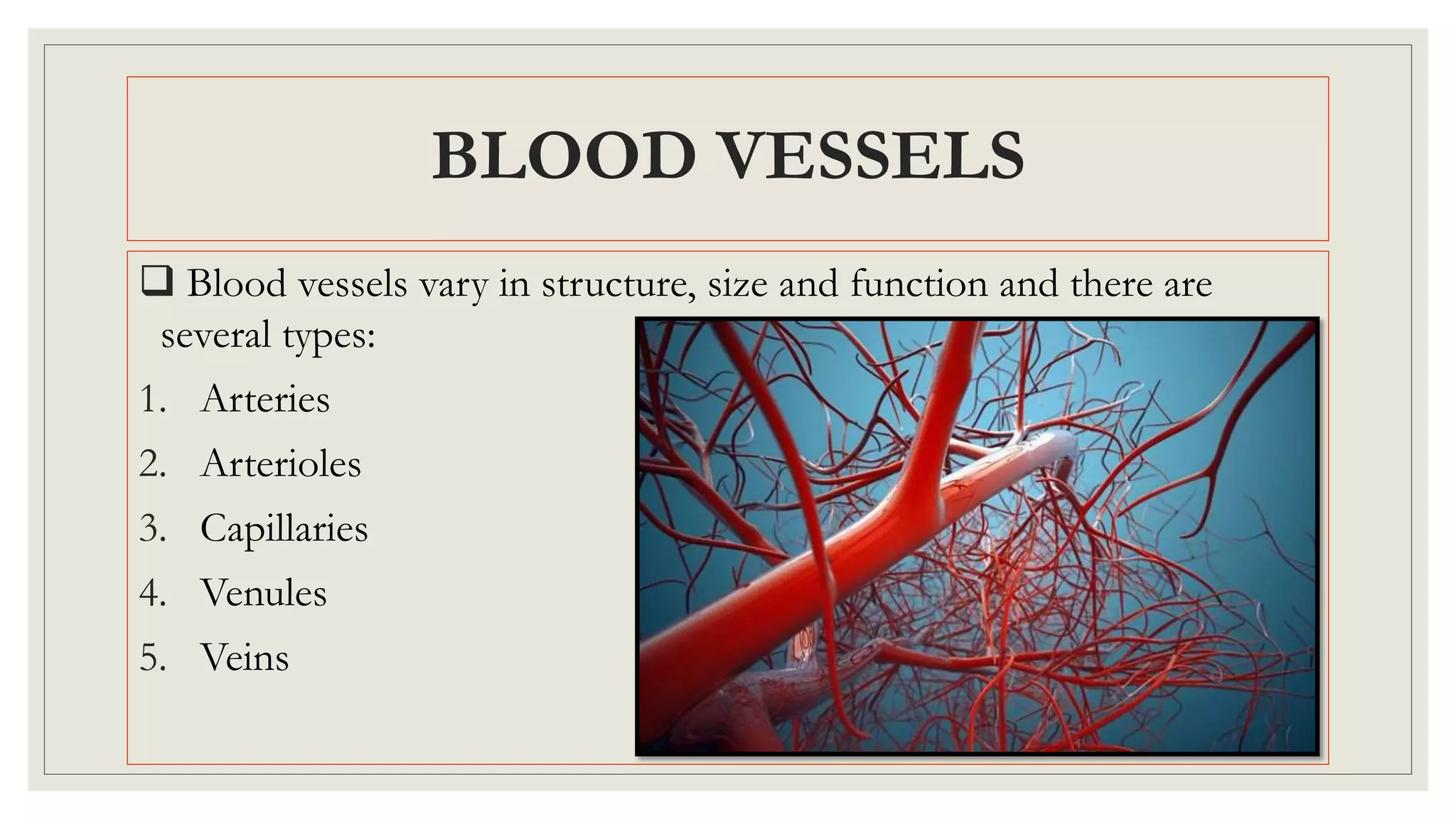
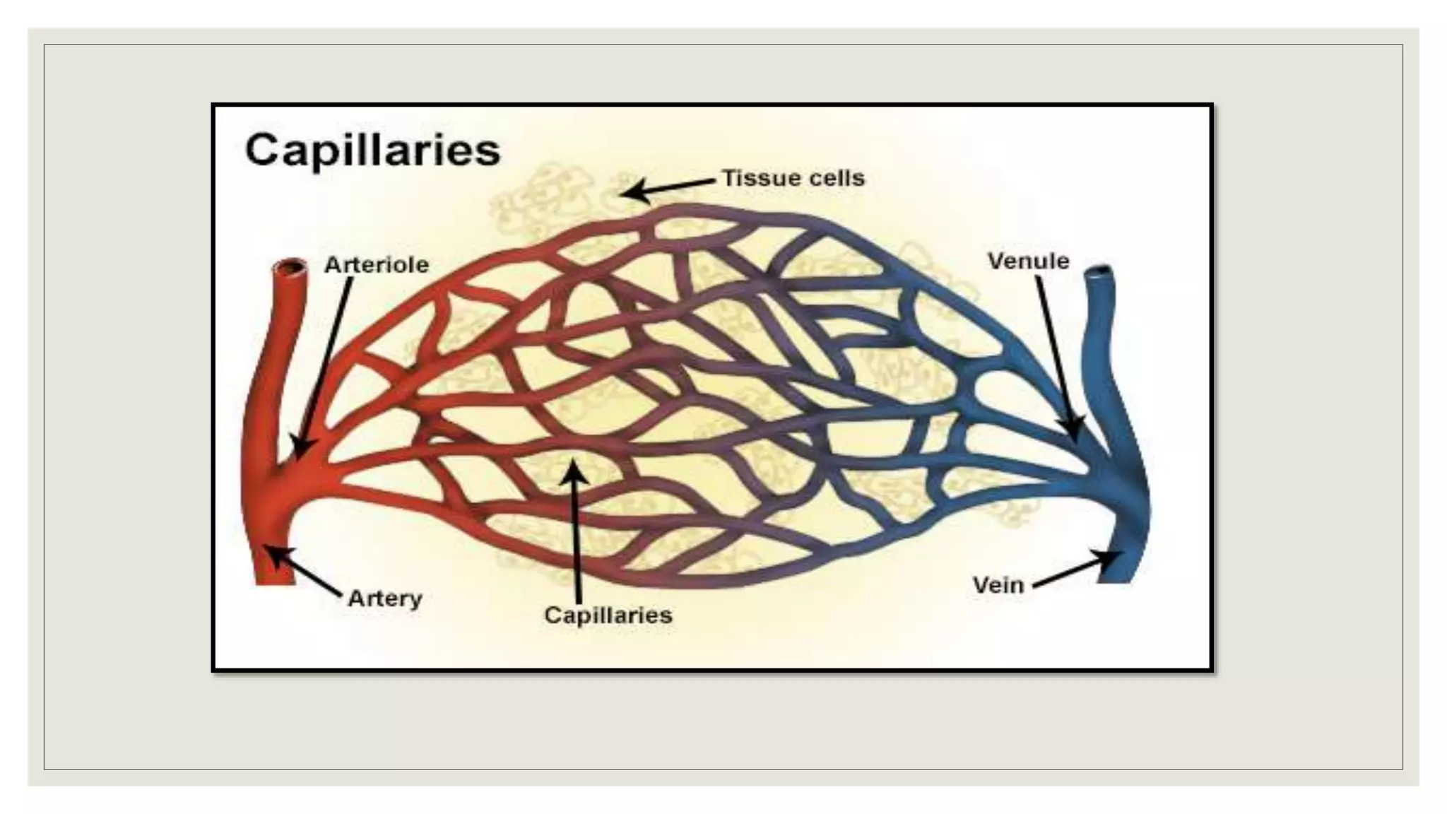
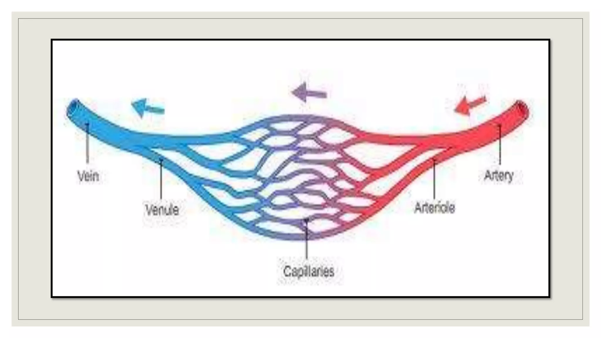
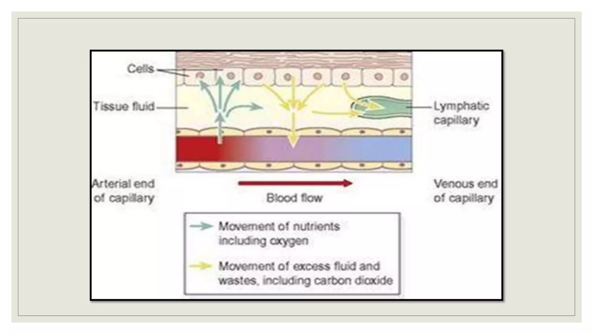
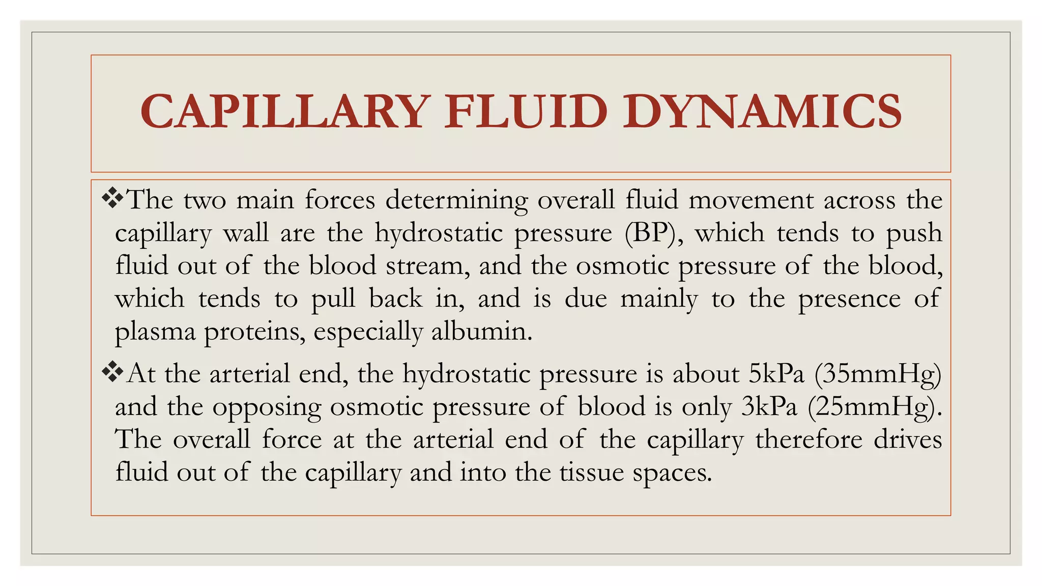
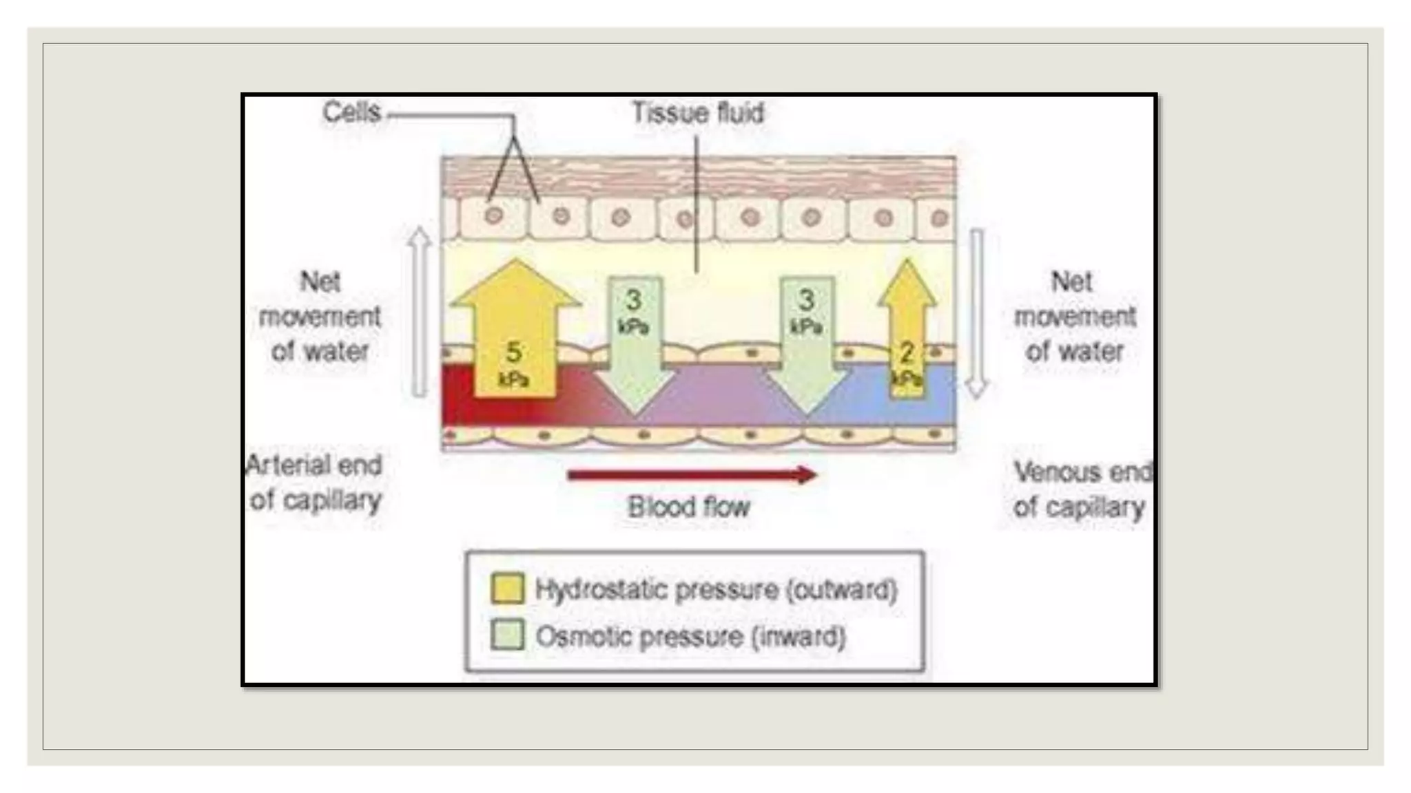
CARDIOVASCULAR
SYSTEM
NIKITA SHARMA
NURSING TUTOR
BECON, JAMMU
BLOOD VESSELS
 Blood vessels vary in structure, size and function and there are
several types:
1. Arteries
2. Arterioles
3. Capillaries
4. Venules
5. Veins
ARTERIES & ARTERIOLES
These blood vessels transport blood away from the heart.
They vary in size
Their wall consist of 3 layers of tissues:
1. Outer layer: fibrous tissue (tunica adventitia)
2. Middle layer: smooth muscle & elastic tissue (tunica media)
3. Inner layer: squamous epithelium called endothelium (tunica
intima)
The amount of muscular and elastic tissue varies in the arteries
depends upon its size & function.
In the large arteries, including the aorta, sometimes called elastic
arteries, the tunica media contains more elastic tissues and less smooth
muscles.
This allows the vessel wall to stretch, absorbing the pressure wave
generated by the heart as it beats.
These proportions gradually change as the arteries branch many
times and become smaller until in the arterioles tunica media almost
entirely of smooth muscle.
ARTERIES & ARTERIOLES
This enables their diameter to be precisely controlled, which
regulates the pressure with in them.
Systemic blood pressure is mainly determined by the resistance these
tiny arteries offer to blood flow, & for this reason they are called
resistance vessels.
Arteries have thicker walls than veins to withstand the high pressure
of arterial blood.
ARTERIES & ARTERIOLES
ANASTOMOSES & END-ARTERIES
Anastomoses are arteries that form a link between main arteries
supplying an area, e.g. the arterial supply to the palms of the hand &
soles of the feet, the brain, the joints & to a limited extent, the heart
muscle.
If one artery supplying the area is occluded, anastomotic arteries
provide a collateral circulation.
This is most likely to provide an adequate blood supply when the
occlusion occurs gradually, giving the anastomotic arteries time to
dilate.
An end-artery is an artery that is the sole source of blood to a tissue,
e.g. the branches from the circulus arteriosus (circle of willis) in the
brain or the central artery to the retina of the eye.
When an end-artery is occluded the tissues it supplies die because
there is no alternative blood supply.
ANASTOMOSES & END-ARTERIES
CAPILLARIES & SINUSOIDS
 The smallest arterioles break up into a number of minute vessels
called capillaries.
Capillary wall consist of a single layer of endothelial cells sitting on a very
thin basement membrane, through which water and other small molecules
can pass.
The capillaries form a vast network of tiny vessels that link the smallest
arterioles to the smallest venules; having 7 micrometer diameter.
Capillary bed is the site of exchange of substances between the blood and
the tissue fluid, which bathes the body cells & with the exception of those
On the skin surface & in the cornea of the eye, everybody cell lies close
to the capillaries.
Entry to capillary bed is guarded by rings of smooth muscle
(precapillary sphincters) that direct blood flow.
Hypoxia and more tissue wastes leads to dilatation of these sphincters
and increase blood flow through the affected bed.
In certain places like liver and bone marrow; the capillaries are
significantly wider and leakier than normal and known as sinusoids.
CAPILLARIES & SINUSOIDS
CAPILLARY REFILL TIME
VEINS & VENULES
Veins return blood at low pressure to the heart.
The walls of the veins are thinner than arteries but have the same
three layers of tissue.
Some veins possess valves.
Valves are abundant in limbs especially in lower limbs.
Valves are absent in very small and very large veins in the thorax and
abdomen.
The smallest veins are called venules.
Veins are called capacitance vessels.
At any one time, about 2/3rd of the blood is in the venous system.
This allows the vascular system to absorb the much extent of the
blood during sudden change in the blood volume such as in
haemorrhage; veins can constrict, helping to prevent an sudden fall in
BP.
VEINS & VENULES
BLOOD SUPPLY
 The outer layer of the tissue of thick walled blood vessel receive
their blood supply via a network of blood vessels called vasa
vasorum.
CONTROL OF BLOOD VESSEL
DIAMETER
oThe smooth muscle in the tunica media of veins and arteries is
supplied by sympathetic nerves of the automatic nervous system.
oThese nerves arise from the vasomotor centre in the medulla
oblongata and they change the diameter of blood vessels, controlling
the volume of blood they contain.
oSympathetic activity generally constricts blood vessel smooth muscle
and therefore narrows the vessels (vasoconstriction), increasing
the pressure inside.
CONTROL OF BLOOD VESSEL
DIAMETER
◦ A degree of resting sympathetic activity maintains a constant baseline
tone in the vessel wall and prevents pressure falling too low.
◦ Decreased nerve stimulation relaxes the smooth muscle, thinning the
vessel wall and enlarging the lumen (vasodilation), increased blood
pressure under low pressure.
◦ Constant adjustment of blood vessel diameter helps to regulate
peripheral blood resistance and systemic blood pressure.
LOCAL REGUALTION OF BLOOD
FLOW
◦ The ability of an organ to control its own blood flow according to
need is called autoregulation.
e.g. Blood flow through the GIT increasing after a meal to allow for
increased activity in the tract, & adjustments to blood flow through the
skin in the control of body temperature.
◦ Blood flow through individual organ is increased by vasodilation of
the vessels supplying it & decreased through vasoconstriction.
◦ The main mechanisms associated with this local control of blood flow
include:
Release of
metabolic waste
products
Tissue
temperature
Hypoxia
Release of
vasodilator
chemicals
Action of
vasoconstrictors
LOCAL REGUALTION OF BLOOD
FLOW
CAPILLARY EXCHANGE
EXCHANGE OF GASES
Internal respiration is the process by which gases are exchanged
between capillary blood and local body cells.
Oxygen is carried from the lungs to the tissues in combination with
haemoglobin as oxyhaemoglobin.
Exchange in the tissues takes place between blood at the arterial end
of the capillaries and the tissue fluid and then between the tissue fluid
and the cells.
EXCHANGE OF GASES
Oxygen diffuses down its concentration gradient, from the oxygen-
rich arterial blood, into the tissues, where oxygen levels are lower
because of constant tissue consumption.
Blood transports carbon dioxide to the lungs for excretion by 3
different mechanisms;
1.Dissolved in the water of blood plasma- 7%
2.In chemical combination with sodium in the form of sodium
carbonate-70%
3.Remainder in combination with haemoglobin- 23%
EXCHANGE OF OTHER
SUBSTANCES
The nutrients, including glucose, amino acids, fatty acids, vitamins
and mineral salts required by all body cells are transported round the
body in the blood plasma.
They diffuse through the semipermeable capillary walls into the
tissues.
Water exchanges freely between the plasma and tissue fluid by
osmosis.
CAPILLARY FLUID DYNAMICS
The two main forces determining overall fluid movement across the
capillary wall are the hydrostatic pressure (BP), which tends to push
fluid out of the blood stream, and the osmotic pressure of the blood,
which tends to pull back in, and is due mainly to the presence of
plasma proteins, especially albumin.
At the arterial end, the hydrostatic pressure is about 5kPa (35mmHg)
and the opposing osmotic pressure of blood is only 3kPa (25mmHg).
The overall force at the arterial end of the capillary therefore drives
fluid out of the capillary and into the tissue spaces.
◦ At the venous end of the capillary, the situation is reversed.
◦ Blood flow is slower than at the arterial end because the hydrostatic
pressure drops along the capillary to only 2kPa.
◦ The osmotic pressure remains unchanged at 3kPa, because of this
now exceeds hydrostatic pressure, fluid moves back into the capillary.
CAPILLARY FLUID DYNAMICS
arteries, arterioles, veins, venules, capillary and fluid or gaseous exchange in tissues.

arteries, arterioles, veins, venules, capillary and fluid or gaseous exchange in tissues.

  • 1.
  • 2.
    BLOOD VESSELS  Bloodvessels vary in structure, size and function and there are several types: 1. Arteries 2. Arterioles 3. Capillaries 4. Venules 5. Veins
  • 3.
    ARTERIES & ARTERIOLES Theseblood vessels transport blood away from the heart. They vary in size Their wall consist of 3 layers of tissues: 1. Outer layer: fibrous tissue (tunica adventitia) 2. Middle layer: smooth muscle & elastic tissue (tunica media) 3. Inner layer: squamous epithelium called endothelium (tunica intima)
  • 5.
    The amount ofmuscular and elastic tissue varies in the arteries depends upon its size & function. In the large arteries, including the aorta, sometimes called elastic arteries, the tunica media contains more elastic tissues and less smooth muscles. This allows the vessel wall to stretch, absorbing the pressure wave generated by the heart as it beats. These proportions gradually change as the arteries branch many times and become smaller until in the arterioles tunica media almost entirely of smooth muscle. ARTERIES & ARTERIOLES
  • 6.
    This enables theirdiameter to be precisely controlled, which regulates the pressure with in them. Systemic blood pressure is mainly determined by the resistance these tiny arteries offer to blood flow, & for this reason they are called resistance vessels. Arteries have thicker walls than veins to withstand the high pressure of arterial blood. ARTERIES & ARTERIOLES
  • 7.
    ANASTOMOSES & END-ARTERIES Anastomosesare arteries that form a link between main arteries supplying an area, e.g. the arterial supply to the palms of the hand & soles of the feet, the brain, the joints & to a limited extent, the heart muscle. If one artery supplying the area is occluded, anastomotic arteries provide a collateral circulation. This is most likely to provide an adequate blood supply when the occlusion occurs gradually, giving the anastomotic arteries time to dilate.
  • 9.
    An end-artery isan artery that is the sole source of blood to a tissue, e.g. the branches from the circulus arteriosus (circle of willis) in the brain or the central artery to the retina of the eye. When an end-artery is occluded the tissues it supplies die because there is no alternative blood supply. ANASTOMOSES & END-ARTERIES
  • 11.
    CAPILLARIES & SINUSOIDS The smallest arterioles break up into a number of minute vessels called capillaries. Capillary wall consist of a single layer of endothelial cells sitting on a very thin basement membrane, through which water and other small molecules can pass. The capillaries form a vast network of tiny vessels that link the smallest arterioles to the smallest venules; having 7 micrometer diameter. Capillary bed is the site of exchange of substances between the blood and the tissue fluid, which bathes the body cells & with the exception of those
  • 13.
    On the skinsurface & in the cornea of the eye, everybody cell lies close to the capillaries. Entry to capillary bed is guarded by rings of smooth muscle (precapillary sphincters) that direct blood flow. Hypoxia and more tissue wastes leads to dilatation of these sphincters and increase blood flow through the affected bed. In certain places like liver and bone marrow; the capillaries are significantly wider and leakier than normal and known as sinusoids. CAPILLARIES & SINUSOIDS
  • 15.
  • 16.
    VEINS & VENULES Veinsreturn blood at low pressure to the heart. The walls of the veins are thinner than arteries but have the same three layers of tissue. Some veins possess valves. Valves are abundant in limbs especially in lower limbs. Valves are absent in very small and very large veins in the thorax and abdomen. The smallest veins are called venules.
  • 17.
    Veins are calledcapacitance vessels. At any one time, about 2/3rd of the blood is in the venous system. This allows the vascular system to absorb the much extent of the blood during sudden change in the blood volume such as in haemorrhage; veins can constrict, helping to prevent an sudden fall in BP. VEINS & VENULES
  • 20.
    BLOOD SUPPLY  Theouter layer of the tissue of thick walled blood vessel receive their blood supply via a network of blood vessels called vasa vasorum.
  • 22.
    CONTROL OF BLOODVESSEL DIAMETER oThe smooth muscle in the tunica media of veins and arteries is supplied by sympathetic nerves of the automatic nervous system. oThese nerves arise from the vasomotor centre in the medulla oblongata and they change the diameter of blood vessels, controlling the volume of blood they contain. oSympathetic activity generally constricts blood vessel smooth muscle and therefore narrows the vessels (vasoconstriction), increasing the pressure inside.
  • 24.
    CONTROL OF BLOODVESSEL DIAMETER ◦ A degree of resting sympathetic activity maintains a constant baseline tone in the vessel wall and prevents pressure falling too low. ◦ Decreased nerve stimulation relaxes the smooth muscle, thinning the vessel wall and enlarging the lumen (vasodilation), increased blood pressure under low pressure. ◦ Constant adjustment of blood vessel diameter helps to regulate peripheral blood resistance and systemic blood pressure.
  • 25.
    LOCAL REGUALTION OFBLOOD FLOW ◦ The ability of an organ to control its own blood flow according to need is called autoregulation. e.g. Blood flow through the GIT increasing after a meal to allow for increased activity in the tract, & adjustments to blood flow through the skin in the control of body temperature. ◦ Blood flow through individual organ is increased by vasodilation of the vessels supplying it & decreased through vasoconstriction. ◦ The main mechanisms associated with this local control of blood flow include:
  • 26.
    Release of metabolic waste products Tissue temperature Hypoxia Releaseof vasodilator chemicals Action of vasoconstrictors LOCAL REGUALTION OF BLOOD FLOW
  • 27.
    CAPILLARY EXCHANGE EXCHANGE OFGASES Internal respiration is the process by which gases are exchanged between capillary blood and local body cells. Oxygen is carried from the lungs to the tissues in combination with haemoglobin as oxyhaemoglobin. Exchange in the tissues takes place between blood at the arterial end of the capillaries and the tissue fluid and then between the tissue fluid and the cells.
  • 29.
    EXCHANGE OF GASES Oxygendiffuses down its concentration gradient, from the oxygen- rich arterial blood, into the tissues, where oxygen levels are lower because of constant tissue consumption. Blood transports carbon dioxide to the lungs for excretion by 3 different mechanisms; 1.Dissolved in the water of blood plasma- 7% 2.In chemical combination with sodium in the form of sodium carbonate-70% 3.Remainder in combination with haemoglobin- 23%
  • 30.
    EXCHANGE OF OTHER SUBSTANCES Thenutrients, including glucose, amino acids, fatty acids, vitamins and mineral salts required by all body cells are transported round the body in the blood plasma. They diffuse through the semipermeable capillary walls into the tissues. Water exchanges freely between the plasma and tissue fluid by osmosis.
  • 32.
    CAPILLARY FLUID DYNAMICS Thetwo main forces determining overall fluid movement across the capillary wall are the hydrostatic pressure (BP), which tends to push fluid out of the blood stream, and the osmotic pressure of the blood, which tends to pull back in, and is due mainly to the presence of plasma proteins, especially albumin. At the arterial end, the hydrostatic pressure is about 5kPa (35mmHg) and the opposing osmotic pressure of blood is only 3kPa (25mmHg). The overall force at the arterial end of the capillary therefore drives fluid out of the capillary and into the tissue spaces.
  • 34.
    ◦ At thevenous end of the capillary, the situation is reversed. ◦ Blood flow is slower than at the arterial end because the hydrostatic pressure drops along the capillary to only 2kPa. ◦ The osmotic pressure remains unchanged at 3kPa, because of this now exceeds hydrostatic pressure, fluid moves back into the capillary. CAPILLARY FLUID DYNAMICS