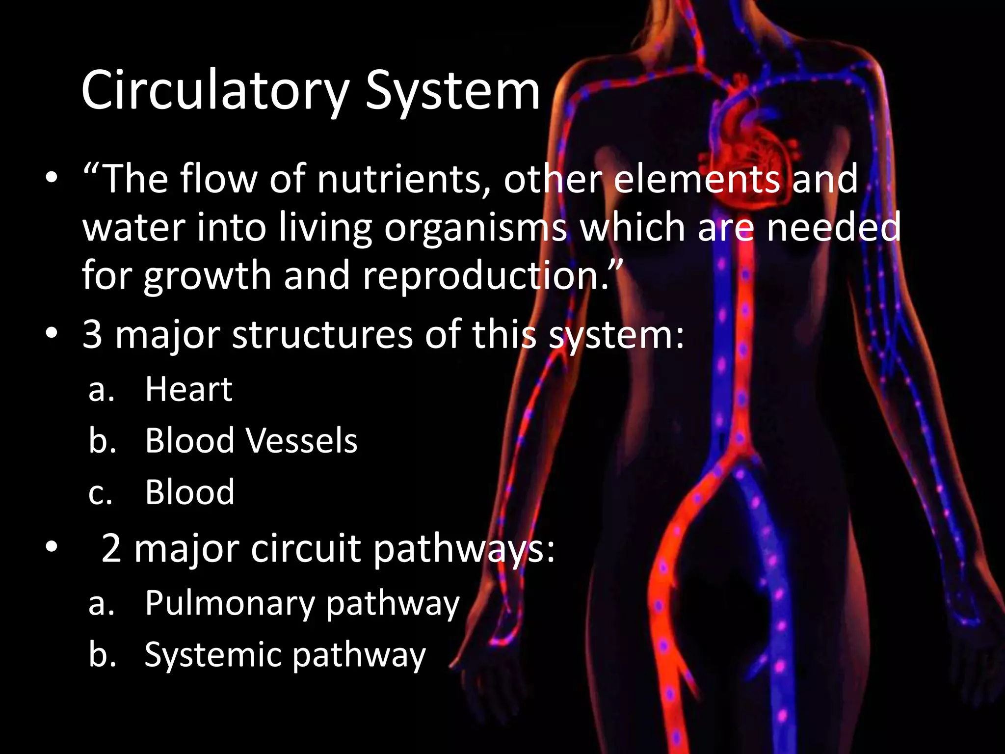
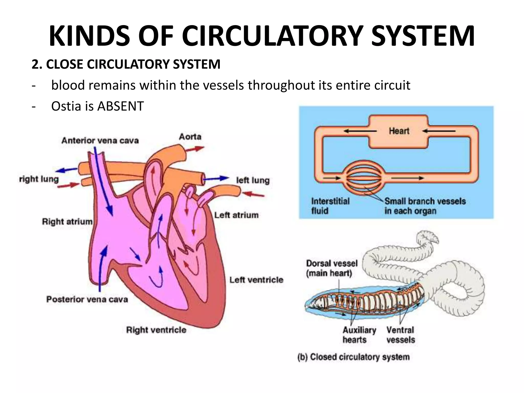
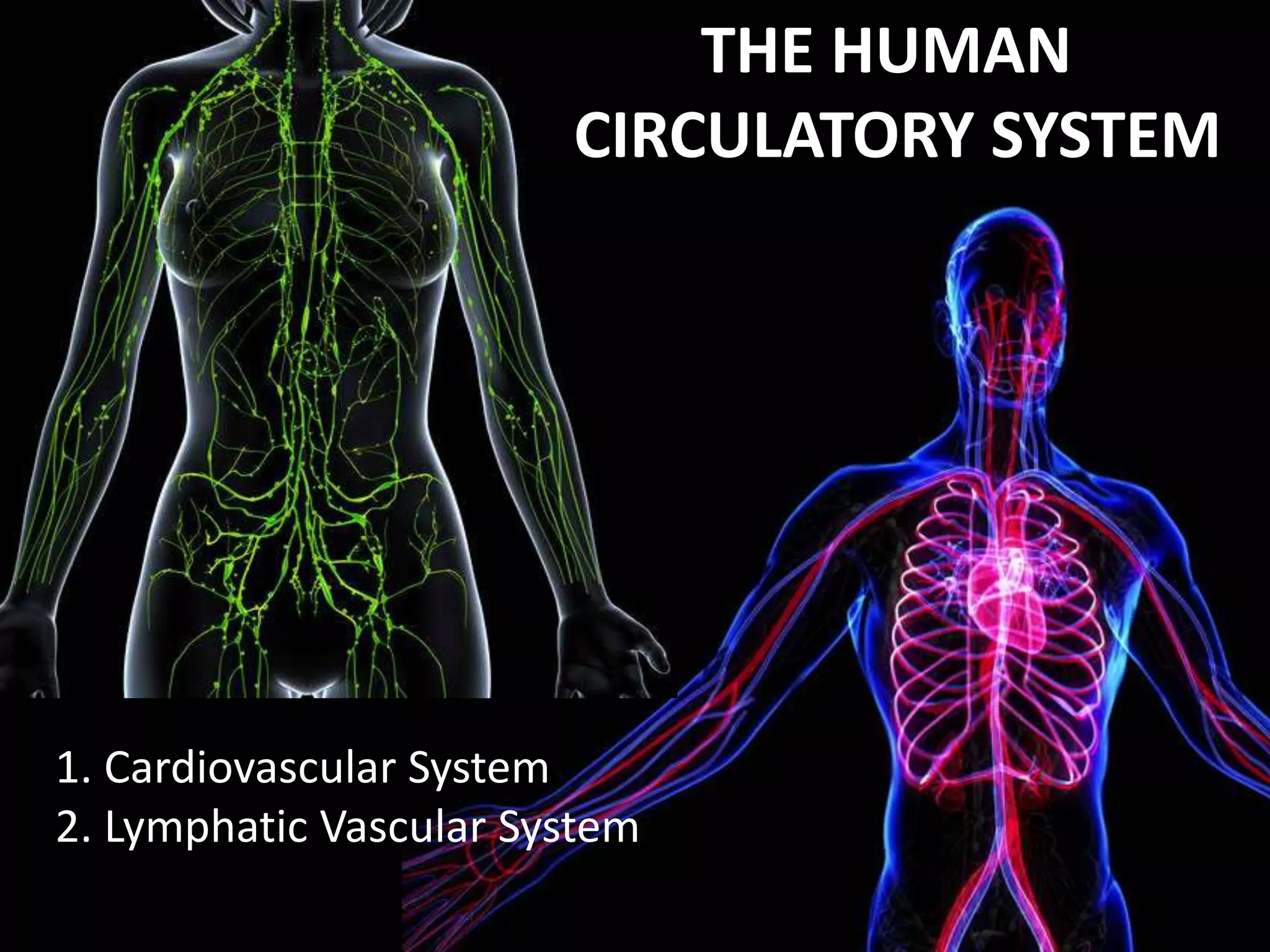
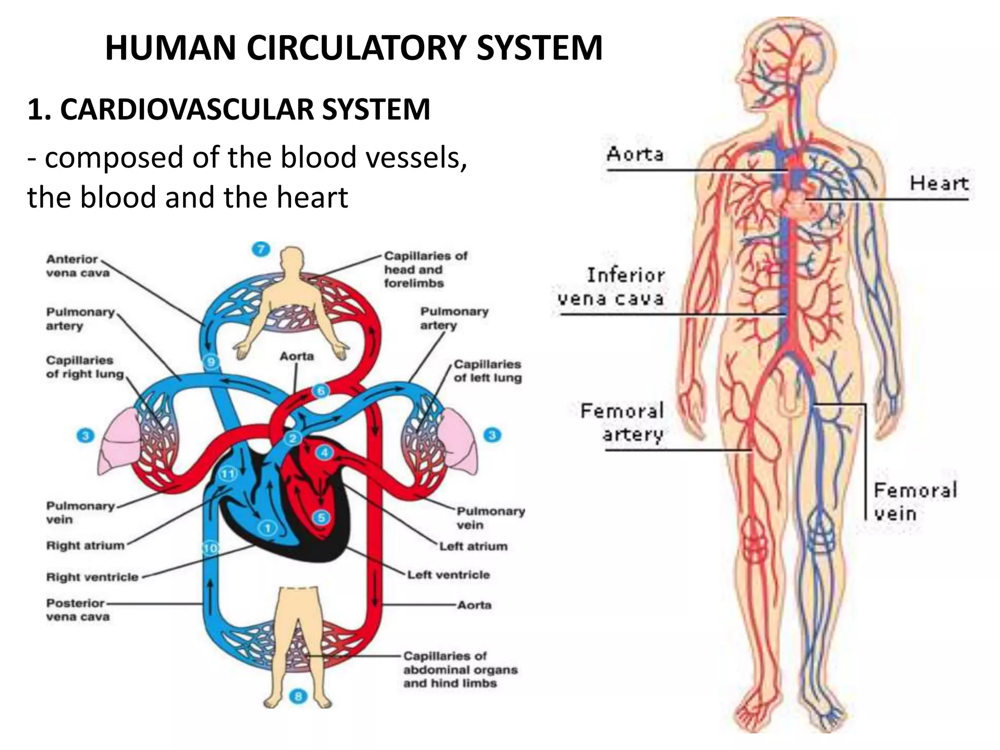
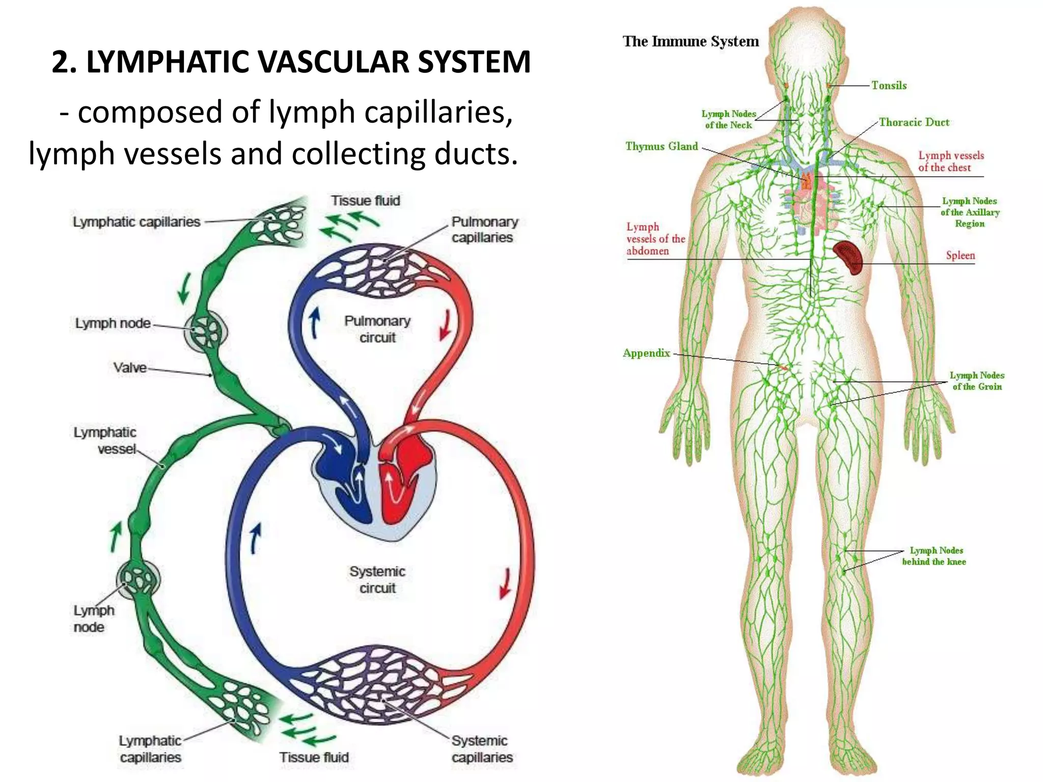
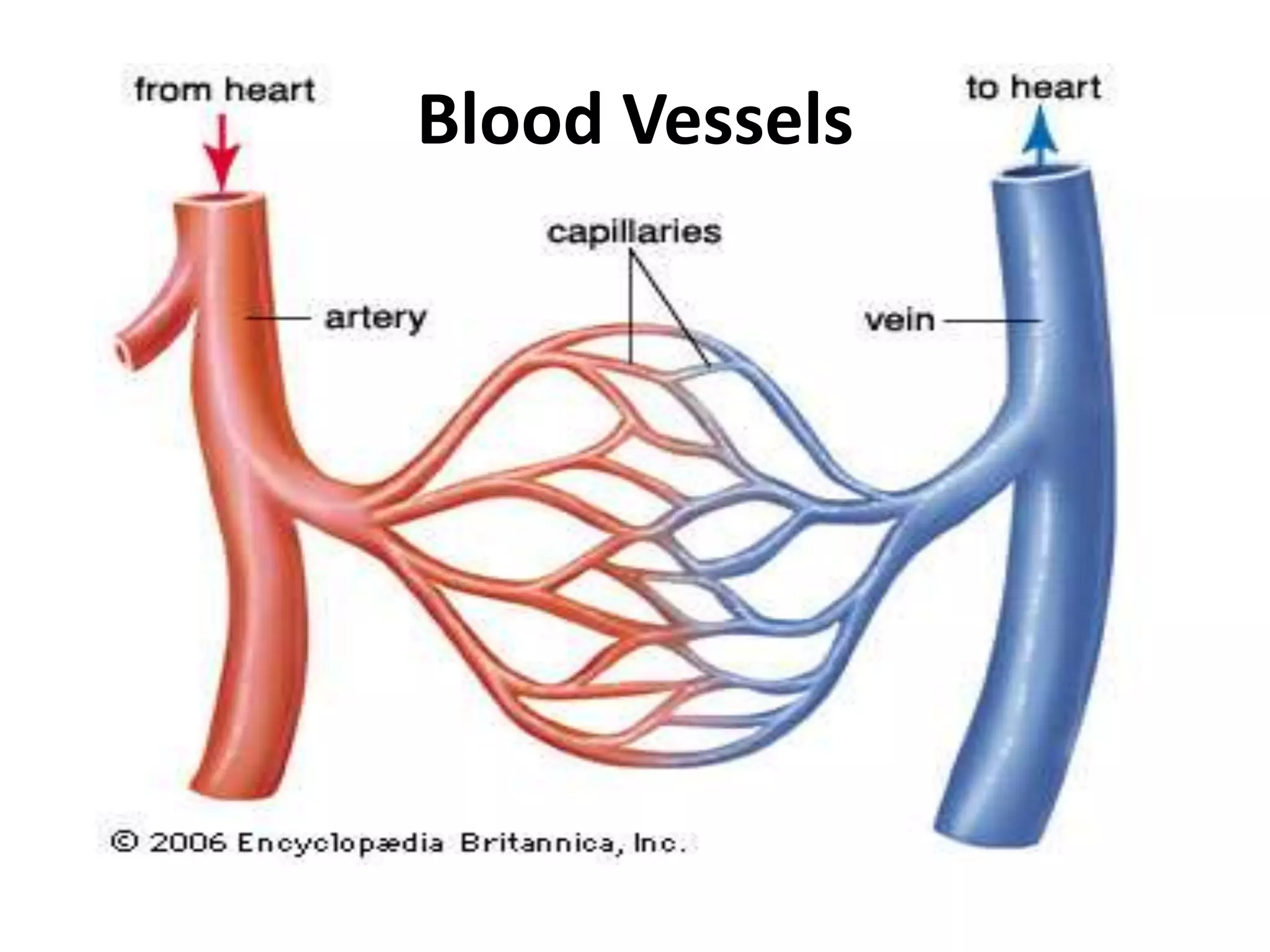
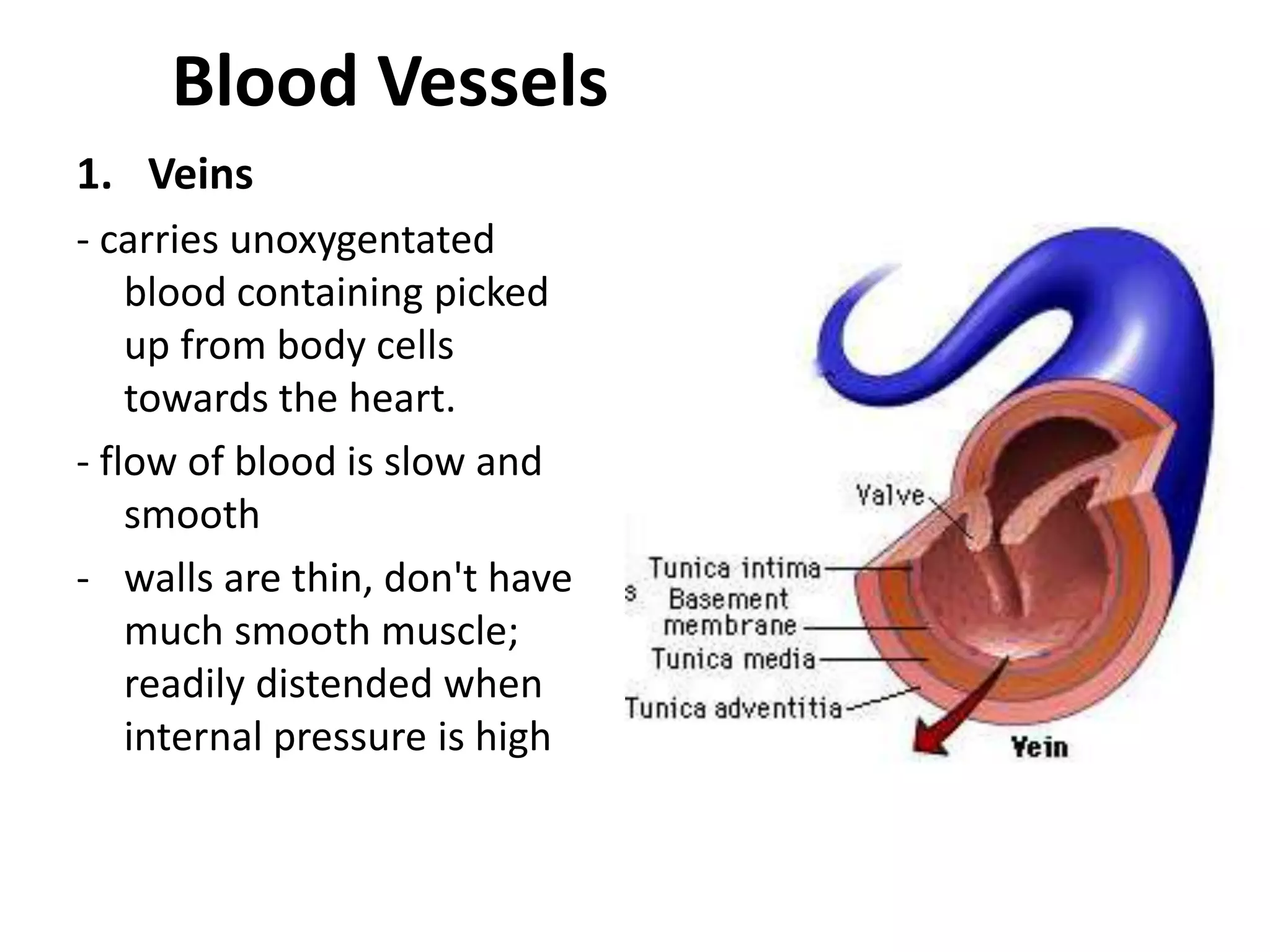
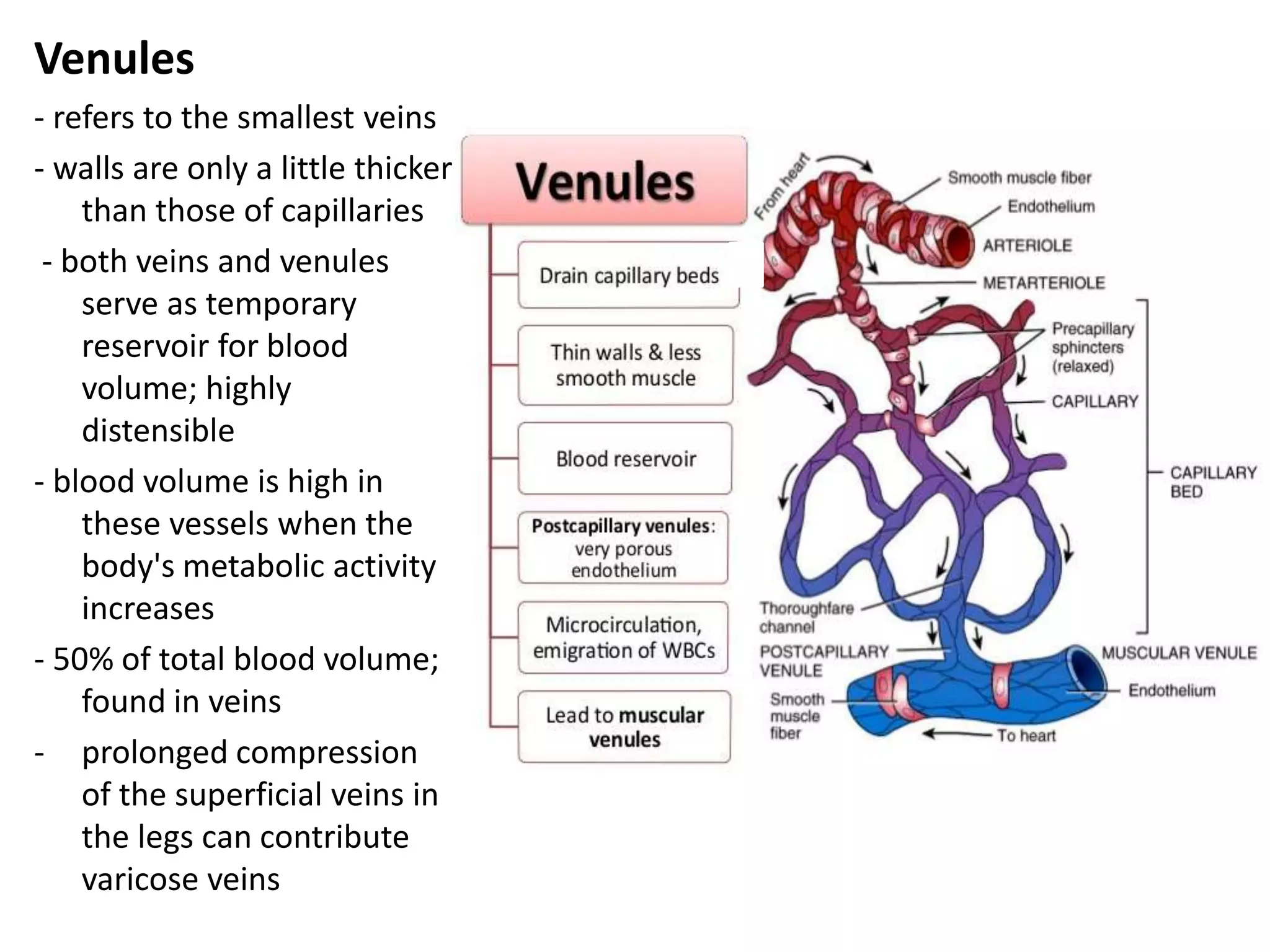
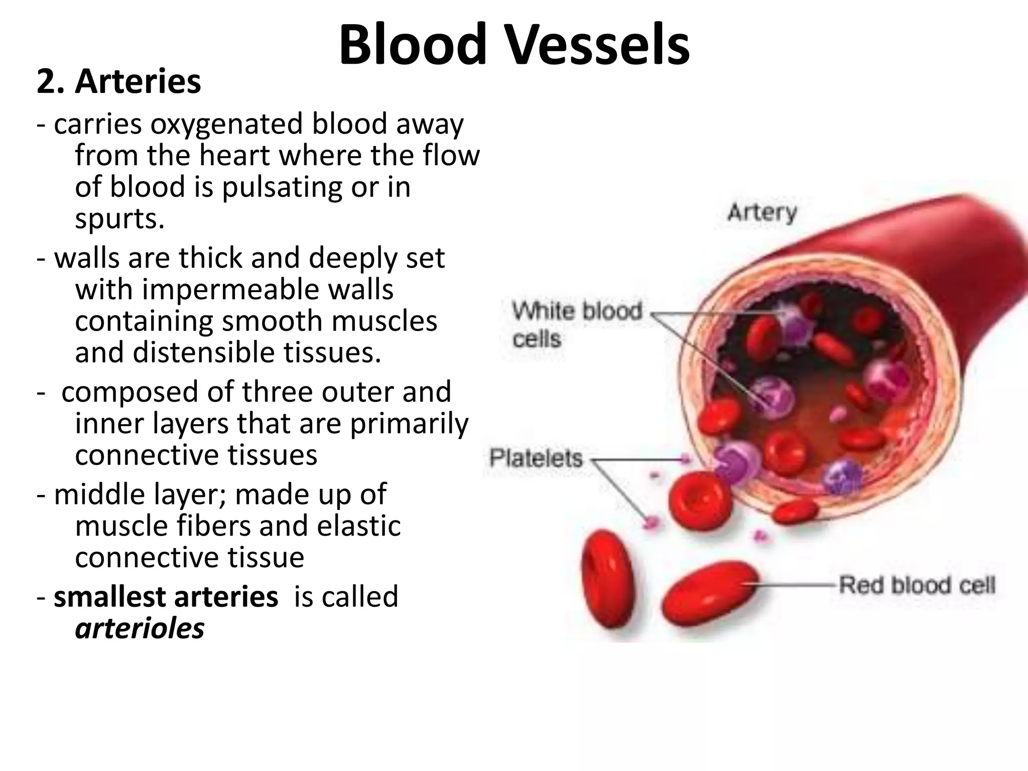
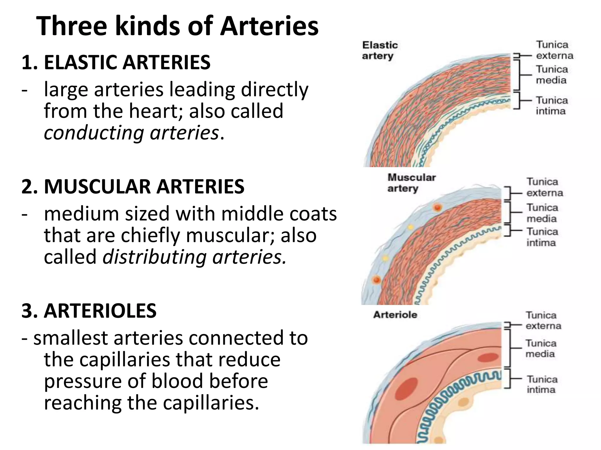
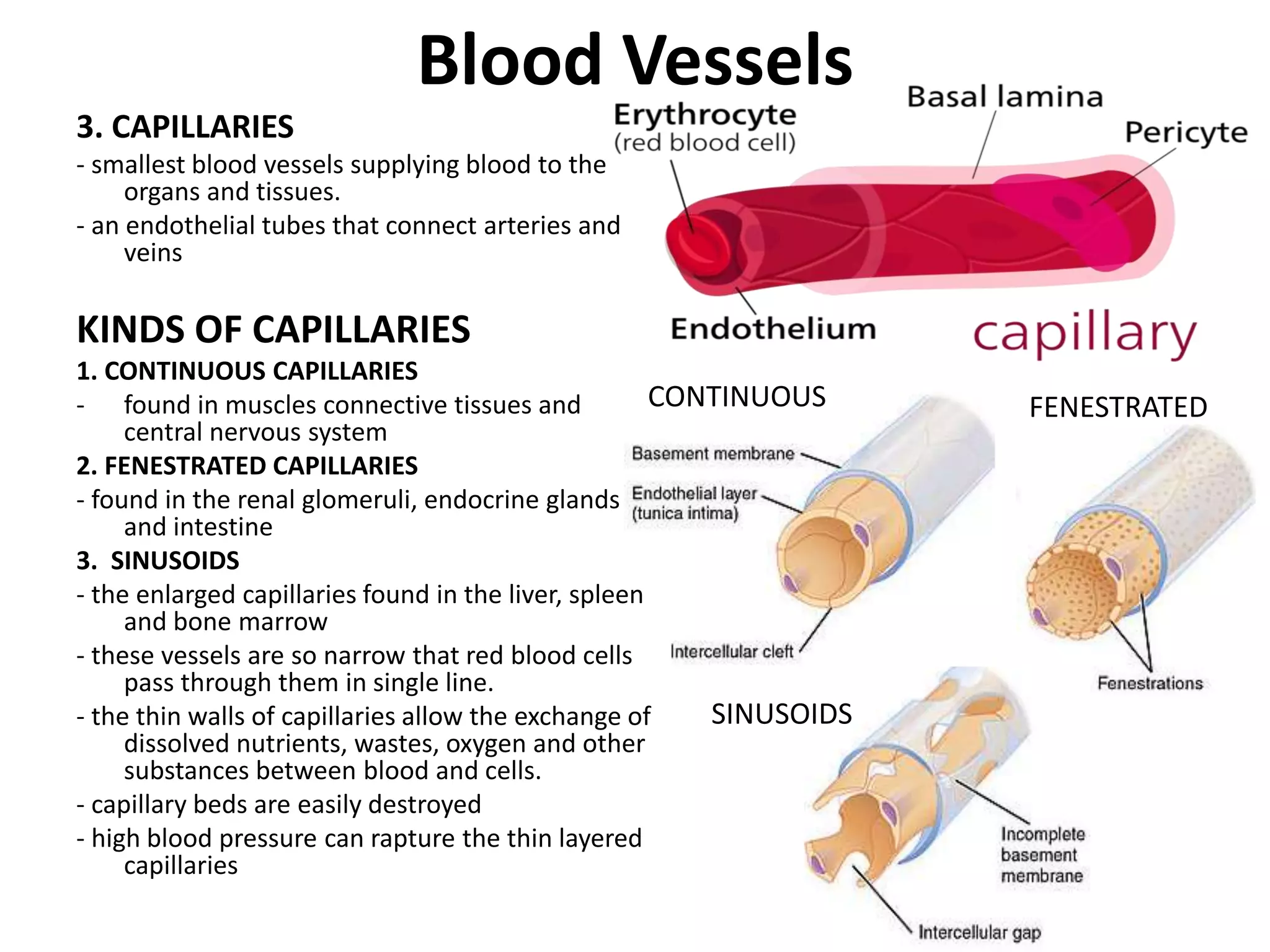
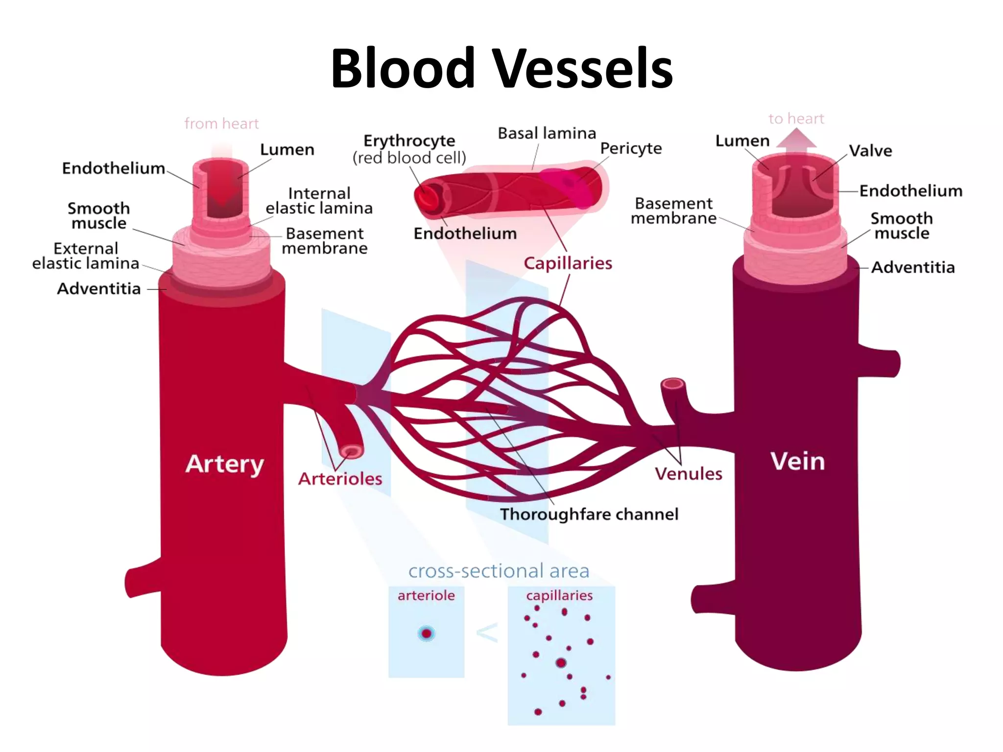
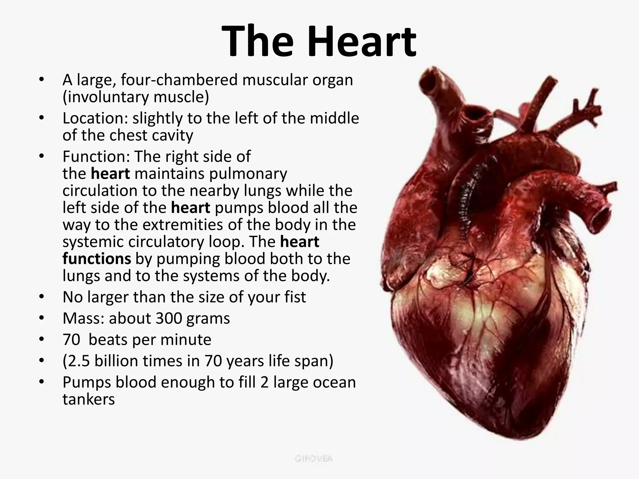
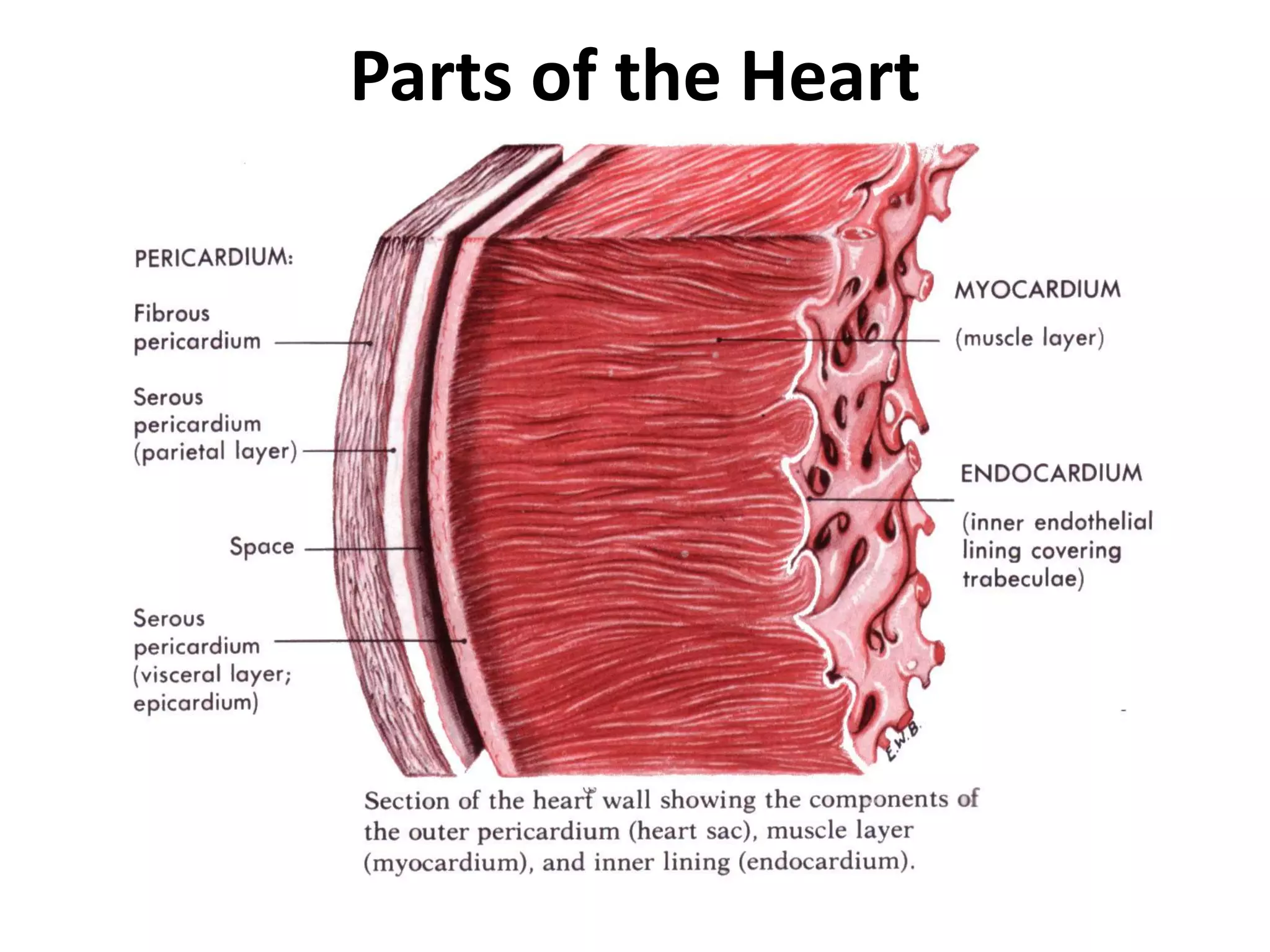
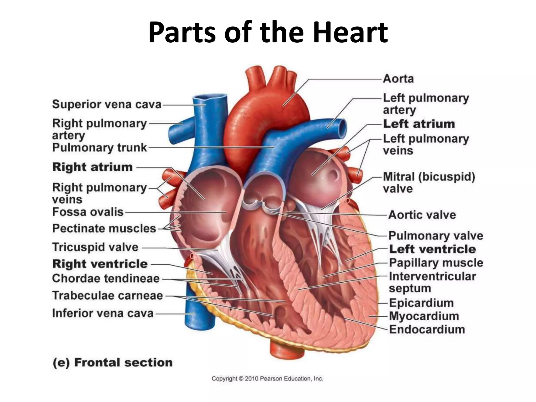
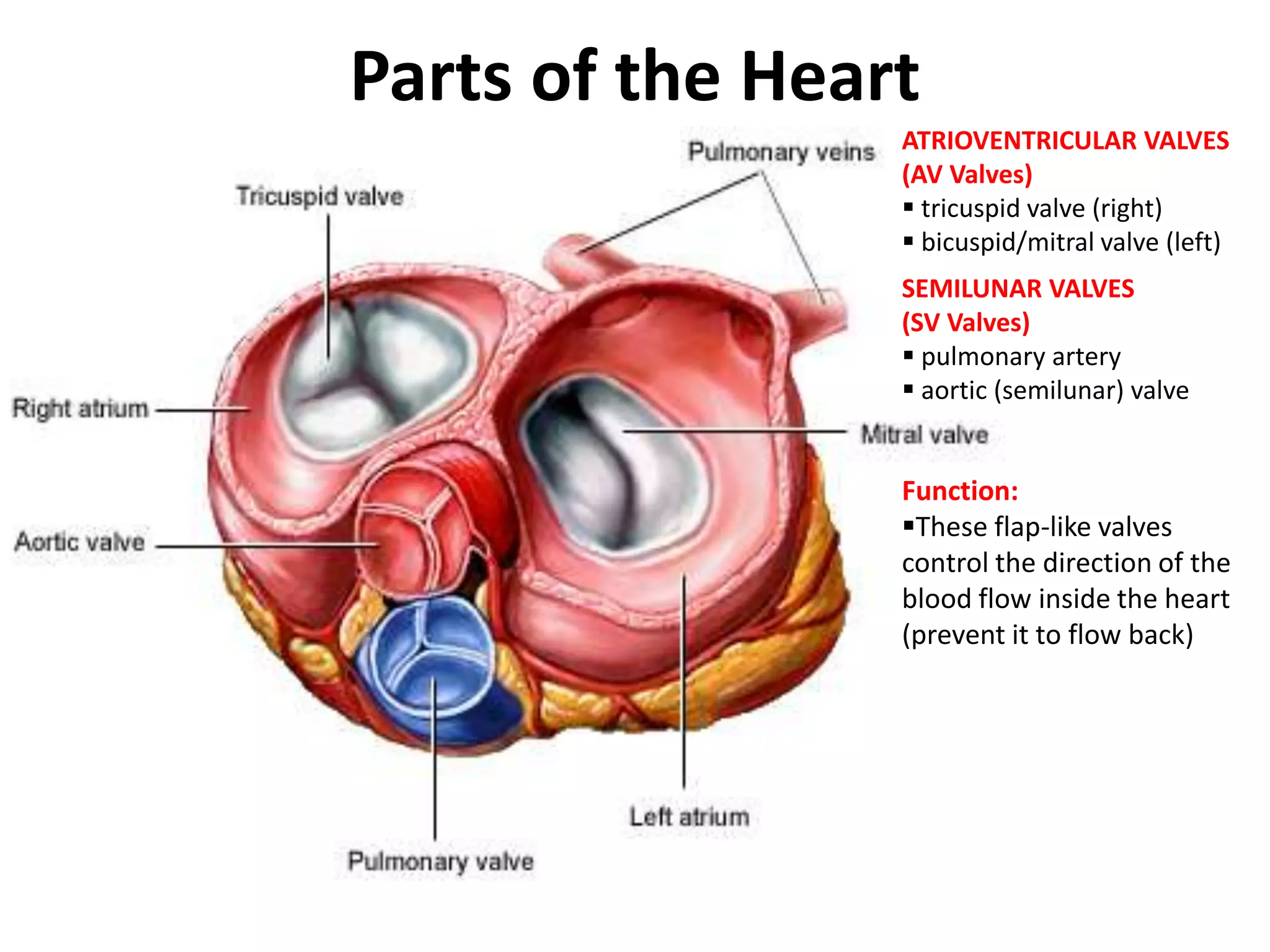
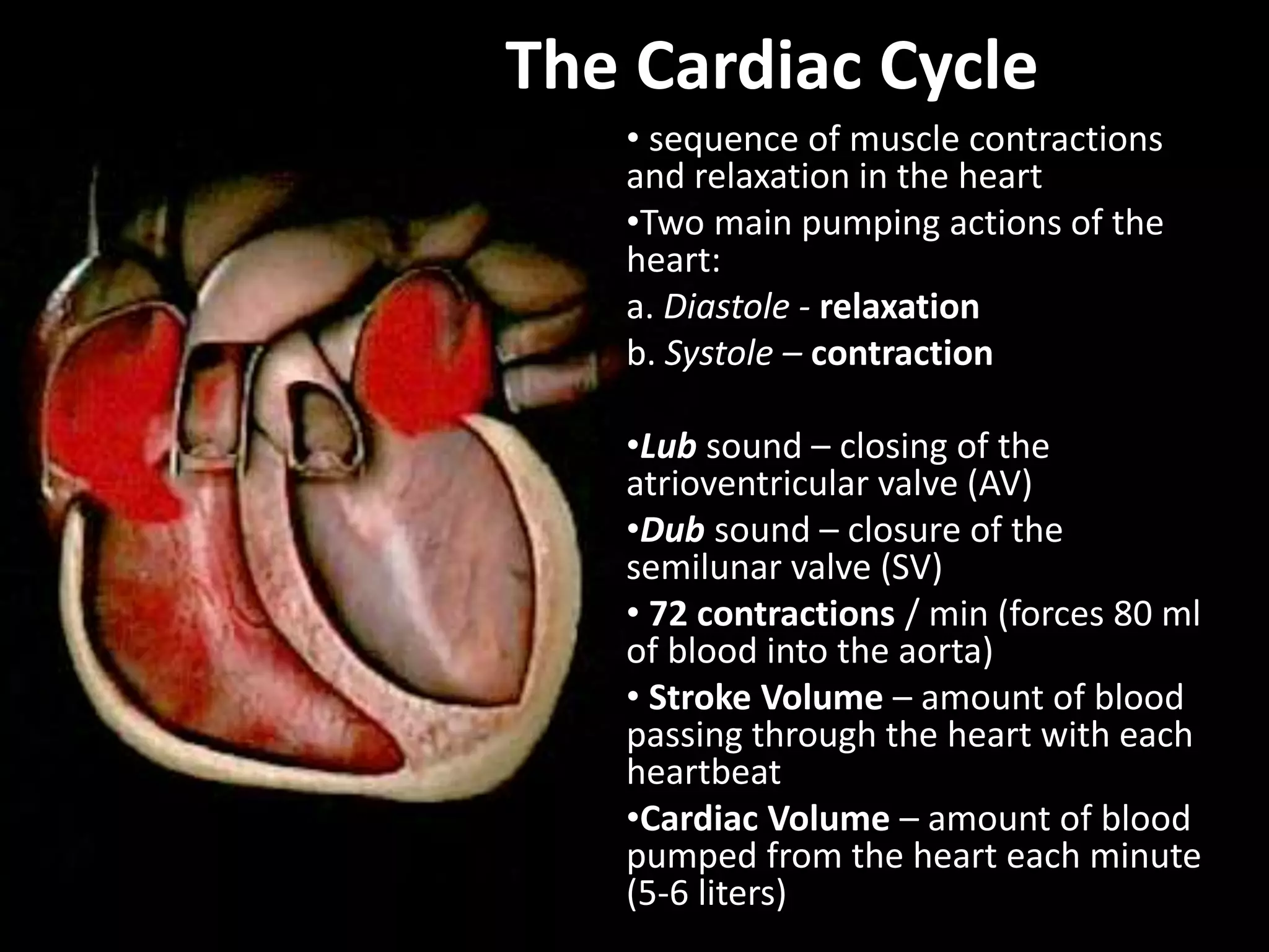
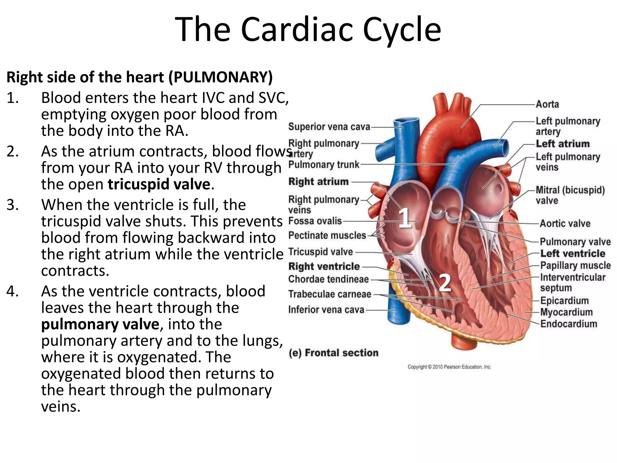
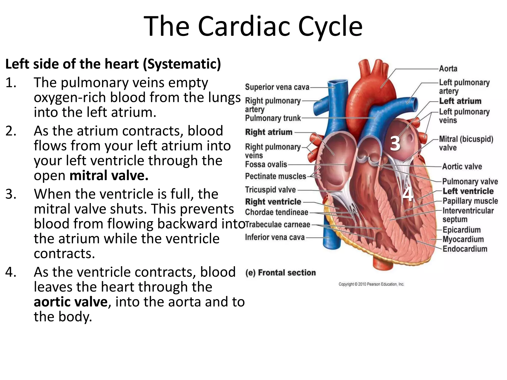
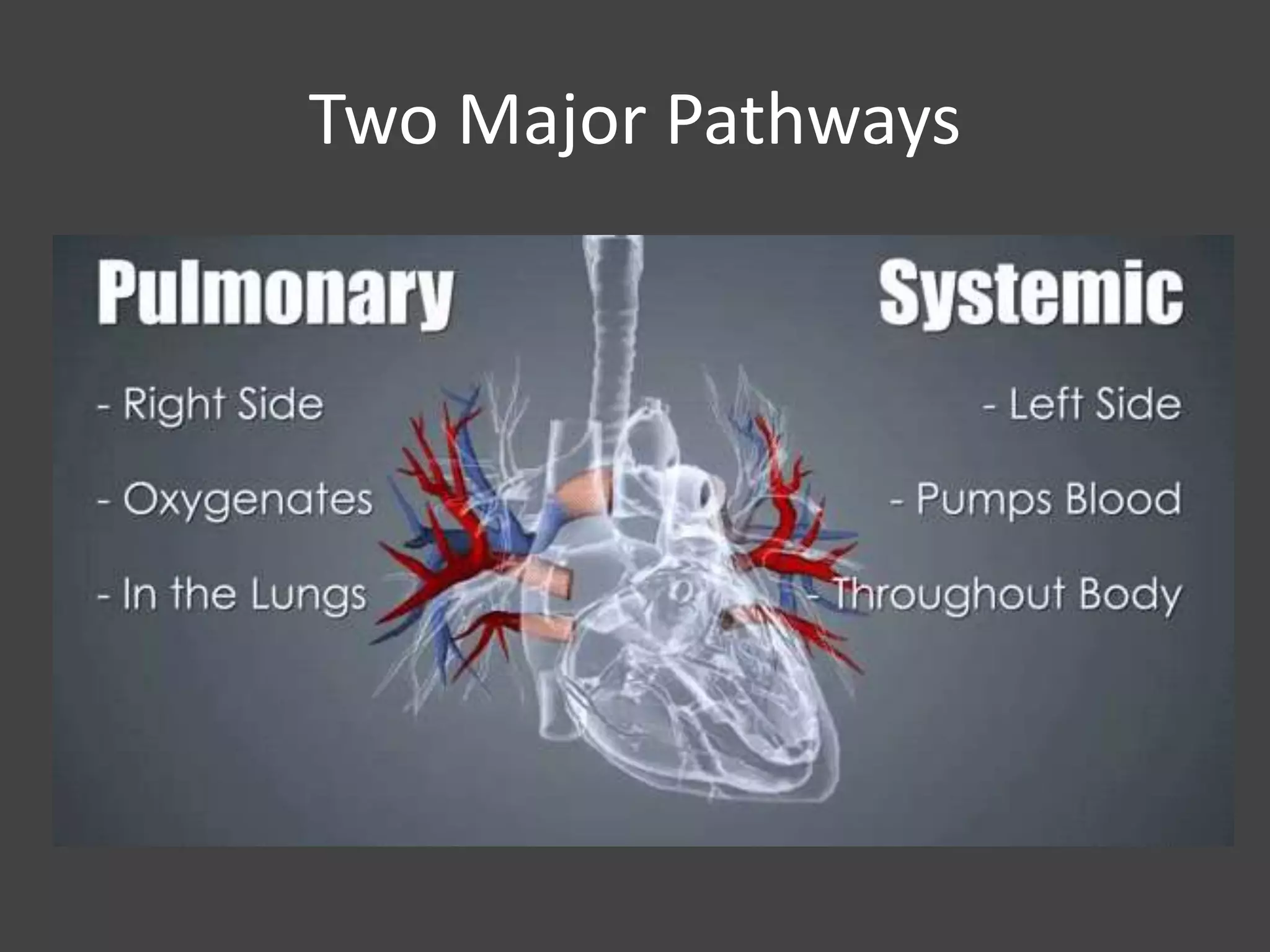
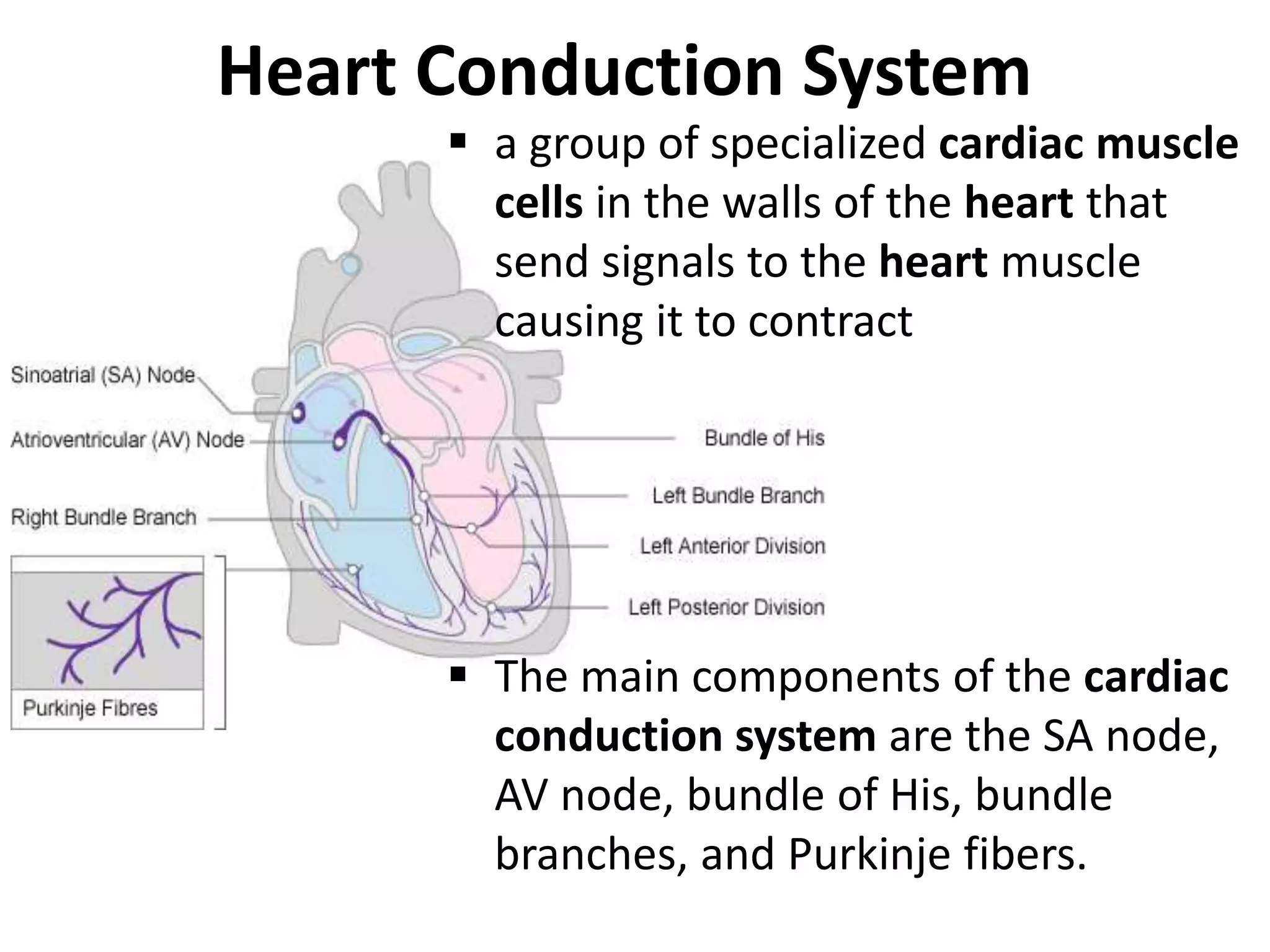
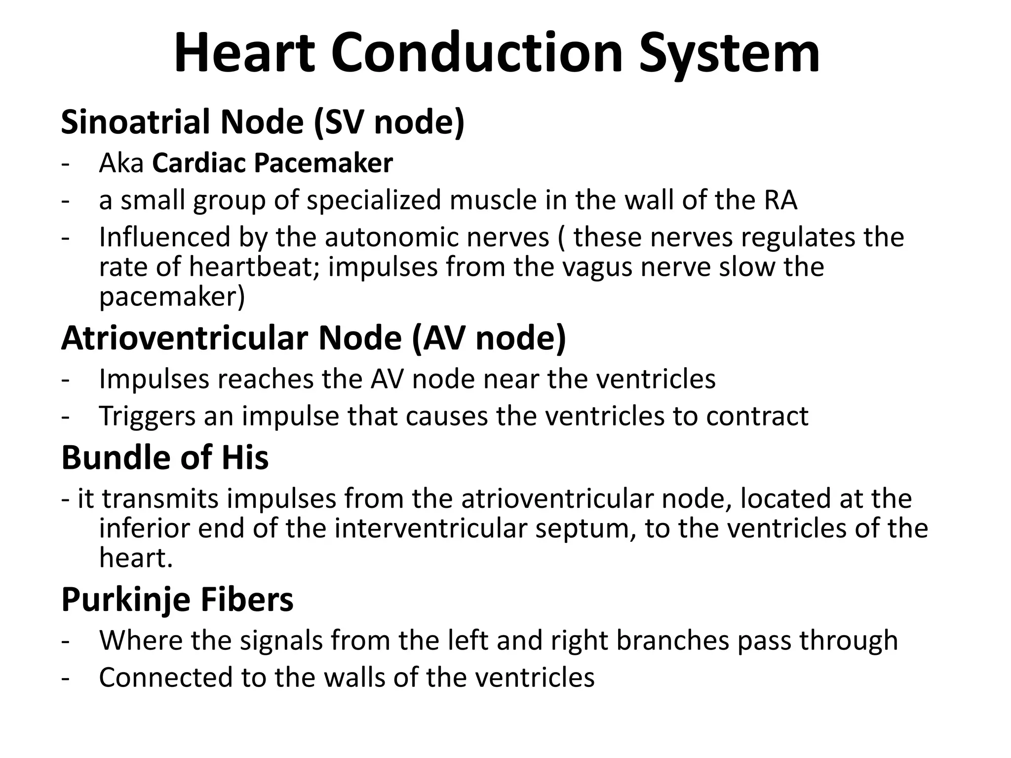
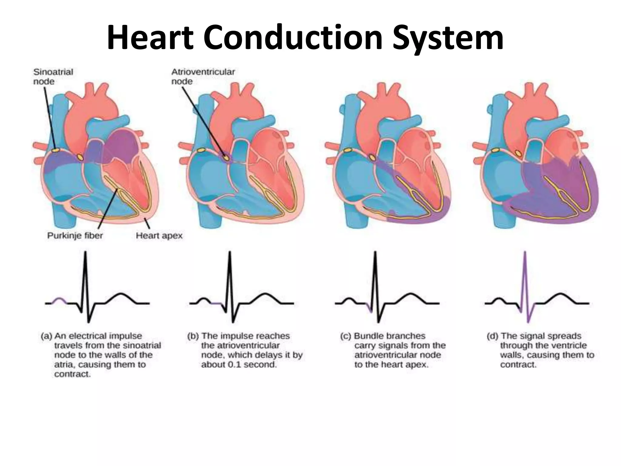
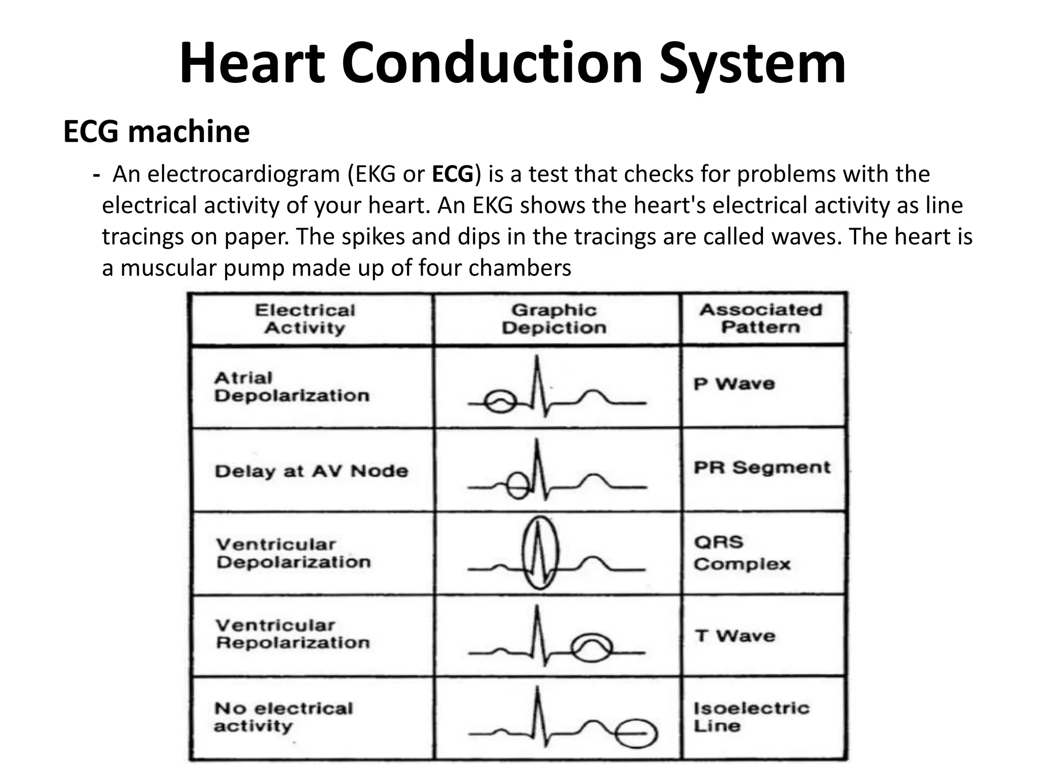
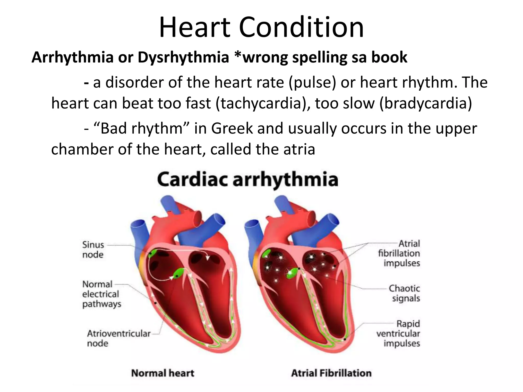
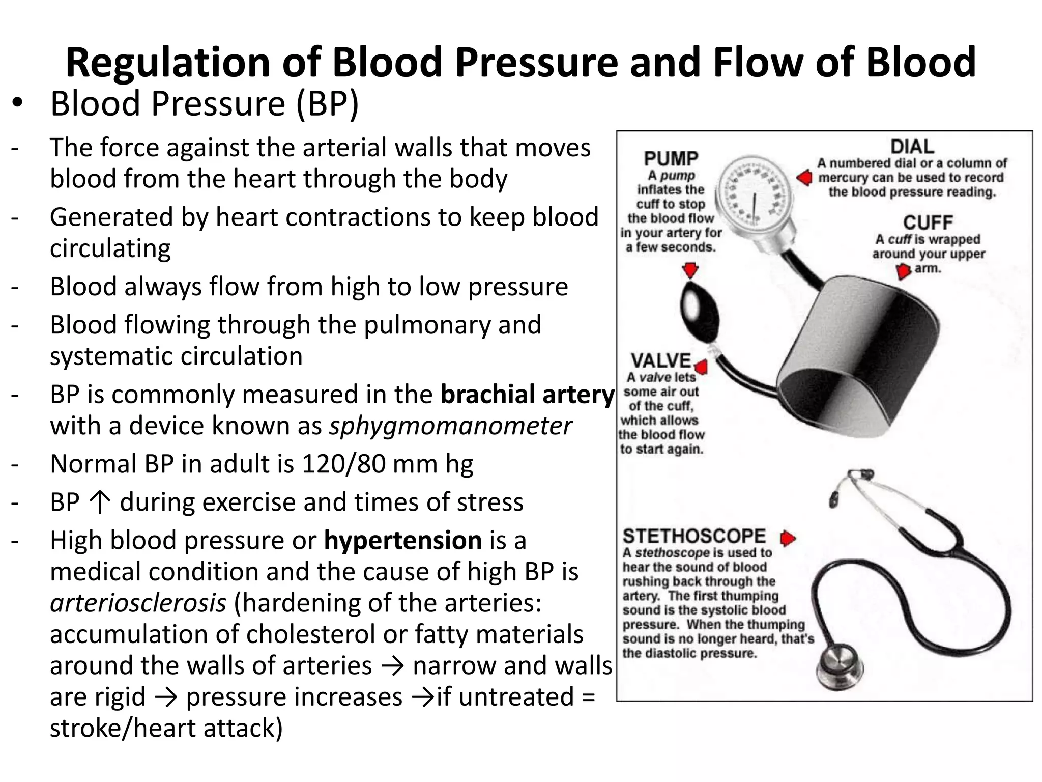
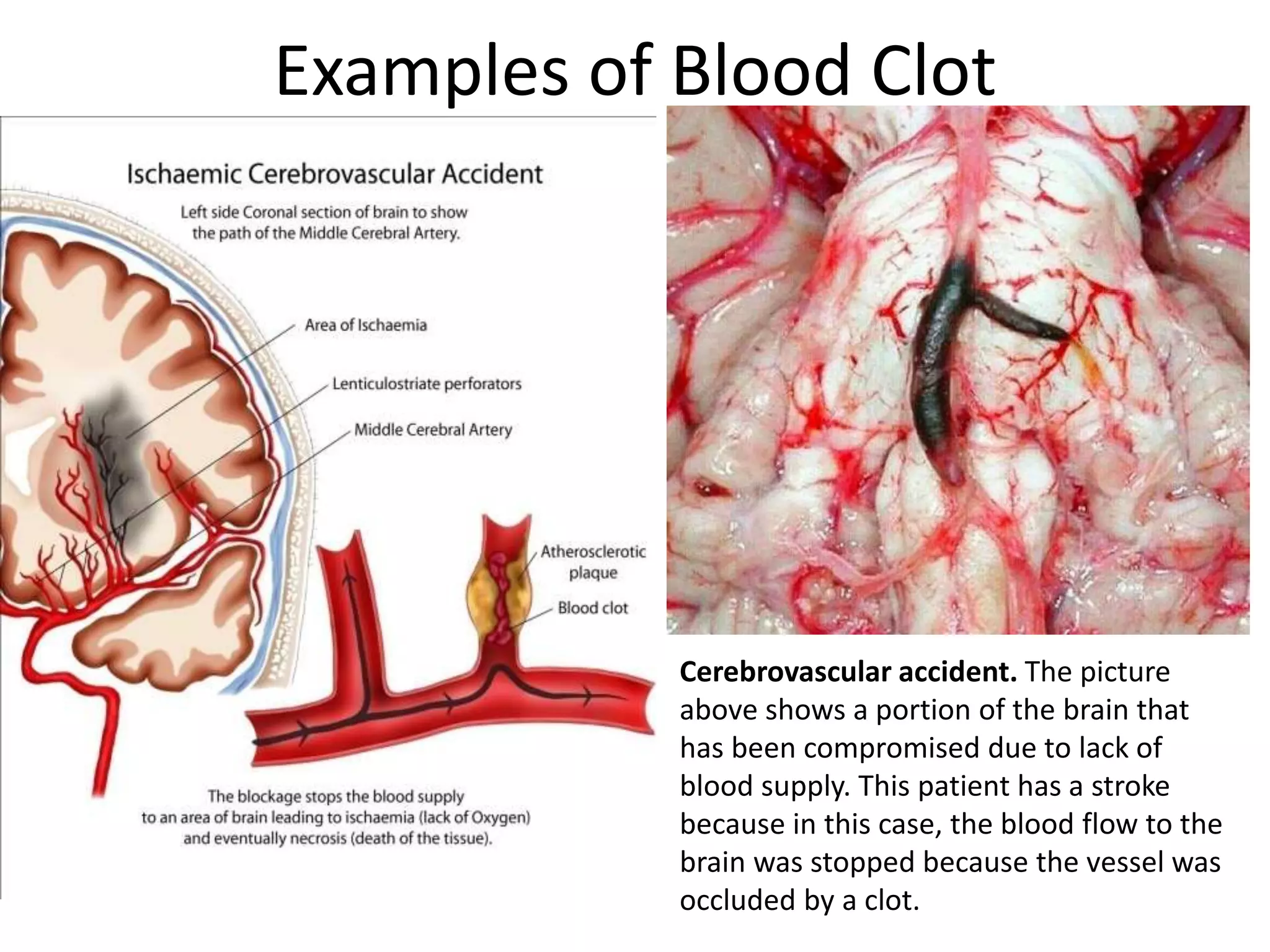
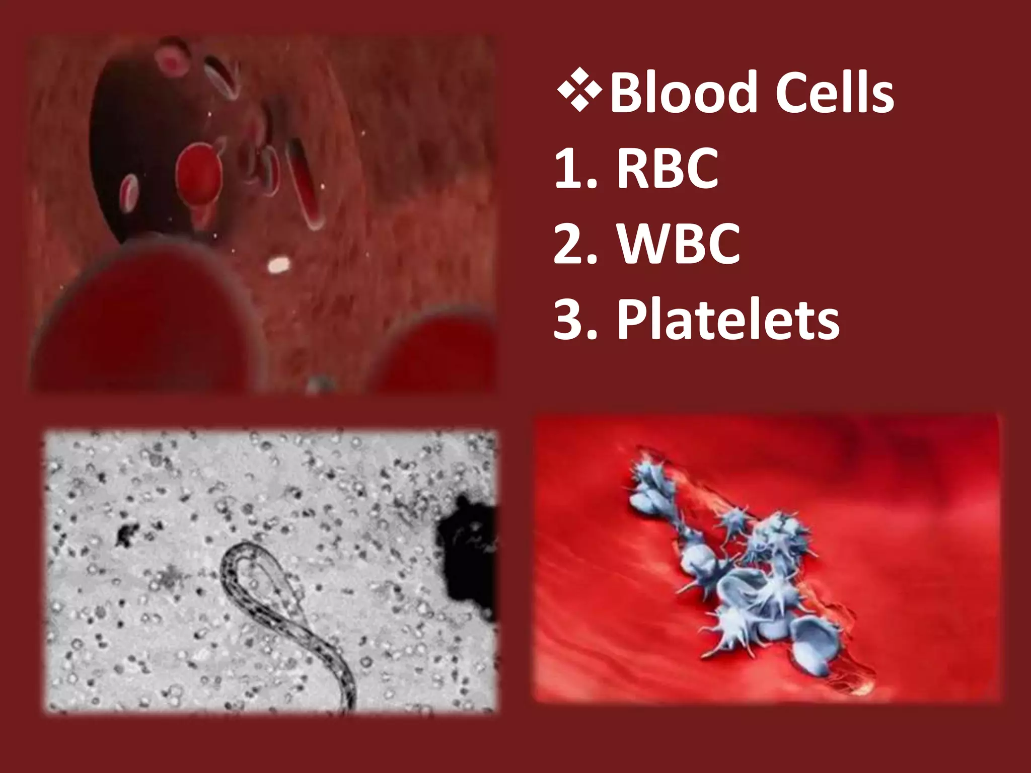
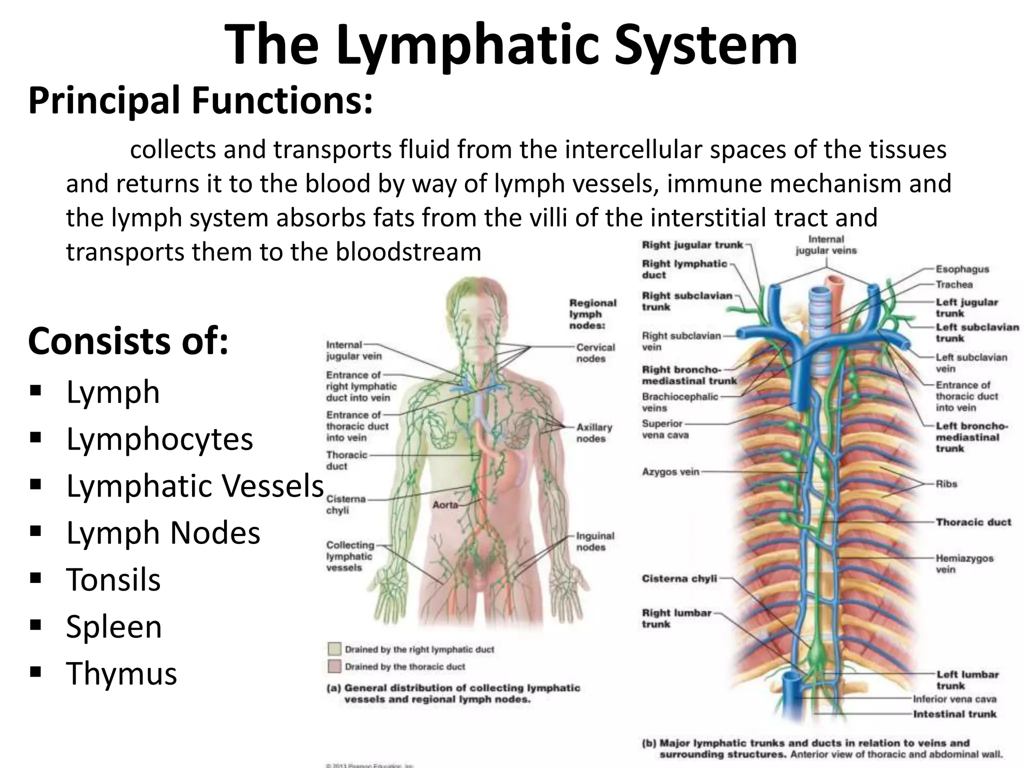
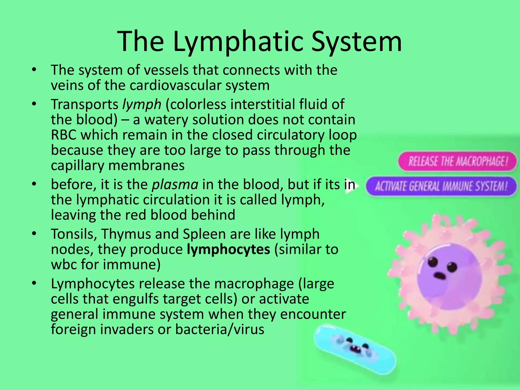
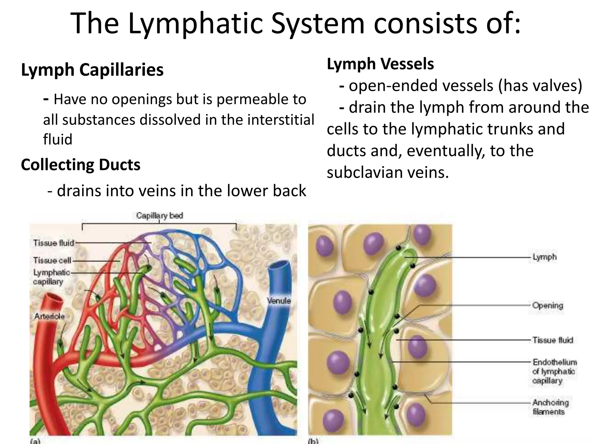
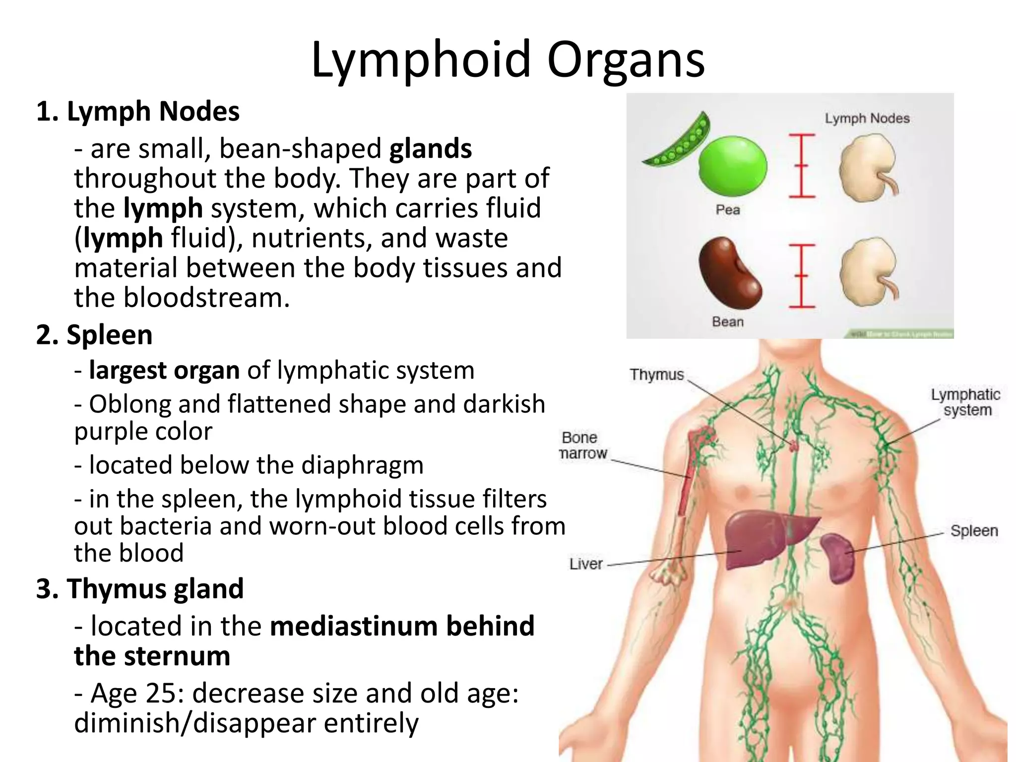
Chapter 11
The Circulatory System
Group 5
Toñacao, Christine
Castillo, Melecia Megan
Cal, Kesler
Calustre, Reian
Circulatory System
• “The flow of nutrients, other elements and
water into living organisms which are needed
for growth and reproduction.”
• 3 major structures of this system:
a. Heart
b. Blood Vessels
c. Blood
• 2 major circuit pathways:
a. Pulmonary pathway
b. Systemic pathway
Circulation
Two Kinds of Circulatory System:
1. Open Circulatory System
2. Close Circulatory System
KINDS OF CIRCULATORY SYSTEM
1. OPEN CIRCULATORY SYSTEM (insects and other invertebrates)
- blood does not remain in the vessels but instead is pumped from the
heart into that open directly into tissue
- OSTIA (openings in heart) is present
- common to molluscs and arthropods
(evolved in crustaceans, insects, mollusks
and other invertebrates)
KINDS OF CIRCULATORY SYSTEM
2. CLOSE CIRCULATORY SYSTEM
- blood remains within the vessels throughout its entire circuit
- Ostia is ABSENT
THE HUMAN
CIRCULATORY SYSTEM
1. Cardiovascular System
2. Lymphatic Vascular System
HUMAN CIRCULATORY SYSTEM
1. CARDIOVASCULAR SYSTEM
- composed of the blood vessels,
the blood and the heart
2. LYMPHATIC VASCULAR SYSTEM
- composed of lymph capillaries,
lymph vessels and collecting ducts.
BLOOD VESSELS
VEINS, ARTERIES & CAPILLARIES
Blood Vessels
Blood Vessels
1. Veins
- carries unoxygentated
blood containing picked
up from body cells
towards the heart.
- flow of blood is slow and
smooth
- walls are thin, don't have
much smooth muscle;
readily distended when
internal pressure is high
Venules
- refers to the smallest veins
- walls are only a little thicker
than those of capillaries
- both veins and venules
serve as temporary
reservoir for blood
volume; highly
distensible
- blood volume is high in
these vessels when the
body's metabolic activity
increases
- 50% of total blood volume;
found in veins
- prolonged compression
of the superficial veins in
the legs can contribute
varicose veins
Blood Vessels2. Arteries
- carries oxygenated blood away
from the heart where the flow
of blood is pulsating or in
spurts.
- walls are thick and deeply set
with impermeable walls
containing smooth muscles
and distensible tissues.
- composed of three outer and
inner layers that are primarily
connective tissues
- middle layer; made up of
muscle fibers and elastic
connective tissue
- smallest arteries is called
arterioles
CONDITIONS
Aneurism
- a birth defect or
injury can cause
the inner wall of
the artery to bulge
Stroke
- a weakened
artery in the brain
Three kinds of Arteries
1. ELASTIC ARTERIES
- large arteries leading directly
from the heart; also called
conducting arteries.
2. MUSCULAR ARTERIES
- medium sized with middle coats
that are chiefly muscular; also
called distributing arteries.
3. ARTERIOLES
- smallest arteries connected to
the capillaries that reduce
pressure of blood before
reaching the capillaries.
Blood Vessels
3. CAPILLARIES
- smallest blood vessels supplying blood to the
organs and tissues.
- an endothelial tubes that connect arteries and
veins
KINDS OF CAPILLARIES
1. CONTINUOUS CAPILLARIES
- found in muscles connective tissues and
central nervous system
2. FENESTRATED CAPILLARIES
- found in the renal glomeruli, endocrine glands
and intestine
3. SINUSOIDS
- the enlarged capillaries found in the liver, spleen
and bone marrow
- these vessels are so narrow that red blood cells
pass through them in single line.
- the thin walls of capillaries allow the exchange of
dissolved nutrients, wastes, oxygen and other
substances between blood and cells.
- capillary beds are easily destroyed
- high blood pressure can rapture the thin layered
capillaries
CONTINUOUS FENESTRATED
SINUSOIDS
Blood Vessels
The Heart
• A large, four-chambered muscular organ
(involuntary muscle)
• Location: slightly to the left of the middle
of the chest cavity
• Function: The right side of
the heart maintains pulmonary
circulation to the nearby lungs while the
left side of the heart pumps blood all the
way to the extremities of the body in the
systemic circulatory loop. The heart
functions by pumping blood both to the
lungs and to the systems of the body.
• No larger than the size of your fist
• Mass: about 300 grams
• 70 beats per minute
• (2.5 billion times in 70 years life span)
• Pumps blood enough to fill 2 large ocean
tankers
Parts of the Heart
Parts of the Heart
Parts of the Heart
ATRIOVENTRICULAR VALVES
(AV Valves)
 tricuspid valve (right)
 bicuspid/mitral valve (left)
SEMILUNAR VALVES
(SV Valves)
 pulmonary artery
 aortic (semilunar) valve
Function:
These flap-like valves
control the direction of the
blood flow inside the heart
(prevent it to flow back)
The Cardiac Cycle
• sequence of muscle contractions
and relaxation in the heart
•Two main pumping actions of the
heart:
a. Diastole - relaxation
b. Systole – contraction
•Lub sound – closing of the
atrioventricular valve (AV)
•Dub sound – closure of the
semilunar valve (SV)
• 72 contractions / min (forces 80 ml
of blood into the aorta)
• Stroke Volume – amount of blood
passing through the heart with each
heartbeat
•Cardiac Volume – amount of blood
pumped from the heart each minute
(5-6 liters)
The Cardiac Cycle
Right side of the heart (PULMONARY)
1. Blood enters the heart IVC and SVC,
emptying oxygen poor blood from
the body into the RA.
2. As the atrium contracts, blood flows
from your RA into your RV through
the open tricuspid valve.
3. When the ventricle is full, the
tricuspid valve shuts. This prevents
blood from flowing backward into
the right atrium while the ventricle
contracts.
4. As the ventricle contracts, blood
leaves the heart through the
pulmonary valve, into the
pulmonary artery and to the lungs,
where it is oxygenated. The
oxygenated blood then returns to
the heart through the pulmonary
veins.
1
2
The Cardiac Cycle
Left side of the heart (Systematic)
1. The pulmonary veins empty
oxygen-rich blood from the lungs
into the left atrium.
2. As the atrium contracts, blood
flows from your left atrium into
your left ventricle through the
open mitral valve.
3. When the ventricle is full, the
mitral valve shuts. This prevents
blood from flowing backward into
the atrium while the ventricle
contracts.
4. As the ventricle contracts, blood
leaves the heart through the
aortic valve, into the aorta and to
the body.
3
4
Two Major Pathways
Heart Conduction System
 a group of specialized cardiac muscle
cells in the walls of the heart that
send signals to the heart muscle
causing it to contract
 The main components of the cardiac
conduction system are the SA node,
AV node, bundle of His, bundle
branches, and Purkinje fibers.
Heart Conduction System
Sinoatrial Node (SV node)
- Aka Cardiac Pacemaker
- a small group of specialized muscle in the wall of the RA
- Influenced by the autonomic nerves ( these nerves regulates the
rate of heartbeat; impulses from the vagus nerve slow the
pacemaker)
Atrioventricular Node (AV node)
- Impulses reaches the AV node near the ventricles
- Triggers an impulse that causes the ventricles to contract
Bundle of His
- it transmits impulses from the atrioventricular node, located at the
inferior end of the interventricular septum, to the ventricles of the
heart.
Purkinje Fibers
- Where the signals from the left and right branches pass through
- Connected to the walls of the ventricles
Heart Conduction System
ECG machine
- An electrocardiogram (EKG or ECG) is a test that checks for problems with the
electrical activity of your heart. An EKG shows the heart's electrical activity as line
tracings on paper. The spikes and dips in the tracings are called waves. The heart is
a muscular pump made up of four chambers
Heart Condition
Arrhythmia or Dysrhythmia *wrong spelling sa book
- a disorder of the heart rate (pulse) or heart rhythm. The
heart can beat too fast (tachycardia), too slow (bradycardia)
- “Bad rhythm” in Greek and usually occurs in the upper
chamber of the heart, called the atria
Regulation of Blood Pressure and Flow of Blood
• Blood Pressure (BP)
- The force against the arterial walls that moves
blood from the heart through the body
- Generated by heart contractions to keep blood
circulating
- Blood always flow from high to low pressure
- Blood flowing through the pulmonary and
systematic circulation
- BP is commonly measured in the brachial artery
with a device known as sphygmomanometer
- Normal BP in adult is 120/80 mm hg
- BP ↑ during exercise and times of stress
- High blood pressure or hypertension is a
medical condition and the cause of high BP is
arteriosclerosis (hardening of the arteries:
accumulation of cholesterol or fatty materials
around the walls of arteries → narrow and walls
are rigid → pressure increases →if untreated =
stroke/heart attack)
Examples of Blood Clot
Cerebrovascular accident. The picture
above shows a portion of the brain that
has been compromised due to lack of
blood supply. This patient has a stroke
because in this case, the blood flow to the
brain was stopped because the vessel was
occluded by a clot.
Blood Cells
1. RBC
2. WBC
3. Platelets
Differences between RBC, WBC & Platelet
THE CLOTTING PROCESS
Platelets, Thromboplastin, Fibrin
The Clotting Process
• Aka Blood Coagulation
• an important process that prevents excessive
bleeding when a blood vessel is injured.
• Platelets (a type of blood cell) and proteins in
your plasma (the liquid part of blood) work
together to stop the bleeding by forming
a clot over the injury.
• The expected range for clotting time 4 – 10
mins
Blood Type
• Four types: O, A, B and AB
• Variation are due to two antigens on the surface
of the red cells, symbolized as A and B
• Antigens are proteins that increase the
manufacture of the anti-body (one or two other
types of proteins found in the blood plasma; Anti-
A and Anti-B)
• Blood Type O are universal donors because they
have no antigens
• Blood Type AB are universal recipients because of
having no antibodies in the plasma
Blood Clot illustration
Examples of Blood Clot
Venous thrombus. A blood clot (thrombus) that forms within the vein. The pictures
above is a condition called deep vein thrombosis (DVT) or sa bisaya “Barikos”. There
is blood clot in the deep veins of the leg.
The Lymphatic System
Principal Functions:
collects and transports fluid from the intercellular spaces of the tissues
and returns it to the blood by way of lymph vessels, immune mechanism and
the lymph system absorbs fats from the villi of the interstitial tract and
transports them to the bloodstream
Consists of:
 Lymph
 Lymphocytes
 Lymphatic Vessels
 Lymph Nodes
 Tonsils
 Spleen
 Thymus
The Lymphatic System
• The system of vessels that connects with the
veins of the cardiovascular system
• Transports lymph (colorless interstitial fluid of
the blood) – a watery solution does not contain
RBC which remain in the closed circulatory loop
because they are too large to pass through the
capillary membranes
• before, it is the plasma in the blood, but if its in
the lymphatic circulation it is called lymph,
leaving the red blood behind
• Tonsils, Thymus and Spleen are like lymph
nodes, they produce lymphocytes (similar to
wbc for immune)
• Lymphocytes release the macrophage (large
cells that engulfs target cells) or activate
general immune system when they encounter
foreign invaders or bacteria/virus
The Lymphatic System consists of:
Lymph Capillaries
- Have no openings but is permeable to
all substances dissolved in the interstitial
fluid
Collecting Ducts
- drains into veins in the lower back
Lymph Vessels
- open-ended vessels (has valves)
- drain the lymph from around the
cells to the lymphatic trunks and
ducts and, eventually, to the
subclavian veins.
Lymphoid Organs
1. Lymph Nodes
- are small, bean-shaped glands
throughout the body. They are part of
the lymph system, which carries fluid
(lymph fluid), nutrients, and waste
material between the body tissues and
the bloodstream.
2. Spleen
- largest organ of lymphatic system
- Oblong and flattened shape and darkish
purple color
- located below the diaphragm
- in the spleen, the lymphoid tissue filters
out bacteria and worn-out blood cells from
the blood
3. Thymus gland
- located in the mediastinum behind
the sternum
- Age 25: decrease size and old age:
diminish/disappear entirely
Lymphoid Organs
4. Tonsil
- masses of lymphoid tissues embedded in
mucous membrane
- disk-like shape and become smaller
during puberty
5. Patches of lymphoid tissues in the small
intestine
- The lymph nodes that become inflamed
are in a membrane that attaches
the intestine to the abdominal wall.
These lymph nodes are among the
hundreds that help your body fight
disease. They trap and destroy
microscopic "invaders" like viruses or
bacteria. Mesenteric lymphadenitis often
causes abdominal pain.
Thank you for listening!
Now lets play a game 

Cardiovascular System Part 1

  • 1.
    Chapter 11 The CirculatorySystem Group 5 Toñacao, Christine Castillo, Melecia Megan Cal, Kesler Calustre, Reian
  • 2.
    Circulatory System • “Theflow of nutrients, other elements and water into living organisms which are needed for growth and reproduction.” • 3 major structures of this system: a. Heart b. Blood Vessels c. Blood • 2 major circuit pathways: a. Pulmonary pathway b. Systemic pathway
  • 3.
    Circulation Two Kinds ofCirculatory System: 1. Open Circulatory System 2. Close Circulatory System
  • 4.
    KINDS OF CIRCULATORYSYSTEM 1. OPEN CIRCULATORY SYSTEM (insects and other invertebrates) - blood does not remain in the vessels but instead is pumped from the heart into that open directly into tissue - OSTIA (openings in heart) is present - common to molluscs and arthropods (evolved in crustaceans, insects, mollusks and other invertebrates)
  • 5.
    KINDS OF CIRCULATORYSYSTEM 2. CLOSE CIRCULATORY SYSTEM - blood remains within the vessels throughout its entire circuit - Ostia is ABSENT
  • 6.
    THE HUMAN CIRCULATORY SYSTEM 1.Cardiovascular System 2. Lymphatic Vascular System
  • 7.
    HUMAN CIRCULATORY SYSTEM 1.CARDIOVASCULAR SYSTEM - composed of the blood vessels, the blood and the heart
  • 8.
    2. LYMPHATIC VASCULARSYSTEM - composed of lymph capillaries, lymph vessels and collecting ducts.
  • 9.
  • 10.
  • 11.
    Blood Vessels 1. Veins -carries unoxygentated blood containing picked up from body cells towards the heart. - flow of blood is slow and smooth - walls are thin, don't have much smooth muscle; readily distended when internal pressure is high
  • 12.
    Venules - refers tothe smallest veins - walls are only a little thicker than those of capillaries - both veins and venules serve as temporary reservoir for blood volume; highly distensible - blood volume is high in these vessels when the body's metabolic activity increases - 50% of total blood volume; found in veins - prolonged compression of the superficial veins in the legs can contribute varicose veins
  • 13.
    Blood Vessels2. Arteries -carries oxygenated blood away from the heart where the flow of blood is pulsating or in spurts. - walls are thick and deeply set with impermeable walls containing smooth muscles and distensible tissues. - composed of three outer and inner layers that are primarily connective tissues - middle layer; made up of muscle fibers and elastic connective tissue - smallest arteries is called arterioles
  • 14.
    CONDITIONS Aneurism - a birthdefect or injury can cause the inner wall of the artery to bulge Stroke - a weakened artery in the brain
  • 15.
    Three kinds ofArteries 1. ELASTIC ARTERIES - large arteries leading directly from the heart; also called conducting arteries. 2. MUSCULAR ARTERIES - medium sized with middle coats that are chiefly muscular; also called distributing arteries. 3. ARTERIOLES - smallest arteries connected to the capillaries that reduce pressure of blood before reaching the capillaries.
  • 16.
    Blood Vessels 3. CAPILLARIES -smallest blood vessels supplying blood to the organs and tissues. - an endothelial tubes that connect arteries and veins KINDS OF CAPILLARIES 1. CONTINUOUS CAPILLARIES - found in muscles connective tissues and central nervous system 2. FENESTRATED CAPILLARIES - found in the renal glomeruli, endocrine glands and intestine 3. SINUSOIDS - the enlarged capillaries found in the liver, spleen and bone marrow - these vessels are so narrow that red blood cells pass through them in single line. - the thin walls of capillaries allow the exchange of dissolved nutrients, wastes, oxygen and other substances between blood and cells. - capillary beds are easily destroyed - high blood pressure can rapture the thin layered capillaries CONTINUOUS FENESTRATED SINUSOIDS
  • 17.
  • 18.
    The Heart • Alarge, four-chambered muscular organ (involuntary muscle) • Location: slightly to the left of the middle of the chest cavity • Function: The right side of the heart maintains pulmonary circulation to the nearby lungs while the left side of the heart pumps blood all the way to the extremities of the body in the systemic circulatory loop. The heart functions by pumping blood both to the lungs and to the systems of the body. • No larger than the size of your fist • Mass: about 300 grams • 70 beats per minute • (2.5 billion times in 70 years life span) • Pumps blood enough to fill 2 large ocean tankers
  • 19.
  • 20.
  • 21.
    Parts of theHeart ATRIOVENTRICULAR VALVES (AV Valves)  tricuspid valve (right)  bicuspid/mitral valve (left) SEMILUNAR VALVES (SV Valves)  pulmonary artery  aortic (semilunar) valve Function: These flap-like valves control the direction of the blood flow inside the heart (prevent it to flow back)
  • 22.
    The Cardiac Cycle •sequence of muscle contractions and relaxation in the heart •Two main pumping actions of the heart: a. Diastole - relaxation b. Systole – contraction •Lub sound – closing of the atrioventricular valve (AV) •Dub sound – closure of the semilunar valve (SV) • 72 contractions / min (forces 80 ml of blood into the aorta) • Stroke Volume – amount of blood passing through the heart with each heartbeat •Cardiac Volume – amount of blood pumped from the heart each minute (5-6 liters)
  • 23.
    The Cardiac Cycle Rightside of the heart (PULMONARY) 1. Blood enters the heart IVC and SVC, emptying oxygen poor blood from the body into the RA. 2. As the atrium contracts, blood flows from your RA into your RV through the open tricuspid valve. 3. When the ventricle is full, the tricuspid valve shuts. This prevents blood from flowing backward into the right atrium while the ventricle contracts. 4. As the ventricle contracts, blood leaves the heart through the pulmonary valve, into the pulmonary artery and to the lungs, where it is oxygenated. The oxygenated blood then returns to the heart through the pulmonary veins. 1 2
  • 24.
    The Cardiac Cycle Leftside of the heart (Systematic) 1. The pulmonary veins empty oxygen-rich blood from the lungs into the left atrium. 2. As the atrium contracts, blood flows from your left atrium into your left ventricle through the open mitral valve. 3. When the ventricle is full, the mitral valve shuts. This prevents blood from flowing backward into the atrium while the ventricle contracts. 4. As the ventricle contracts, blood leaves the heart through the aortic valve, into the aorta and to the body. 3 4
  • 25.
  • 26.
    Heart Conduction System a group of specialized cardiac muscle cells in the walls of the heart that send signals to the heart muscle causing it to contract  The main components of the cardiac conduction system are the SA node, AV node, bundle of His, bundle branches, and Purkinje fibers.
  • 27.
    Heart Conduction System SinoatrialNode (SV node) - Aka Cardiac Pacemaker - a small group of specialized muscle in the wall of the RA - Influenced by the autonomic nerves ( these nerves regulates the rate of heartbeat; impulses from the vagus nerve slow the pacemaker) Atrioventricular Node (AV node) - Impulses reaches the AV node near the ventricles - Triggers an impulse that causes the ventricles to contract Bundle of His - it transmits impulses from the atrioventricular node, located at the inferior end of the interventricular septum, to the ventricles of the heart. Purkinje Fibers - Where the signals from the left and right branches pass through - Connected to the walls of the ventricles
  • 28.
  • 29.
    Heart Conduction System ECGmachine - An electrocardiogram (EKG or ECG) is a test that checks for problems with the electrical activity of your heart. An EKG shows the heart's electrical activity as line tracings on paper. The spikes and dips in the tracings are called waves. The heart is a muscular pump made up of four chambers
  • 30.
    Heart Condition Arrhythmia orDysrhythmia *wrong spelling sa book - a disorder of the heart rate (pulse) or heart rhythm. The heart can beat too fast (tachycardia), too slow (bradycardia) - “Bad rhythm” in Greek and usually occurs in the upper chamber of the heart, called the atria
  • 31.
    Regulation of BloodPressure and Flow of Blood • Blood Pressure (BP) - The force against the arterial walls that moves blood from the heart through the body - Generated by heart contractions to keep blood circulating - Blood always flow from high to low pressure - Blood flowing through the pulmonary and systematic circulation - BP is commonly measured in the brachial artery with a device known as sphygmomanometer - Normal BP in adult is 120/80 mm hg - BP ↑ during exercise and times of stress - High blood pressure or hypertension is a medical condition and the cause of high BP is arteriosclerosis (hardening of the arteries: accumulation of cholesterol or fatty materials around the walls of arteries → narrow and walls are rigid → pressure increases →if untreated = stroke/heart attack)
  • 32.
    Examples of BloodClot Cerebrovascular accident. The picture above shows a portion of the brain that has been compromised due to lack of blood supply. This patient has a stroke because in this case, the blood flow to the brain was stopped because the vessel was occluded by a clot.
  • 33.
    Blood Cells 1. RBC 2.WBC 3. Platelets
  • 35.
  • 36.
    THE CLOTTING PROCESS Platelets,Thromboplastin, Fibrin
  • 37.
    The Clotting Process •Aka Blood Coagulation • an important process that prevents excessive bleeding when a blood vessel is injured. • Platelets (a type of blood cell) and proteins in your plasma (the liquid part of blood) work together to stop the bleeding by forming a clot over the injury. • The expected range for clotting time 4 – 10 mins
  • 38.
    Blood Type • Fourtypes: O, A, B and AB • Variation are due to two antigens on the surface of the red cells, symbolized as A and B • Antigens are proteins that increase the manufacture of the anti-body (one or two other types of proteins found in the blood plasma; Anti- A and Anti-B) • Blood Type O are universal donors because they have no antigens • Blood Type AB are universal recipients because of having no antibodies in the plasma
  • 39.
  • 40.
    Examples of BloodClot Venous thrombus. A blood clot (thrombus) that forms within the vein. The pictures above is a condition called deep vein thrombosis (DVT) or sa bisaya “Barikos”. There is blood clot in the deep veins of the leg.
  • 41.
    The Lymphatic System PrincipalFunctions: collects and transports fluid from the intercellular spaces of the tissues and returns it to the blood by way of lymph vessels, immune mechanism and the lymph system absorbs fats from the villi of the interstitial tract and transports them to the bloodstream Consists of:  Lymph  Lymphocytes  Lymphatic Vessels  Lymph Nodes  Tonsils  Spleen  Thymus
  • 42.
    The Lymphatic System •The system of vessels that connects with the veins of the cardiovascular system • Transports lymph (colorless interstitial fluid of the blood) – a watery solution does not contain RBC which remain in the closed circulatory loop because they are too large to pass through the capillary membranes • before, it is the plasma in the blood, but if its in the lymphatic circulation it is called lymph, leaving the red blood behind • Tonsils, Thymus and Spleen are like lymph nodes, they produce lymphocytes (similar to wbc for immune) • Lymphocytes release the macrophage (large cells that engulfs target cells) or activate general immune system when they encounter foreign invaders or bacteria/virus
  • 43.
    The Lymphatic Systemconsists of: Lymph Capillaries - Have no openings but is permeable to all substances dissolved in the interstitial fluid Collecting Ducts - drains into veins in the lower back Lymph Vessels - open-ended vessels (has valves) - drain the lymph from around the cells to the lymphatic trunks and ducts and, eventually, to the subclavian veins.
  • 44.
    Lymphoid Organs 1. LymphNodes - are small, bean-shaped glands throughout the body. They are part of the lymph system, which carries fluid (lymph fluid), nutrients, and waste material between the body tissues and the bloodstream. 2. Spleen - largest organ of lymphatic system - Oblong and flattened shape and darkish purple color - located below the diaphragm - in the spleen, the lymphoid tissue filters out bacteria and worn-out blood cells from the blood 3. Thymus gland - located in the mediastinum behind the sternum - Age 25: decrease size and old age: diminish/disappear entirely
  • 45.
    Lymphoid Organs 4. Tonsil -masses of lymphoid tissues embedded in mucous membrane - disk-like shape and become smaller during puberty 5. Patches of lymphoid tissues in the small intestine - The lymph nodes that become inflamed are in a membrane that attaches the intestine to the abdominal wall. These lymph nodes are among the hundreds that help your body fight disease. They trap and destroy microscopic "invaders" like viruses or bacteria. Mesenteric lymphadenitis often causes abdominal pain.
  • 46.
    Thank you forlistening! Now lets play a game 