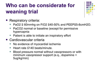
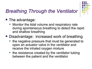
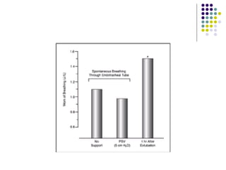
ICU book reading Chap 27 Discontinuing mechanical ventilation
Outline Readiness criteria Rapid-shallow breathing index Maximum inspiratory pressure The spontaneous breathing trial Breathing through the ventilator Breathing through the T-piece Protocol An approach to Rapid breathing Failure of spontaneous breathing Low cardiac output Overfeeding Respiratory muscle weakness Tracheal decannulation Protect airway Laryngeal edema Postextubation period
Who can be considerate for weaning trial Respiratory criteria: PaO2 ≧ 60mmhg on FiO2 ≦40-50% and PEEP≦5-8cmH2O. PaCO2 normal or baseline (except for permissive hypercapnia Patient is able to initiate an inspiratory effort Cardiovascular criteria No evidence of myocardial ischemia Heart rate ≦140 beats/minute. Blood pressure normal without vasopressors or with minimum vasopressor support (e.q., dopamine < 5ug/kg/min)
Who can be considerated for weaning trial Adequate mental status  Patient is arousable or Glosgow coma score≧13. Absence of correctible comorbid condition Patient is afebrile There are no significant electrolyte abnormalities
Measurements Used to Identify Patients Who Will Tolerate a Spontaneous Breathing Trial (SBT)
The predictive value
The Spontaneous Breathing Trial Breathing Through the Ventilator Breathing Through the T-Piece
Breathing Through the Ventilator The advantage: Monitor the tidal volume and respiratory rate during spontaneous breathing to detect the rapid and shallow breathing  Disadvantage:  increased work of breathing the negative pressure that must be generated to open an actuator valve in the ventilator and receive the inhaled oxygen mixture the resistance created by the ventilator tubing between the patient and the ventilator
Pressure-Support Ventilation add enough inspiratory pressure to reduce the work of breathing through the endotracheal tube and ventilator circuit without augmenting the spontaneous tidal volume A positive pressure of 5–7 cm H2O is routinely used for this purpose.
 
inspiratory pressure support is not necessary during spontaneous breathing trials Breathing through endotracheal tubes and ventilator circuits is not associated with an increased work of breathing, at least in comparison to the early time period following extubation
Breathing Through the T-Piece
Breathing Through the T-Piece Advantage:  it creates a “suction effect” that carries the exhaled gas out of the apparatus and prevents rebreathing of exhaled gas.  it prevents the patient from inhaling room air from the exhalation side of the apparatus. The disadvantage inability to monitor the patient's spontaneous tidal volume and respiratory rate less work of breathing when compared to spontaneous breathing while connected to the ventilator
Protocol Allow 30 to 120 minutes for the initial trial Increase the FiO2 by 10% for the period of spontaneous breathing Judgement: Combination of patient appearance (comfortable versus labored breathing), breathing pattern (i.e., the presence or absence of rapid, shallow breathing) and gas exchange (e.g., ability to maintain SaO2 =90% and end-tidal PCO2 normal or constant throughout the trial).
Protocol Our practice for patients who have been ventilator-dependent for one week or longer is to permit  at least 8 hours (and sometimes up to 24 hours)  of spontaneous breathing before deciding to remove the ventilator from the room.
An Approach to Rapid Breathing Anxiety versus ventilatory failure Abdominal Paradox
Rapid breath Tidal volume Decreased No Decreased Aterial PaCO2 Decreased No Decreased Resume ventilator support Sedation Resume ventilator support
Low Cardiac Output positive-pressure ventilation to negative-pressure spontaneous breathing  => increase in left-ventricular afterload => decrease in cardiac output  => promoting pulmonary congestion (which reduces lung compliance)  => impairing diaphragm function (Diaphram depend heavily on Cardiac output)
Low Cardiac Output O2 EXTRACTION: (SaO2 - SvO2) Arterial-End Tidal PCO2 Gradient: (PaCO2 – PetCO2) increase in the (PaCO2 – PetCO2) difference => A decrease in cardiac output => increase in dead-space ventilation from lung disease Myocardial Ischemia
Continuous Positive Airway Pressure CPAP can help by eliminating the increased afterload caused by negative intrathoracic pressures CPAP has been used effectively in patients with acute cardiogenic pulmonary edema,
Overfeeding An increase in the daily intake of calories is associated with an increase in metabolic CO2 production
Respiratory Muscle Weakness the diaphragm becomes weak during mechanical ventilation the diaphragm is not a voluntary muscle  that will stop contracting during mechanical ventilation. The diaphragm is controlled by the respiratory neurons in the lower brainstem, and these cells fire automatically throughout life.
Critical Illness Polyneuropathy and Myopathy These conditions often accompany cases of severe systemic sepsis with multiorgan failure, and they can prolong the need for mechanical ventilation The diagnosis is usually made by exclusion (although nerve conduction studies and electromyography can secure the diagnosis). There is no treatment for these disorders, and recovery takes weeks to months.
Electrolyte Depletion Depletion of magnesium and phosphorous can impair respiratory muscle strength
Tracheal Decannulation Protecting the Airway the gag and cough reflexes,  the volume of respiratory secretions. Laryngeal Edema reported in as many as 40% of cases of prolonged translaryngeal intubation
Protecting the Airway The ability to protect the airway =  the gag and cough reflexes,  the volume of respiratory secretions. Cough strength  can be assessed by placing a file card or piece of paper 1-2 cm from the end of the tracheal tube and asking the patient to cough. If wetness appears on the card, the cough strength is considered adequate
The Cuff-Leak Test The test is performed while the patient is receiving volume-cycled ventilation, and it involves measuring the volume of gas exhaled through the endotracheal tube before and after deflating the cuff a decrease in exhaled volume after cuff deflation is used as evidence against the presence of a significant obstruction at the level of the larynx .
The Cuff-Leak Test Different studies have used volumes of 110-140 mL, and a 25% change in volume as the threshold. volume leak during positive-pressure lung inflation, related to lung compliance and airways resistance the diameter of the endotracheal tube relative to the diameter of the trachea.
Fenestrated Tracheostomy Tubes Laryngeal injury maybe related to prior endotracheal tube or ischemic injury.
Steroids for Anything That Swells? Controversial it seems unlikely that one dose of steroids (or one day of therapy) will reverse the cumulative effects of days of trauma to the larynx
The Postextubation Period The Work of Breathing Endotracheal tube is smaller in diameter. But work of breathing is less endotracheal tubes should never be removed based on the assumption that it will be easier for the patient to breathe. Posrtextubation Stridor Postextubation stridor is not always an indication for immediate reintubation aerosolized epinephrine  (2.5 mL of 1% epinephrine) Breathing a helium-oxygen (heliox) gas mixture
A Final Word recognizing when a patient is ready to try spontaneous breathing A trial of spontaneous breathing If the patient does well, then consider extubation,

9710 Icu

  • 1.
    ICU book readingChap 27 Discontinuing mechanical ventilation
  • 2.
    Outline Readiness criteriaRapid-shallow breathing index Maximum inspiratory pressure The spontaneous breathing trial Breathing through the ventilator Breathing through the T-piece Protocol An approach to Rapid breathing Failure of spontaneous breathing Low cardiac output Overfeeding Respiratory muscle weakness Tracheal decannulation Protect airway Laryngeal edema Postextubation period
  • 3.
    Who can beconsiderate for weaning trial Respiratory criteria: PaO2 ≧ 60mmhg on FiO2 ≦40-50% and PEEP≦5-8cmH2O. PaCO2 normal or baseline (except for permissive hypercapnia Patient is able to initiate an inspiratory effort Cardiovascular criteria No evidence of myocardial ischemia Heart rate ≦140 beats/minute. Blood pressure normal without vasopressors or with minimum vasopressor support (e.q., dopamine < 5ug/kg/min)
  • 4.
    Who can beconsiderated for weaning trial Adequate mental status Patient is arousable or Glosgow coma score≧13. Absence of correctible comorbid condition Patient is afebrile There are no significant electrolyte abnormalities
  • 5.
    Measurements Used toIdentify Patients Who Will Tolerate a Spontaneous Breathing Trial (SBT)
  • 6.
  • 7.
    The Spontaneous BreathingTrial Breathing Through the Ventilator Breathing Through the T-Piece
  • 8.
    Breathing Through theVentilator The advantage: Monitor the tidal volume and respiratory rate during spontaneous breathing to detect the rapid and shallow breathing Disadvantage: increased work of breathing the negative pressure that must be generated to open an actuator valve in the ventilator and receive the inhaled oxygen mixture the resistance created by the ventilator tubing between the patient and the ventilator
  • 9.
    Pressure-Support Ventilation addenough inspiratory pressure to reduce the work of breathing through the endotracheal tube and ventilator circuit without augmenting the spontaneous tidal volume A positive pressure of 5–7 cm H2O is routinely used for this purpose.
  • 10.
  • 11.
    inspiratory pressure supportis not necessary during spontaneous breathing trials Breathing through endotracheal tubes and ventilator circuits is not associated with an increased work of breathing, at least in comparison to the early time period following extubation
  • 12.
  • 13.
    Breathing Through theT-Piece Advantage: it creates a “suction effect” that carries the exhaled gas out of the apparatus and prevents rebreathing of exhaled gas. it prevents the patient from inhaling room air from the exhalation side of the apparatus. The disadvantage inability to monitor the patient's spontaneous tidal volume and respiratory rate less work of breathing when compared to spontaneous breathing while connected to the ventilator
  • 14.
    Protocol Allow 30to 120 minutes for the initial trial Increase the FiO2 by 10% for the period of spontaneous breathing Judgement: Combination of patient appearance (comfortable versus labored breathing), breathing pattern (i.e., the presence or absence of rapid, shallow breathing) and gas exchange (e.g., ability to maintain SaO2 =90% and end-tidal PCO2 normal or constant throughout the trial).
  • 15.
    Protocol Our practicefor patients who have been ventilator-dependent for one week or longer is to permit at least 8 hours (and sometimes up to 24 hours) of spontaneous breathing before deciding to remove the ventilator from the room.
  • 16.
    An Approach toRapid Breathing Anxiety versus ventilatory failure Abdominal Paradox
  • 17.
    Rapid breath Tidalvolume Decreased No Decreased Aterial PaCO2 Decreased No Decreased Resume ventilator support Sedation Resume ventilator support
  • 18.
    Low Cardiac Outputpositive-pressure ventilation to negative-pressure spontaneous breathing => increase in left-ventricular afterload => decrease in cardiac output => promoting pulmonary congestion (which reduces lung compliance) => impairing diaphragm function (Diaphram depend heavily on Cardiac output)
  • 19.
    Low Cardiac OutputO2 EXTRACTION: (SaO2 - SvO2) Arterial-End Tidal PCO2 Gradient: (PaCO2 – PetCO2) increase in the (PaCO2 – PetCO2) difference => A decrease in cardiac output => increase in dead-space ventilation from lung disease Myocardial Ischemia
  • 20.
    Continuous Positive AirwayPressure CPAP can help by eliminating the increased afterload caused by negative intrathoracic pressures CPAP has been used effectively in patients with acute cardiogenic pulmonary edema,
  • 21.
    Overfeeding An increasein the daily intake of calories is associated with an increase in metabolic CO2 production
  • 22.
    Respiratory Muscle Weaknessthe diaphragm becomes weak during mechanical ventilation the diaphragm is not a voluntary muscle that will stop contracting during mechanical ventilation. The diaphragm is controlled by the respiratory neurons in the lower brainstem, and these cells fire automatically throughout life.
  • 23.
    Critical Illness Polyneuropathyand Myopathy These conditions often accompany cases of severe systemic sepsis with multiorgan failure, and they can prolong the need for mechanical ventilation The diagnosis is usually made by exclusion (although nerve conduction studies and electromyography can secure the diagnosis). There is no treatment for these disorders, and recovery takes weeks to months.
  • 24.
    Electrolyte Depletion Depletionof magnesium and phosphorous can impair respiratory muscle strength
  • 25.
    Tracheal Decannulation Protectingthe Airway the gag and cough reflexes, the volume of respiratory secretions. Laryngeal Edema reported in as many as 40% of cases of prolonged translaryngeal intubation
  • 26.
    Protecting the AirwayThe ability to protect the airway = the gag and cough reflexes, the volume of respiratory secretions. Cough strength can be assessed by placing a file card or piece of paper 1-2 cm from the end of the tracheal tube and asking the patient to cough. If wetness appears on the card, the cough strength is considered adequate
  • 27.
    The Cuff-Leak TestThe test is performed while the patient is receiving volume-cycled ventilation, and it involves measuring the volume of gas exhaled through the endotracheal tube before and after deflating the cuff a decrease in exhaled volume after cuff deflation is used as evidence against the presence of a significant obstruction at the level of the larynx .
  • 28.
    The Cuff-Leak TestDifferent studies have used volumes of 110-140 mL, and a 25% change in volume as the threshold. volume leak during positive-pressure lung inflation, related to lung compliance and airways resistance the diameter of the endotracheal tube relative to the diameter of the trachea.
  • 29.
    Fenestrated Tracheostomy TubesLaryngeal injury maybe related to prior endotracheal tube or ischemic injury.
  • 30.
    Steroids for AnythingThat Swells? Controversial it seems unlikely that one dose of steroids (or one day of therapy) will reverse the cumulative effects of days of trauma to the larynx
  • 31.
    The Postextubation PeriodThe Work of Breathing Endotracheal tube is smaller in diameter. But work of breathing is less endotracheal tubes should never be removed based on the assumption that it will be easier for the patient to breathe. Posrtextubation Stridor Postextubation stridor is not always an indication for immediate reintubation aerosolized epinephrine (2.5 mL of 1% epinephrine) Breathing a helium-oxygen (heliox) gas mixture
  • 32.
    A Final Wordrecognizing when a patient is ready to try spontaneous breathing A trial of spontaneous breathing If the patient does well, then consider extubation,