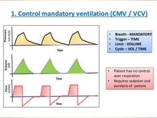
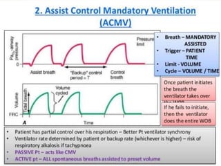
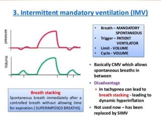
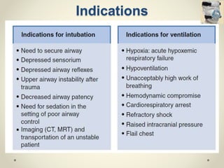
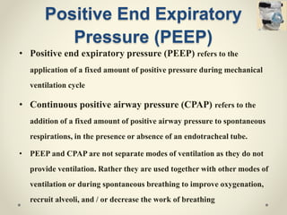
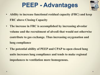
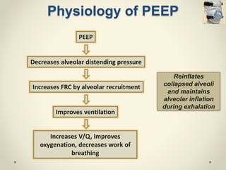
Ventilator in
critical care
Presenter : Dr. Dhileeban Maharajan
Moderator : Prof. Lk. Sharatchandra
Contents
1. History
2. Types of Ventilators
3. Modes of Mechanical Ventilation
4. Indications
5. Complications
6. Ventilator setting in Specific diseases
7. Weaning
8. Newer methods
Introduction
• Mechanical Ventilation - Common life saving intervention
• Required for assisting and replacing spontaneous
breathing
• Poor ventilatory management can inflict serious pulmonary
and extra-pulmonary damage
• Optimizing ventilatory parameters reduces overall duration
of mechanical ventilation and organ failure
• Upsurge in utilization of non-invasive ventilation has
History
• 5th century – Hippocrates (Treatise on air)
• 1530 – Paracelus – Pump air into patients mouth
• 1653 – Andreas Vesalius – Tracheostomy in dog
• 1744 – John Fothergill – Mouth to mouth breathing
• 1880 – First endotracheal intubation tried
• 1929 – Drinker and Shaw – Negative pressure ventilation
• 1963 – Positive pressure ventilation
• 1971 – Gregory et al – CPAP
1. Iron lung
2. Cuirass
3. Body suit
Negative Pressure Ventilation
Negative pressure outside thorax
Expansion of thoracic cage
Fall in pleural pressure
Air enter into the lungs
Examples:
Positive Pressure
Ventilation
Airway pressure is applied at the patient's airway through an
endotracheal or tracheostomy tube. The positive nature of the
pressure causes the gas to flow into the lungs until the ventilator
breath is terminated. As the airway pressure drops to zero, elastic
recoil of the chest accomplishes passive exhalation by pushing the
tidal volume out.
• Polio epidemic in Scandinavia – 1950
• Manual positive pressure ventilation
• Mortality reduced 80 to 25%
Positive Pressure
Ventilation - Cont
• Mode: (Flow, Pressure, Time)
o Manner in which ventilator breaths are triggered, cycled and
limited
• Trigger: (Flow, Pressure)
o Either an inspiratory effort or time based signal, defines
what the ventilator senses to initiate an assisted breath
• Cycle: (Volume, Flow, Pressure, Time)
o Factors that determine the end of inspiration
• Limiting factors:
o Operator specified values such as airway pressure, to
prevent injury to lungs
Types of support
• Control mode
o Delivers preset tidal volume once it triggered
regardless of patient effort
• Support mode
o Provides inspiratory assistance (Requires adequate
respiratory drive)
Methods of ventilatory
support
• Continuous mandatory ventilation (CMV)
• Assist – Control ventilation (A/C)
• Intermittent mandatory ventilation (IMV)
• Synchronous intermittent mandatory ventilation
(SIMV)
• Pressure support ventilation (PSV)
• Noninvasive ventilation
o CPAP
o BIPAP
Continuous Mandatory
Ventilation (CMV)
• Preset interval breath regardless of patients effort
• Patients should be sedated and paralysed (NM blockers)
• Used for patients fights the ventilator during initial
stages, tetanus, seizures, complete rest for 24 hours,
crushed chest injury.
Assist Control
Ventilation (A/C)
• Preset breath in coordination with respiratory effort
• Delivers full assisted tidal volume
• Spontaneous breathing not allowed
• If patient had appropriate ventilator drive, allows patient to
control frequency and minute volume to normalize PaCO2
• To provide full ventilatory support for patients (full work of
breathing)
• Used for patients having stable respiratory drive (at least 10
/min)
Intermittent Mandatory
Ventilation (IMV)
• Breaths at preset interval
• Spontaneous breath allowed
• Allow patients breath in addition to ventilator delivered
breaths.
• Breath stacking – increased risk of barotrauma
Synchronous Intermittent
Mandatory Ventilation (SIMV)
• Preset breaths in coordination with respiratory effort
• Spontaneous breathing allowed
• Time interval just prior to time triggering in which
ventilator is responsive to patients spontaneous
inspiratory efforts is called as synchronization window
mostly 0.5 sec (manufacturer set)
• SIMV maintains respiratory muscle strength, reduces V/Q
mismatch, decreases mean airway pressure, facilitates
weaning
Pressure support
ventilation• For spontaneously breathing patient
• To limit barotrauma and work of breathing
• Pressure support according to inspiratory flow
• when the patient triggers ,pressure supported breath is
delivered by demand valves which generates high flow
to increase airway pressure to pressure limit and
maintains pressure plateau (demand and servo valves)
for the duration of patients spontaneous inspiratory
efforts.
• PSV used with SIMV to facilitate weaning
Noninvasive ventilation
(NPPV)
• Ventilatory support through mask
• Decreases the intubation by 20%
• CPAP and BiPAP
• Used in
o COPD acute exacerbation
o Cardiogenic pulmonary edema
o Post-extubation respiratory
failure in hypercapnic patients
o Pneumonia in
immunocompromised patients
Contraindications :
• Cardiac or Respiratory arrest
• Severe encephalopathy
• Hemodynamic instability
• Facial trauma or surgery
• Upper airway obstruction
• High risk aspiration
• Inability to clear secretions
Noninvasive ventilation –
Cont..
• Place the patient in upright or sitting position
• Choose a proper fitting mask
• Attach the interface and circuit to ventilator
• Allow the patient to hold the mask
• Monitor oxygen saturation
• Secure the mask
• Titrate the pressure settings
• Check for any leaks
• Monitor RR, HR, Saturation, Minute ventilation, Exhaled Tt
• Obtain ABG within one hour
(Protocol for initiating NIV)
• Higher level consciousness
• Younger age
• Less severe abnormalities
• Minimal air leakage
• Intact dentition
• Synchronous breathing
• Absence of pneumonia
• Positive initial response
o Correction of pH
o Decreased respiratory rate
o Reduced PaCO2
Noninvasive ventilation –
Cont..
• Nasal bridge ulcer and necrosis
• Conjunctival irritation
• Gastric insufflation
• Ventilator asynchrony
• Claustrophobia
• Risk of aspiration
Predictors of success
Disadvantages
Indications for mechanical
ventilation
• Bradypnoea or apnoea with respiratory arrest
• Acute lung injury and acute respiratory distress syndrome
• Tachypnoea (respiratory rate >30 breaths per minute)
• Vital capacity less than 15 mL/kg
• Minute ventilation greater than 10 L/min
• PaO2 with a supplemental FIO2 of less than 55 mm Hg
• Alveolar-arterial gradient of oxygen tension (A-a DO2) with
100% oxygenation of greater than 450 mm Hg
Indication – Cont…
• Clinical deterioration
• Respiratory muscle fatigue
• Obtundation or coma
• Hypotension
• Acute partial pressure of carbon dioxide (PaCO2) greater than
50 mm Hg with an arterial pH less than 7.25
• Neuromuscular disease
Indications
Positive End Expiratory
Pressure (PEEP)
• Positive end expiratory pressure (PEEP) refers to the
application of a fixed amount of positive pressure during mechanical
ventilation cycle
• Continuous positive airway pressure (CPAP) refers to the
addition of a fixed amount of positive airway pressure to spontaneous
respirations, in the presence or absence of an endotracheal tube.
• PEEP and CPAP are not separate modes of ventilation as they do not
provide ventilation. Rather they are used together with other modes of
ventilation or during spontaneous breathing to improve oxygenation,
recruit alveoli, and / or decrease the work of breathing
PEEP - Advantages
• Ability to increase functional residual capacity (FRC) and keep
FRC above Closing Capacity
• The increase in FRC is accomplished by increasing alveolar
volume and the recruitment of alveoli that would not otherwise
contribute to gas exchange. Thus increasing oxygenation and
lung compliance
• The potential ability of PEEP and CPAP to open closed lung
units increases lung compliance and tends to make regional
impedances to ventilation more homogenous.
Physiology of PEEP
Reinflates
collapsed alveoli
and maintains
alveolar inflation
during exhalation
PEEP
Decreases alveolar distending pressure
Increases FRC by alveolar recruitment
Improves ventilation
Increases V/Q, improves
oxygenation, decreases work of
breathing
Complications of Mechanical
Ventilation
• Laryngeal injury
• Pharyngeal laceration
o Infection of the retro
pharyngeal space
o Mediastinitis
o Pneumothorax
• Tracheal rupture
• Nasal injury and epistaxis
• Dental trauma
• Cervical spine injury
• Esophageal intubation
• Right mainstem intubation
• Cardiac arrhythmia
• Aspiration
• Bronchospasm
Peri- intubation complication
• Endotracheal tube obstruction
• Endotracheal tube migration
• Self-extubation
• Cuff leak
• Barotrauma and Volutrauma
o Subpleural air cyst
o Pneumothorax
o Pneumomediastinum
o Pneumoperitoneum
o Subcutaneous emphysema
Acute complications that can occur at any time
during mechanical ventilation
• Biotrauma
• Hypotension
• Dynamic hyperinflation
• Atelectasis
• Alveolar hypoventilation or
hyperventilation
• Gastric dilation
• Inadvertent disconnection
from ventilator
• Machine failure
• Peri-oral pressure sores
• Nasal necrosis
• Sinusitis
• Serous or purulent otitis media
• Pneumonia
• Tracheomalacia
• Tracheo-esophageal fistula
• Oxygen toxicity
Mechanical Ventilation - Delayed complications
Ventilator induced lung injury
(Barotrauma)
• Prevalence – 10%
• Large Tidal volume and elevated peak inspiratory and plateau
pressure – Risk factors
• Barotrauma refers to rupture of the alveolus with subsequent
entry of air into the pleural space (pneumothorax) and/or the
tracking or air along the vascular bundle to the mediastinum
(pneumomediastinum)
• Decrease the inspiratory-to expiratory ratio is important to
prevent barotrauma
• local overdistention of normal alveoli
• lung-protective ventilation strategy is recommended
Ventilator induced lung injury
(Volutrauma)
Oxygen Toxicity
• Oxygen toxicity is due to the production of oxygen free radicals
such as superoxide anion, hydroxyl radical, and hydrogen
Peroxide
• Reported in patients given a maintenance FIO2 of 50% or
greater
• Clinician should attempt to attain an FIO2 of 60% or less within
the first 24 hours of mechanical ventilation
• PEEP should be considered a means to improve oxygenation
while a safe FIO2 is maintained
Ventilator Associated
Pneumonia (VAP)
• Incidence 1 to 4 cases per 1000 ventilator days
• Rate of 3% per day for the first 5 days, 2% per day for next 5
days
• VAP is defined as a new infection of the lung parenchyma that
develops within 48 hours after intubation
• Fever, leukocytosis, and purulent tracheobronchial secretions.
• Qualitative and quantitative cultures of protected brush and
bronchoalveolar lavage specimens may help
• First 48 hours after intubation - Flora of the upper airway,
including Haemophilus influenza and Streptococcus pneumonia
VAP – cont..
• After this early period, gram-negative bacilli such as
Pseudomonas aeruginosa, Escherichia coli, Acinetobacter,
Proteus, and Klebsiella species predominate
• Staphylococcus aureus, especially methicillin-resistant S aureus
(MRSA), typically becomes a major infective agent after 7 days
• Initial therapy - with broad-spectrum antibiotics and change the
antibiotic after the culture sensitivity report
Intrinsic PEEP
• Most frequently occurs in patients with COPD or asthma who
require prolonged expiratory phase of respiration
• This problem occurs, a portion of each subsequent tidal volume
may be retained in the patient's lungs, a phenomenon sometimes
referred to as breath stacking
• Results in barotrauma, volutrauma
• The normal inspiratory to expiratory ratio (I:E ratio) is 1:2. In
patients with obstructive airway disease, the target I:E ratio
should be 1:3 to 1:4.
Breath stacking
Ventilator Setup
• Assist – Control mode
• Tidal volume depending upon the lung status
o Normal - 12mL/kg; COPD – 10mL/kg; ARDS – 6-8mL/kg
• Rate of 10 – 12 breaths/min
• FIO2 of 100%
• PEEP only as indicated
Ventilator setting in COPD
and asthma
• Permissive hypercapnia
• Reduce the expiratory rate
• Low set tidal volume
• Increase the expiratory flow time
• NPPV reduced the endotracheal
intubation by 59%
High airway pressure
Breath stacking
Intrinsic PEEP
Barotrauma
• Compliance reduced and dead space increased
• Lung protective ventilation
o Low tidal volume ( Close to 6ml/kg BW)
o Prevent plateau pressure exceeding 30cmH2O
o Lowest possible FIo2 to keep the Spo2 at >90%
o Adjust the PEEP to maintain alveolar patency
• Early prone positioning
Ventilator setting in ARDS
• To decrease the work of breathing and oxygen demand of
the respiratory muscles
• Arrhythmia common with hypocapnia
• In CHF - CPAP or BiPAP reduce preload
• Potential effects of PEEP in LV dysfunction
o Reduce venous return and preload
o Increased FRC lead to increased pulmonary patency
o Compression may increase afterload
Ventilator setting in Heart
Disease
• Prevent aspiration
• Hyperventilation  Reduce the intracranial pressure
• Maintain PaCO2 between 30 to 39mmHg
• Recent studies suggest poor outcome
Ventilator setting in Traumatic
Brain Injury
Weaning
• Unsupported spontaneous breathing trials(SBT)
o The machine support is withdrawn
o T-Piece (or CPAP) circuit can be attached
• Intermittent mandatory ventilation (IMV) weaning
o The ventilator delivers a pre-set minimum minute volume
o Synchronized (SIMV) to the patient's own respiratory efforts
• Pressure support weaning
o Patient initiates all breaths and these are 'boosted' by the ventilator.
o Gradually reducing the level of pressure support,
o Once the level of pressure support is low (5-10 cmH2O above PEEP), a
trial of T-Piece or CPAP weaning should be started
Weaning- Cont…
• Lung injury is stable or resolving
• Gas exchange adequate
o Low PEEP (<8cmH2O)
o Low FIO2 (<0.5)
• Stable hemodynamic variables
• Able to initiate spontaneous breath
• Absence of infection or fever
Indications Failed SBT
• Resp. Rate >35 for >5min
• Heart Rate >140/min
• SPO2 <90% for >30sec
• Systolic BP >180 or <90
• Sustained increased WOB
• Cardiac dysrhythmia
• pH <7.32
10 to 15% patient require reintubation
Newer Modes
• Dual modes
o Pressure regulated volume control(PRVC) ventilation
o Volume support ventilation (VSV)
o Variable pressure control mode
o Volume assured pressure support ventilation
• Proportional assist ventilation
• Inverse ratio ventilation
• High frequency jet ventilation
• Neurally adjusted ventilatory assist ventilation (NAV)
• Extracorporeal membrane oxygenation (ECMO)
Summary
• Technological advances added new methods in MV
• NPPV is an important treatment modality prevents the
need for endotracheal intubation
• Recent guidelines suggest that the use of low tidal
volume in ARDS associated with favorable outcome
• Weaning guidelines have shown improved outcome
• Although MV is life saving, it is fraught with a plethora of
complications
Thank you
REFERENCE
• Pittsburgh critical care medicine – Mechanical Ventilation; Second Edition
• Ashfaq Hasan – Understanding mechanical ventilation;
• Harrison’s Principles of internal medicine; 20th Edition
• API Textbook of Medicine; 11th Edition
• Medicine Update 2019; Volume 29

Ventilator in Critical Care

  • 1.
    Ventilator in critical care Presenter: Dr. Dhileeban Maharajan Moderator : Prof. Lk. Sharatchandra
  • 2.
    Contents 1. History 2. Typesof Ventilators 3. Modes of Mechanical Ventilation 4. Indications 5. Complications 6. Ventilator setting in Specific diseases 7. Weaning 8. Newer methods
  • 3.
    Introduction • Mechanical Ventilation- Common life saving intervention • Required for assisting and replacing spontaneous breathing • Poor ventilatory management can inflict serious pulmonary and extra-pulmonary damage • Optimizing ventilatory parameters reduces overall duration of mechanical ventilation and organ failure • Upsurge in utilization of non-invasive ventilation has
  • 4.
    History • 5th century– Hippocrates (Treatise on air) • 1530 – Paracelus – Pump air into patients mouth • 1653 – Andreas Vesalius – Tracheostomy in dog • 1744 – John Fothergill – Mouth to mouth breathing • 1880 – First endotracheal intubation tried • 1929 – Drinker and Shaw – Negative pressure ventilation • 1963 – Positive pressure ventilation • 1971 – Gregory et al – CPAP
  • 5.
    1. Iron lung 2.Cuirass 3. Body suit Negative Pressure Ventilation Negative pressure outside thorax Expansion of thoracic cage Fall in pleural pressure Air enter into the lungs Examples:
  • 6.
    Positive Pressure Ventilation Airway pressureis applied at the patient's airway through an endotracheal or tracheostomy tube. The positive nature of the pressure causes the gas to flow into the lungs until the ventilator breath is terminated. As the airway pressure drops to zero, elastic recoil of the chest accomplishes passive exhalation by pushing the tidal volume out. • Polio epidemic in Scandinavia – 1950 • Manual positive pressure ventilation • Mortality reduced 80 to 25%
  • 7.
    Positive Pressure Ventilation -Cont • Mode: (Flow, Pressure, Time) o Manner in which ventilator breaths are triggered, cycled and limited • Trigger: (Flow, Pressure) o Either an inspiratory effort or time based signal, defines what the ventilator senses to initiate an assisted breath • Cycle: (Volume, Flow, Pressure, Time) o Factors that determine the end of inspiration • Limiting factors: o Operator specified values such as airway pressure, to prevent injury to lungs
  • 8.
    Types of support •Control mode o Delivers preset tidal volume once it triggered regardless of patient effort • Support mode o Provides inspiratory assistance (Requires adequate respiratory drive)
  • 9.
    Methods of ventilatory support •Continuous mandatory ventilation (CMV) • Assist – Control ventilation (A/C) • Intermittent mandatory ventilation (IMV) • Synchronous intermittent mandatory ventilation (SIMV) • Pressure support ventilation (PSV) • Noninvasive ventilation o CPAP o BIPAP
  • 10.
    Continuous Mandatory Ventilation (CMV) •Preset interval breath regardless of patients effort • Patients should be sedated and paralysed (NM blockers) • Used for patients fights the ventilator during initial stages, tetanus, seizures, complete rest for 24 hours, crushed chest injury.
  • 12.
    Assist Control Ventilation (A/C) •Preset breath in coordination with respiratory effort • Delivers full assisted tidal volume • Spontaneous breathing not allowed • If patient had appropriate ventilator drive, allows patient to control frequency and minute volume to normalize PaCO2 • To provide full ventilatory support for patients (full work of breathing) • Used for patients having stable respiratory drive (at least 10 /min)
  • 14.
    Intermittent Mandatory Ventilation (IMV) •Breaths at preset interval • Spontaneous breath allowed • Allow patients breath in addition to ventilator delivered breaths. • Breath stacking – increased risk of barotrauma
  • 16.
    Synchronous Intermittent Mandatory Ventilation(SIMV) • Preset breaths in coordination with respiratory effort • Spontaneous breathing allowed • Time interval just prior to time triggering in which ventilator is responsive to patients spontaneous inspiratory efforts is called as synchronization window mostly 0.5 sec (manufacturer set) • SIMV maintains respiratory muscle strength, reduces V/Q mismatch, decreases mean airway pressure, facilitates weaning
  • 18.
    Pressure support ventilation• Forspontaneously breathing patient • To limit barotrauma and work of breathing • Pressure support according to inspiratory flow • when the patient triggers ,pressure supported breath is delivered by demand valves which generates high flow to increase airway pressure to pressure limit and maintains pressure plateau (demand and servo valves) for the duration of patients spontaneous inspiratory efforts. • PSV used with SIMV to facilitate weaning
  • 20.
    Noninvasive ventilation (NPPV) • Ventilatorysupport through mask • Decreases the intubation by 20% • CPAP and BiPAP • Used in o COPD acute exacerbation o Cardiogenic pulmonary edema o Post-extubation respiratory failure in hypercapnic patients o Pneumonia in immunocompromised patients Contraindications : • Cardiac or Respiratory arrest • Severe encephalopathy • Hemodynamic instability • Facial trauma or surgery • Upper airway obstruction • High risk aspiration • Inability to clear secretions
  • 21.
    Noninvasive ventilation – Cont.. •Place the patient in upright or sitting position • Choose a proper fitting mask • Attach the interface and circuit to ventilator • Allow the patient to hold the mask • Monitor oxygen saturation • Secure the mask • Titrate the pressure settings • Check for any leaks • Monitor RR, HR, Saturation, Minute ventilation, Exhaled Tt • Obtain ABG within one hour (Protocol for initiating NIV)
  • 22.
    • Higher levelconsciousness • Younger age • Less severe abnormalities • Minimal air leakage • Intact dentition • Synchronous breathing • Absence of pneumonia • Positive initial response o Correction of pH o Decreased respiratory rate o Reduced PaCO2 Noninvasive ventilation – Cont.. • Nasal bridge ulcer and necrosis • Conjunctival irritation • Gastric insufflation • Ventilator asynchrony • Claustrophobia • Risk of aspiration Predictors of success Disadvantages
  • 23.
    Indications for mechanical ventilation •Bradypnoea or apnoea with respiratory arrest • Acute lung injury and acute respiratory distress syndrome • Tachypnoea (respiratory rate >30 breaths per minute) • Vital capacity less than 15 mL/kg • Minute ventilation greater than 10 L/min • PaO2 with a supplemental FIO2 of less than 55 mm Hg • Alveolar-arterial gradient of oxygen tension (A-a DO2) with 100% oxygenation of greater than 450 mm Hg
  • 24.
    Indication – Cont… •Clinical deterioration • Respiratory muscle fatigue • Obtundation or coma • Hypotension • Acute partial pressure of carbon dioxide (PaCO2) greater than 50 mm Hg with an arterial pH less than 7.25 • Neuromuscular disease
  • 25.
  • 26.
    Positive End Expiratory Pressure(PEEP) • Positive end expiratory pressure (PEEP) refers to the application of a fixed amount of positive pressure during mechanical ventilation cycle • Continuous positive airway pressure (CPAP) refers to the addition of a fixed amount of positive airway pressure to spontaneous respirations, in the presence or absence of an endotracheal tube. • PEEP and CPAP are not separate modes of ventilation as they do not provide ventilation. Rather they are used together with other modes of ventilation or during spontaneous breathing to improve oxygenation, recruit alveoli, and / or decrease the work of breathing
  • 27.
    PEEP - Advantages •Ability to increase functional residual capacity (FRC) and keep FRC above Closing Capacity • The increase in FRC is accomplished by increasing alveolar volume and the recruitment of alveoli that would not otherwise contribute to gas exchange. Thus increasing oxygenation and lung compliance • The potential ability of PEEP and CPAP to open closed lung units increases lung compliance and tends to make regional impedances to ventilation more homogenous.
  • 28.
    Physiology of PEEP Reinflates collapsedalveoli and maintains alveolar inflation during exhalation PEEP Decreases alveolar distending pressure Increases FRC by alveolar recruitment Improves ventilation Increases V/Q, improves oxygenation, decreases work of breathing
  • 29.
    Complications of Mechanical Ventilation •Laryngeal injury • Pharyngeal laceration o Infection of the retro pharyngeal space o Mediastinitis o Pneumothorax • Tracheal rupture • Nasal injury and epistaxis • Dental trauma • Cervical spine injury • Esophageal intubation • Right mainstem intubation • Cardiac arrhythmia • Aspiration • Bronchospasm Peri- intubation complication
  • 30.
    • Endotracheal tubeobstruction • Endotracheal tube migration • Self-extubation • Cuff leak • Barotrauma and Volutrauma o Subpleural air cyst o Pneumothorax o Pneumomediastinum o Pneumoperitoneum o Subcutaneous emphysema Acute complications that can occur at any time during mechanical ventilation • Biotrauma • Hypotension • Dynamic hyperinflation • Atelectasis • Alveolar hypoventilation or hyperventilation • Gastric dilation • Inadvertent disconnection from ventilator • Machine failure
  • 31.
    • Peri-oral pressuresores • Nasal necrosis • Sinusitis • Serous or purulent otitis media • Pneumonia • Tracheomalacia • Tracheo-esophageal fistula • Oxygen toxicity Mechanical Ventilation - Delayed complications
  • 32.
    Ventilator induced lunginjury (Barotrauma) • Prevalence – 10% • Large Tidal volume and elevated peak inspiratory and plateau pressure – Risk factors • Barotrauma refers to rupture of the alveolus with subsequent entry of air into the pleural space (pneumothorax) and/or the tracking or air along the vascular bundle to the mediastinum (pneumomediastinum) • Decrease the inspiratory-to expiratory ratio is important to prevent barotrauma
  • 33.
    • local overdistentionof normal alveoli • lung-protective ventilation strategy is recommended Ventilator induced lung injury (Volutrauma)
  • 34.
    Oxygen Toxicity • Oxygentoxicity is due to the production of oxygen free radicals such as superoxide anion, hydroxyl radical, and hydrogen Peroxide • Reported in patients given a maintenance FIO2 of 50% or greater • Clinician should attempt to attain an FIO2 of 60% or less within the first 24 hours of mechanical ventilation • PEEP should be considered a means to improve oxygenation while a safe FIO2 is maintained
  • 35.
    Ventilator Associated Pneumonia (VAP) •Incidence 1 to 4 cases per 1000 ventilator days • Rate of 3% per day for the first 5 days, 2% per day for next 5 days • VAP is defined as a new infection of the lung parenchyma that develops within 48 hours after intubation • Fever, leukocytosis, and purulent tracheobronchial secretions. • Qualitative and quantitative cultures of protected brush and bronchoalveolar lavage specimens may help • First 48 hours after intubation - Flora of the upper airway, including Haemophilus influenza and Streptococcus pneumonia
  • 36.
    VAP – cont.. •After this early period, gram-negative bacilli such as Pseudomonas aeruginosa, Escherichia coli, Acinetobacter, Proteus, and Klebsiella species predominate • Staphylococcus aureus, especially methicillin-resistant S aureus (MRSA), typically becomes a major infective agent after 7 days • Initial therapy - with broad-spectrum antibiotics and change the antibiotic after the culture sensitivity report
  • 37.
    Intrinsic PEEP • Mostfrequently occurs in patients with COPD or asthma who require prolonged expiratory phase of respiration • This problem occurs, a portion of each subsequent tidal volume may be retained in the patient's lungs, a phenomenon sometimes referred to as breath stacking • Results in barotrauma, volutrauma • The normal inspiratory to expiratory ratio (I:E ratio) is 1:2. In patients with obstructive airway disease, the target I:E ratio should be 1:3 to 1:4.
  • 38.
  • 39.
    Ventilator Setup • Assist– Control mode • Tidal volume depending upon the lung status o Normal - 12mL/kg; COPD – 10mL/kg; ARDS – 6-8mL/kg • Rate of 10 – 12 breaths/min • FIO2 of 100% • PEEP only as indicated
  • 40.
    Ventilator setting inCOPD and asthma • Permissive hypercapnia • Reduce the expiratory rate • Low set tidal volume • Increase the expiratory flow time • NPPV reduced the endotracheal intubation by 59% High airway pressure Breath stacking Intrinsic PEEP Barotrauma
  • 41.
    • Compliance reducedand dead space increased • Lung protective ventilation o Low tidal volume ( Close to 6ml/kg BW) o Prevent plateau pressure exceeding 30cmH2O o Lowest possible FIo2 to keep the Spo2 at >90% o Adjust the PEEP to maintain alveolar patency • Early prone positioning Ventilator setting in ARDS
  • 42.
    • To decreasethe work of breathing and oxygen demand of the respiratory muscles • Arrhythmia common with hypocapnia • In CHF - CPAP or BiPAP reduce preload • Potential effects of PEEP in LV dysfunction o Reduce venous return and preload o Increased FRC lead to increased pulmonary patency o Compression may increase afterload Ventilator setting in Heart Disease
  • 43.
    • Prevent aspiration •Hyperventilation  Reduce the intracranial pressure • Maintain PaCO2 between 30 to 39mmHg • Recent studies suggest poor outcome Ventilator setting in Traumatic Brain Injury
  • 44.
    Weaning • Unsupported spontaneousbreathing trials(SBT) o The machine support is withdrawn o T-Piece (or CPAP) circuit can be attached • Intermittent mandatory ventilation (IMV) weaning o The ventilator delivers a pre-set minimum minute volume o Synchronized (SIMV) to the patient's own respiratory efforts • Pressure support weaning o Patient initiates all breaths and these are 'boosted' by the ventilator. o Gradually reducing the level of pressure support, o Once the level of pressure support is low (5-10 cmH2O above PEEP), a trial of T-Piece or CPAP weaning should be started
  • 45.
    Weaning- Cont… • Lunginjury is stable or resolving • Gas exchange adequate o Low PEEP (<8cmH2O) o Low FIO2 (<0.5) • Stable hemodynamic variables • Able to initiate spontaneous breath • Absence of infection or fever Indications Failed SBT • Resp. Rate >35 for >5min • Heart Rate >140/min • SPO2 <90% for >30sec • Systolic BP >180 or <90 • Sustained increased WOB • Cardiac dysrhythmia • pH <7.32 10 to 15% patient require reintubation
  • 46.
    Newer Modes • Dualmodes o Pressure regulated volume control(PRVC) ventilation o Volume support ventilation (VSV) o Variable pressure control mode o Volume assured pressure support ventilation • Proportional assist ventilation • Inverse ratio ventilation • High frequency jet ventilation • Neurally adjusted ventilatory assist ventilation (NAV) • Extracorporeal membrane oxygenation (ECMO)
  • 47.
    Summary • Technological advancesadded new methods in MV • NPPV is an important treatment modality prevents the need for endotracheal intubation • Recent guidelines suggest that the use of low tidal volume in ARDS associated with favorable outcome • Weaning guidelines have shown improved outcome • Although MV is life saving, it is fraught with a plethora of complications
  • 48.
    Thank you REFERENCE • Pittsburghcritical care medicine – Mechanical Ventilation; Second Edition • Ashfaq Hasan – Understanding mechanical ventilation; • Harrison’s Principles of internal medicine; 20th Edition • API Textbook of Medicine; 11th Edition • Medicine Update 2019; Volume 29