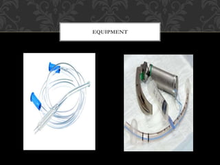
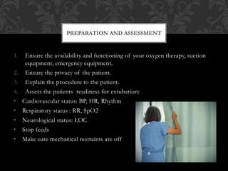
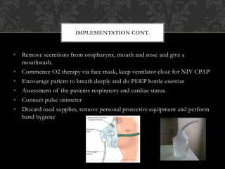
This document discusses the criteria and process for extubating a patient from mechanical ventilation. It begins by defining extubation as the removal of the endotracheal tube, which should only be done once a patient no longer requires ventilation and can protect their airway. Specific criteria are provided for assessing a patient's readiness, including hemodynamic stability, adequate oxygenation, normal blood gases, resolution of underlying conditions, and intact neurological function. Potential complications are also reviewed. The process of extubation involves preparing equipment, explaining to the patient, continuously monitoring their condition, and carefully removing the tube while suctioning and assessing airway patency.
It is the fastest and most commonly practiced approach and allows visual inspection of the supraglottic areas for foreign bodies (e.g., false or loose teeth, aspirated objects) and other obstructions (e.g., tumors). The most important consideration in oral intubation is appropriate head position.
A nasopharyngeal airway, also known as an NPA, nasal trumpet (because of its flared end), or nose hose, is a type of airway adjunct, a tube that is designed to be inserted into the nasal passageway to secure an open airway
An oropharyngeal airway (also known as an oral airway, OPA or Guedel pattern airway) is a medical device called an airway adjunct used in airway management.
The CVP catheter is an important tool used to assess right ventricular function and systemic fluid status. Normal CVP is 2-6 mm Hg. CVP is elevated by : overhydration which increases venous return.
Bag and Mask Ventilation By Sakun Rasaily @Ram K Dhamalaramdhamala11
Bag and mask Ventilation Presented by Sakun Rasaily,
(Pediatric Nurse, Pediatric ward , B.P. Koirala Institute of Health Science
Dharan, Sunsari (Nepal)
ENDOTRACHEAL TUBE INTUBATION II Parts II Details II Clinical DiscussionSwatilekha Das
What is endotracheal intubation?
Endotracheal intubation is a procedure by which a tube is inserted through the mouth down into the trachea (the large airway from the mouth to the lungs). Before surgery, this is often done under deep sedation. In emergency situations, the patient is often unconscious at the time of this procedure.
For detailed information plz watch the slides till end.......
And plz like, share and comment and follow......
Bronchoscopy is an endoscopic technique of visualizing the inside of the airways for diagnostic and therapeutic purposes. An instrument is inserted into the airways, usually through the nose or mouth, or occasionally through a tracheostomy.
A tracheal tube is a catheter that is inserted into the trachea for the primary purpose of establishing and maintaining a patent airway and to ensure the adequate exchange of oxygen and carbon dioxide.
3. Extubation refers to removal of the endotracheal tube
(ETT). It is the final step in liberating a patient from
mechanical ventilation.
At the end of the weaning process, it may be apparent
that a patient no longer requires mechanical ventilation
to maintain sufficient ventilation and oxygenation.
However, extubation should not be ordered until it has
been determined that the patient is able to protect the
airway and the airway is patent.
EXTUBATION
4. Airway protection is the ability to guard against aspiration during spontaneous
breathing. It requires sufficient cough strength and an adequate level of
consciousness, each of which should be assessed prior to extubation. The
amount of secretions should also be considered prior to extubation because
airway protection is significantly more difficult when secretions are increased.
AIRWAY PROTECTION
6. 3. Arterial Blood Gas
• PCO2 < 6kPa.
• PO2 > 8kPa on FIO2 of 40% & PEEP5.
• PH with a normal range (7.35 – 7.45)
4. Other :
• Sedating agents must be stopped for > 24hrs.
• Causative condition resolved/under control.
• Paralysing agents stopped > 24hrs.
• Normal metabolic status. Electrolytes balance must be normal.
• Patient must be neurologically intact. Awake, well motivated, follows verbal
commands & intact gag/ cough reflex.
• Take into consideration aspiration risk and airway edema.
CRITERIA FOR EXTUBATION
7. • Local haemorrhage at tracheostomy site.
• Air embolism
• Infection
• Tracheal necrosis
• Tracheal stenosis
• Tracheoesophageal fistula
• Failure of tracheostomy tube
• Obstruction of tracheostomy
• Accidental extubation
• Tube displacement
• Pneumothorax
• Swallowing dysfunction
COMPLICATIONS ASSOCIATED WITH
ET & TRACHEOSTOMY TUBES
8. • Suctioning equipment
• Personal protective equipment
• Sterile suction catheter
• Self – inflating manual resuscitating bag – valve device connected to 100% O2
source.
• O2 source and tubing.
• Scissors
• Supplemental oxygen
• 10ml syringe
• A rigid pharyngeal suction tip ( yankauer)
• Sterile dressing for stoma
• ET intubation supplies
• Emergency trolley
EQUIPMENT
10. 1. Ensure the availability and functioning of your oxygen therapy, suction
equipment, emergency equipment.
2. Ensure the privacy of the patient.
3. Explain the procedure to the patient.
4. Assess the patients readiness for extubation:
• Cardiovascular status: BP, HR, Rhythm
• Respiratory status : RR, SpO2
• Neurological status: LOC
• Stop feeds
• Make sure mechanical restraints are off
PREPARATION AND ASSESSMENT
11. • Wash hands put on sterile gloves
• Hyper oxygenate the patient and suction the patient via ET / tracheostomy
tube
• Position the patient in high fowlers position
• Cut & remove tracheostomy tapes/ plaster of ET tube
• Deflate the cuff with 10 ml syringe and instruct the patient to breath
• Introduce suction catheter into tube
• Ask patient to cough
• Withdraw the tube and suction simultaneously
• Ask patient to cough again ( to determine laryngeal paralysis)
IMPLEMENTATION
12. • Remove secretions from oropharynx, mouth and nose and give a
mouthwash.
• Commence O2 therapy via face mask, keep ventilator close for NIV CPAP
• Encourage patient to breath deeply and do PEEP bottle exercise
• Assessment of the patients respiratory and cardiac status.
• Connect pulse oximeter
• Discard used supplies, remove personal protective equipment and perform
hand hygiene
IMPLEMENTATION CONT.
14. • Assess air entry and respiratory status
• Auscultate the chest for breathing sounds and for the presence of
secretions
• Do vital observations
• Remain with the patient to determine respiratory stability
• Obtain arterial blood gas within the next hour
EVALUATION
15. • Tidy up after the procedure
• Wash hands!!!!!
• Record all actions taken and chart vital signs on observation chart.
RECORD KEEPING