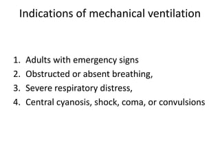
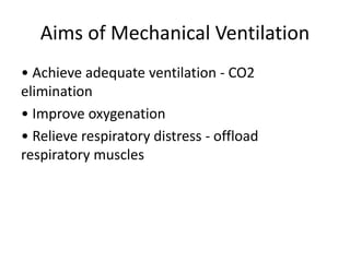
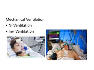
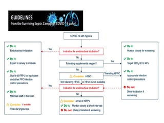
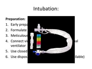
Airway management in COVID 19
Dr Tushar Kumar
Asst professor
Trauma – Anaesthesiology
Trauma center and central emergency, RIMS, Ranchi
Target patients:
• Suspected cases
• Reportable cases
• Confirmed cases
Presentation
• Frequently reported signs and symptoms of
patients admitted to the hospital include
• Fever (77–98%)
• Cough (46%–82%)
• Myalgia or fatigue (11–52%)
• Shortness of breath (3-31%) at illness onset
Huang C, Wang Y, Li X, Ren L,et al. The Lancet. 2020 Jan 24. Wang D, Hu B, Hu C, Zhu F, Liu X et al. Published online February 7, 2020.
Chen N, Zhou M, Dong X, Qu J, Gong F. Lancet. 2020 Jan 30. Holshue ML, DeBolt C, Lindquist S, Lofy KH, Wiesman J et al. N Engl J Med.
2020 Huang C, Wang Y, Li X, Ren L, Zhao J, et al. Lancet. 2020 Jan 24.
Indications of mechanical ventilation
1. Adults with emergency signs
2. Obstructed or absent breathing,
3. Severe respiratory distress,
4. Central cyanosis, shock, coma, or convulsions
Aims of Mechanical Ventilation
• Achieve adequate ventilation - CO2
elimination
• Improve oxygenation
• Relieve respiratory distress - offload
respiratory muscles
Mechanical Ventilation
• NI Ventilation
• Inv. Ventilation
Intubation:
Before :
1. Staff protection
2. Hand hygiene
3. Full PPE
4. Minimize personnel
5. Keep in isolation room if available.
Intubation:
Preparation:
1. Early preparation of drug and equipment
2. Formulate plan early
3. Meticulous airway assessment
4. Connect viral/ bacterial circuits and manual
ventilator
5. Use closed suctioning system
6. Use disposable video laryngoscopy( if available)
During
Team dynamics:
1. Clear assigned roles
2. Clear communication of airway plan
3. Through close loop communication
4. Close monitoring by all team members for
potential contamination.
During
Technical aspects:
Airway management by most experienced
practitioner.
Lowest gas flows, if possible, to maintain
oxygenation.
Tight fitting mask with two hands to minimise leak.
Rapid sequence induction and avoid bag mask
ventilation when possible.
Ensure paralysis to avoid coughing
Positive pressure ventilation only after cuff
inflation.
After
1. Avoid unnecessary circuit disconnection
2. If disconnection needed, wear PPE and put
ventilator on stand by mode and/ or clamp
tube.
3. Strict adherence to proper degowning steps.
4. Hand hygiene
5. Team debriefing
Monitoring:
• Vital monitoring
•Sp02
•ECG
•NIBP
•Urine output, sensorium
•Invasive monitoring
Respiratory mechanics
•Paw
•Pplat
•Driving pressures
•Transpulmonary pressures (Inspiration and expiration)
Maintenance :
• Suctioning
•Close suction only
•As and when required
•Not hourly basis
•PPE precautions if using open suction.
Complications
•Barotrauma
•Acute right ventricle failure
•Hemodynamic instability
•Infections
Weaning
•Normalised I:E ratio
•Reducing FiO2 (usually <0.5)
•No requirement for high PEEP
•Appropriate underlying respiratory rate
•Appropriate tidal volume with moderate
airway pressures
Discharge from ICU to isolation ward
•Depends upon duration of illness
•<2 weeks consider them as infective use all PPE
•>2 weeks may treat as non COVID patients
COVID and Cardiac Arrest
•Secure definitive airway as soon as possible to
avoid aerosol generation
•Use PPE (aerosol generating procedure) at all
time during resuscitation.
•Switch patient from ventilator to AMBU with
viral filter during cardiac arrest.
Loosing is not an option……THANKYOU

Airway covid 19

  • 1.
    Airway management inCOVID 19 Dr Tushar Kumar Asst professor Trauma – Anaesthesiology Trauma center and central emergency, RIMS, Ranchi
  • 2.
    Target patients: • Suspectedcases • Reportable cases • Confirmed cases
  • 3.
    Presentation • Frequently reportedsigns and symptoms of patients admitted to the hospital include • Fever (77–98%) • Cough (46%–82%) • Myalgia or fatigue (11–52%) • Shortness of breath (3-31%) at illness onset Huang C, Wang Y, Li X, Ren L,et al. The Lancet. 2020 Jan 24. Wang D, Hu B, Hu C, Zhu F, Liu X et al. Published online February 7, 2020. Chen N, Zhou M, Dong X, Qu J, Gong F. Lancet. 2020 Jan 30. Holshue ML, DeBolt C, Lindquist S, Lofy KH, Wiesman J et al. N Engl J Med. 2020 Huang C, Wang Y, Li X, Ren L, Zhao J, et al. Lancet. 2020 Jan 24.
  • 5.
    Indications of mechanicalventilation 1. Adults with emergency signs 2. Obstructed or absent breathing, 3. Severe respiratory distress, 4. Central cyanosis, shock, coma, or convulsions
  • 6.
    Aims of MechanicalVentilation • Achieve adequate ventilation - CO2 elimination • Improve oxygenation • Relieve respiratory distress - offload respiratory muscles
  • 7.
    Mechanical Ventilation • NIVentilation • Inv. Ventilation
  • 9.
    Intubation: Before : 1. Staffprotection 2. Hand hygiene 3. Full PPE 4. Minimize personnel 5. Keep in isolation room if available.
  • 10.
    Intubation: Preparation: 1. Early preparationof drug and equipment 2. Formulate plan early 3. Meticulous airway assessment 4. Connect viral/ bacterial circuits and manual ventilator 5. Use closed suctioning system 6. Use disposable video laryngoscopy( if available)
  • 11.
    During Team dynamics: 1. Clearassigned roles 2. Clear communication of airway plan 3. Through close loop communication 4. Close monitoring by all team members for potential contamination.
  • 12.
    During Technical aspects: Airway managementby most experienced practitioner. Lowest gas flows, if possible, to maintain oxygenation. Tight fitting mask with two hands to minimise leak. Rapid sequence induction and avoid bag mask ventilation when possible. Ensure paralysis to avoid coughing Positive pressure ventilation only after cuff inflation.
  • 13.
    After 1. Avoid unnecessarycircuit disconnection 2. If disconnection needed, wear PPE and put ventilator on stand by mode and/ or clamp tube. 3. Strict adherence to proper degowning steps. 4. Hand hygiene 5. Team debriefing
  • 14.
    Monitoring: • Vital monitoring •Sp02 •ECG •NIBP •Urineoutput, sensorium •Invasive monitoring Respiratory mechanics •Paw •Pplat •Driving pressures •Transpulmonary pressures (Inspiration and expiration)
  • 15.
    Maintenance : • Suctioning •Closesuction only •As and when required •Not hourly basis •PPE precautions if using open suction.
  • 16.
    Complications •Barotrauma •Acute right ventriclefailure •Hemodynamic instability •Infections
  • 17.
    Weaning •Normalised I:E ratio •ReducingFiO2 (usually <0.5) •No requirement for high PEEP •Appropriate underlying respiratory rate •Appropriate tidal volume with moderate airway pressures
  • 18.
    Discharge from ICUto isolation ward •Depends upon duration of illness •<2 weeks consider them as infective use all PPE •>2 weeks may treat as non COVID patients
  • 19.
    COVID and CardiacArrest •Secure definitive airway as soon as possible to avoid aerosol generation •Use PPE (aerosol generating procedure) at all time during resuscitation. •Switch patient from ventilator to AMBU with viral filter during cardiac arrest.
  • 20.
    Loosing is notan option……THANKYOU

Editor's Notes

  • #13 • Emptying of the stomach via a gastric tube which is then removed • Pre-oxygenation • Positioning the patient supine with a head-down tilt • Induction of anaesthesia with a barbiturate (e.g. thiopentone) or volatile, and a rapid-acting muscle relaxant (e.g. suxamethonium) • Application of cricoid pressure • Laryngoscopy and intubation of the trachea with a cuffed tube immediately following fasciculations