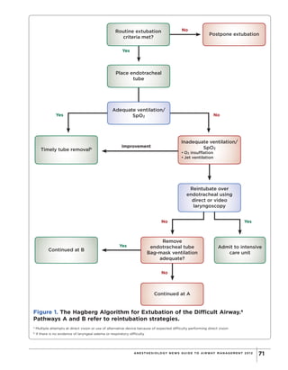
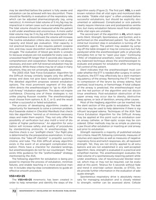
PRINTER-FRIENDLY VERSION AT ANESTHESIOLOGYNEWS.COM
69ANE ST HE S IOLOGY NE WS GUIDE TO AIRWAY MANAGE ME NT 2 012
Extubation: Making
The Unpredictable Safer
RAM ROTH, MD
Assistant Professor of Anesthesiology
Mount Sinai School of Medicine
New York, New York
FAIEJA CHOWDHURY
Student
Stony Brook University
Stony Brook, New York
ELIZABETH A.M. FROST, MD
Professor of Anesthesiology
Mount Sinai School of Medicine
New York, New York
The authors report no relevant financial conflicts.
A
irway management is a fundamental aspect of anesthesiology comprising
mask ventilation, laryngoscopy, endotracheal intubation, and extubation.
Considerable research and development has focused on the first 3 aspects of
airway management, but relatively little consideration has been given to extubation—
despite the fact that many preventable complications result from the improper
extubation of patients.
Copyright©
2012
M
cM
ahon
Publishing
G
roup
unless
otherw
ise
noted.
A
llrights
reserved.Reproduction
in
w
hole
orin
partw
ithoutperm
ission
is
prohibited.
INDEPENDENTLY DEVELOPED BY MCMAHON PU B LIS HING70
Magill and Macintosh popularized endotracheal tube
(ETT) intubation shortly after World War I. It was intro-
duced to protect the patient from aspiration and sus-
tain ventilation while under anesthesia. Clearly, the ETT
should be removed only after surgery is completed and
patients can maintain ventilation.
Quality improvement in the field of anesthesiology
has focused largely on intubation rather than extuba-
tion. Conferences and workshops for difficult intuba-
tion are manifold. Most vendors develop equipment for
improving intubation rather than extubation. Anesthesi-
ology textbooks focus on intubation, not extubation. It
is interesting to note that in a search of peer-reviewed
literature for the 2 keywords “intubation” and “extu-
bation,” about 90% of articles included “intubation”
whereas only 10% mentioned “extubation.”
There are established strategies to perform intuba-
tion and algorithms to aid in other difficult situations
in anesthesiology. A plethora of research has been
conducted to create new guidelines, procedures, and
predictive approaches and management of difficult tra-
cheal intubation. In 1993, the guidelines of the American
Society of Anesthesiologists (ASA) for the manage-
ment of the difficult airway established a protocol for
intubation, but not for extubation. Those guidelines
indeed have had the desired effect. A 2005 analysis
of the Closed Claims database showed that the num-
ber of claims for death and brain death associated with
intubation had decreased since the incorporation of
the guidelines. However, the number of claims linked to
extubation has not fallen.
Indeed, extubation now appears to be more danger-
ous than intubation. A 1998 study by Asai et al found
that the overall rate of complications associated with
extubation was 7.4% greater than that for intuba-
tion. Respiratory events include coughing, desatura-
tion, breath holding, airway obstruction, laryngospasm,
apnea, hypoventilation, inadequate reversal, vomiting,
and masseter spasm. Cardiovascular complications
consist of arterial hypertension, tachycardia, and dys-
rhythmias. Other complications include laryngeal and
supraglottic edema.
Limited Guidance
Extubation quality depends on the anesthesiologist.
The lack of a standard makes it difficult to teach extuba-
tion to residents or to have a meaningful discussion that
would improve the quality of care. What few extuba-
tion guidelines exist, in fact, involve methods for reduc-
ing complications in specific cases in order to treat
or minimize injury. For example, if a patient exhibits
post-extubation stridor, the textbook Miller’s Anesthe-
sia recommends that helium be used to improve oxy-
genation and increase tidal volume in the extubated
patient to prevent reintubation. If a patient develops
severe hypoxemia, succinylcholine and reintubation are
recommended.
The ASA suggests that anesthesiologists should have
a “preformulated” strategy for extubation that includes
constant oxygen management, ventilation, and a rein-
tubation strategy to prevent such complications from
occurring.
Two algorithms for extubation of the difficult air-
way have been proposed, but they may not be practi-
cal for many situations. The recently revised Hagberg
Extubation Algorithm of Patients with a Difficult Airway
(Figure 1) is not a tool for teaching basic extubation.
Routine extubation criteria recently revised by Hag-
berg, call for the patient to be awake and cooperative
(Table). Using these criteria patients who are in coma,
have Alzheimer’s disease, are children, or speak a for-
eign language cannot be extubated. Airway reflexes
Table. Routine Extubation Criteria10
Awake, alert, able to follow commands
• Sustained eye opening for pediatric patients or
patients unable to understand commands
Vital signs stable
• Blood pressure, pulse rate, temperature
• Respiratory rate ≤30 breaths per minute
• O2 saturation
Protective reflexes returned
• Gag
• Swallow
• Cough
Adequate reversal of neuromuscular blockade
• Train-of-4 stimulation 4/4, sustained tetany at
50 Hz
• Strong hand grip
• Usassisted head lift (>5 sec)
Arterial blood gases reasonable with FiO2 40
• pH >7.30
• PaO2 ≥60 mm Hg
• PaCO2 <50 mm Hg
Respiratory mechanics adequate
• Tidal volume >5 mL/kg
• Vital capacity >15 mL/kg
• Negative inspiratory force >–20 cm H2O
For patients at risk for laryngeal edema, consider
cuff leak test and airway inspection
• Evaluation by fiber-optic bronchoscopy
Copyright©
2012
M
cM
ahon
Publishing
G
roup
unless
otherw
ise
noted.
A
llrights
reserved.Reproduction
in
w
hole
orin
partw
ithoutperm
ission
is
prohibited.
ANE ST HE S IOLOGY NE WS GUIDE TO AIRWAY MANAGE ME NT 2 012 71
Routine extubation
criteria met?
Postpone extubation
Inadequate ventilation/
SpO2
• O2 insufflation
• Jet ventilation
Remove
endotracheal tube
Bag-mask ventilation
adequate?
Continued at A
Timely tube removalb
Admit to intensive
care unit
Continued at B
No
No
No
No
Yes
Yes
Yes
Yes
Improvement
Figure 1. The Hagberg Algorithm for Extubation of the Difficult Airway.a
Pathways A and B refer to reintubation strategies.
a
Multiple attempts at direct vision or use of alternative device because of expected difficulty performing direct vision
b
If there is no evidence of laryngeal edema or respiratory difficulty
Place endotracheal
tube
Adequate ventilation/
SpO2
Reintubate over
endotracheal using
direct or video
laryngoscopy
Copyright©
2012
M
cM
ahon
Publishing
G
roup
unless
otherw
ise
noted.
A
llrights
reserved.Reproduction
in
w
hole
orin
partw
ithoutperm
ission
is
prohibited.
INDEPENDENTLY DEVELOPED BY MCMAHON PU B LIS HING72
may be identified before the patient is fully awake and
extubation can be achieved with less discomfort. There
should be flexibility in evaluation of the respiratory rate,
which can be adjusted pharmacologically (eg, using
narcotics). A minimum tidal volume of 5 mL/kg may be
impractical in certain cases such as overweight patients.
The tidal volume should be measured while the patient
is still under anesthesia and unconscious. A more useful
tidal volume may be 3 mL/kg with the expectation that
once anesthesia is discontinued, the volume will increase.
The routine extubation criteria also recommend a vital
capacity and blood gas evaluation. Vital capacity is
not practical because it also requires patient coopera-
tion, and may cause discomfort and lead the patient to
struggle. The evaluation of blood gas levels is unrealis-
tic because analyses are not available for every case. An
unassisted head lift longer than 5 seconds also requires
comprehension and cooperation. Reversal is not always
necessary, and even with full reversal extubation may be
premature. While these criteria may be of value in many
situations, they are not universally applicable.
The 2003 ASA Task Force Extubation Algorithm for
the Difficult Airway similarly targets only the difficult
airway and does not give basic extubation guidance.
The detailed algorithm proposes several extubation
options. The most prominent segment of this algo-
rithm directs the anesthesiologist to “go to ASA Diffi-
cult Airway” intubation algorithm. This does not inspire
confidence. Choosing one of these strategies is not
unlike playing Russian roulette: The anesthesiologist
must choose from options A, B, C, or D, and the result
is either a successful or failed extubation.
The process of developing algorithms creates an
opportunity for teamwork to solve a common problem.
Atul Gawande stated in Checklist Manifesto that check-
lists like algorithms “remind us of minimum necessary
steps and make them explicit. They not only offer the
possibility of verification but also instill a kind of dis-
cipline of higher performance.” An algorithm for extu-
bation will increase safety and quality of procedures
by standardizing protocols. In anesthesiology, the
machine check is our “preflight check.” Our flight plan
is determined by the preoperative examination. In many
instances, the “take-off checklist” is propofol, succinyl-
choline, and tube. The ASA difficult airway algorithm
exists in the event of an emergent complicated intu-
bation. Pilots have a checklist for standard landings,
but anesthesiologists do not for our counterpart. There
exists little guidance for the “preformulated” strategy
mentioned by the ASA.
The following algorithm for extubation is being pro-
posed to improve the process of extubation, minimize
failures, and enable teaching in a more practical man-
ner. It includes step-by-step considerations to guide an
effective smooth procedure.
VSS+4S+2S
The VSS+4S+2S mnemonic has been created in
order to help remember and identify the steps of the
algorithm easily (Figure 2). The first part, VSS, is a well-
known notation that all vital signs and monitored data
are stable. “Stable vital signs” is often unstated before
successful extubations, but should be explicitly doc-
umented or addressed. Complicated or sick patients
clearly require intensive care and postoperative ventila-
tion, but many extubation failures result from attempts
while vital signs are unstable.
The second part of the algorithm is 4S, which repre-
sents Stent, Strength, Spontaneous, and Suction, and is
performed on the anesthetized patient. Too often, the
initial but incorrect step in extubation is turning off all
anesthetic agents. The patient may awaken by jump-
ing off the table enraged or may be conscious but fully
paralyzed while the anesthesiologist yells or physically
stimulates him or her to “open your eyes.” Continuing
1 MAC of inhalational anesthetic, total IV anesthesia, or
any balanced technique allows the anesthesiologist to
evaluate and prepare for extubation while maintaining
full control of the patient.
Stent indicates that the anesthesiologist must con-
sider whether the ETT is needed after surgery. In certain
situations, the ETT may effectively be a stent maintain-
ing patency of the airway. Removing the ETT may lead
to compression of the larynx by airway edema, fluid
overload, local trauma, or hemorrhage. If total collapse
of the airway is predicted, the anesthesiologist must
use the red portion of the algorithm and not discon-
tinue anesthesia. Post-extubation obstruction of the
upper airway may occur due to obesity, obstructive
sleep apnea, or lack of dentition, among other causes.
Most of the Hagberg algorithm can be inserted into
the stent section of this guide to extubation. The leak
test now may be used to help determine if there is sig-
nificant laryngeal edema. Techniques of the ASA Task
Force Extubation Algorithm for the Difficult Airway
may be applied at this point such as extubation over
an airway catheter, or fiber-optic scope may be con-
sidered. Other methods may be as simple as planning
a jaw thrust after extubation or inserting an oral airway
just prior to extubation.
Strength represents a majority of published extuba-
tion criteria. Head lift, following commands, measures of
volumes and capacities as well as measures of negative
inspiratory force and twitch responses, etc, all represent
strength. Yet, they are not strictly applied to all extu-
bations and are not established in any well-accepted
algorithm. Here, strength is evaluated in a practical and
simplified manner. The anesthesiologist evaluates the
tidal volume of a spontaneously respiring patient while
under anesthesia. Use of neuromuscular blocker rever-
sal, which may or may not be required, can be evalu-
ated at this point. Tetanus for 5 seconds without fade
and trend in end-tidal carbon dioxide, or ETCO2, lev-
els provide further information in the evaluation of ade-
quate strength.
Spontaneous respiratory drive is absolutely neces-
sary for managing extubation and is closely related
to strength; neither can be assessed alone. Cellular
Copyright©
2012
M
cM
ahon
Publishing
G
roup
unless
otherw
ise
noted.
A
llrights
reserved.Reproduction
in
w
hole
orin
partw
ithoutperm
ission
is
prohibited.
ANE ST HE S IOLOGY NE WS GUIDE TO AIRWAY MANAGE ME NT 2 012 73
metabolism generates CO2, which stimulates the apneus-
tic center in the pons, creating a respiratory drive. Despite
intraoperative narcotic administration and maintenance
of anesthesia, supernormal levels of CO2 will result in a
spontaneous drive that will lead to respiratory efforts,
only if the patient is not paralyzed. With strength and
spontaneous drive established, the fifth vital sign—pain—
can be evaluated. Rate of respiration may indicate pain
and can be addressed by titrating narcotics.
Suction is the final step in the preparation for extu-
bation performed on the anesthetized patient. The only
acceptable purpose for suction is to clear the airway of
secretions or blood. If suction is applied to a lightly con-
scious or conscious patient, it may cause irritation and
result in bucking, breath holding, and discomfort.
At this point, the anesthetized patient has stable vital
signs, a patent airway, strength with a tidal volume of
about 3 mL/kg, and spontaneous respiratory drive with
a rate of about 18 breaths per minute, and the orophar-
ynx is clear. Extubation, if carried out at this point, is
considered a deep extubation and is classically rec-
ommended in patients with severe asthma. If a deep
extubation is not indicated, the green portion of the
algorithm represents the next phase in which anesthe-
sia is discontinued.
The final part of the algorithm is 2S, consisting of
Swallow and Secure.
Swallow indicates that the anesthesiologist must
observe and detect the return of airway reflexes. Air-
way reflexes require strength to be present which
already has been established earlier in the algorithm.
The patient may begin swallowing, moving the tongue,
gagging or coughing. Commonly, anesthesiologists
delay extubation until after the patient is “fully” awake.
Manage
Consider
continuing
ventilation
Reassess
Stent No potential obstruction
Suction Secretions cleared
Secure Ease of reintubation Awake extubation
Swallow Airway reflexes intact Sedated extubation
DISCONTINUE ANESTHESIA Deep extubation
Spontaneous Normal respiratory rate
Strength Tidal volume, tetanus, ETCO2 appropriate
Vital signs Stable
Vital signs Stable
No
No
No
No
No
No
Yes
Yes
Yes
Yes
Yes
Yes
Figure 2. The “VSS+4S+2S” proposed algorithm for extubation.
Copyright©
2012
M
cM
ahon
Publishing
G
roup
unless
otherw
ise
noted.
A
llrights
reserved.Reproduction
in
w
hole
orin
partw
ithoutperm
ission
is
prohibited.
INDEPENDENTLY DEVELOPED BY MCMAHON PU B LIS HING74
This delay often results in choking or bucking, and may
lead to throat pain or injury.
Now, with anesthesia discontinued, the patient has
stable vital signs, a patent airway, strength demon-
strated with a tidal volume of about 3 mL/kg, spon-
taneous respiratory drive with a rate of 18 breaths per
minute, and airway reflexes. The oropharynx has been
suctioned if necessary. Extubation may be achieved
with the patient still sedated if there are no concerns
for aspiration, such as full stomach. Immediately after
extubation, a few minutes of jaw thrust may be neces-
sary to prevent upper airway obstruction in the sedated
patient.
Secure, the final step in the algorithm, reflects the
comfort level and experience of the anesthesiologist. If
extubation has not occurred up to this point in the algo-
rithm, it may be because it is a known difficult airway.
Perhaps surgery has affected the anatomy of the air-
way. In this situation, the anesthesiologist may not feel
secure about regaining control of the airway in case of
an emergency. Reintubation as a rescue maneuver is
not an option in this algorithm. Therefore, a fully awake
extubation is warranted and the risks for patient dis-
comfort, awareness, or injury are secondary to a safe
extubation.
Within each step of the checklist, if the patient devi-
ates from the ideal conditions, appropriate measures are
recommended (represented in red). Return to full anes-
thesia and assessment of the situation are indicated.
Emergence is commonly defined as the period dur-
ing which maintenance agents are discontinued, the
patient begins to wake up, spontaneous respirations
resume, airway reflexes return, and he/she is prepared
for extubation and transport to the recovery room.
Medications may be given to control pain, decrease or
prevent nausea, and reverse the effects of nondepolar-
izing muscle relaxants. This description of emergence
is inherently flawed and the sequence should be rear-
ranged into a more logical order.
Conclusion
This new extubation algorithm reminds anesthesiolo-
gists of the steps necessary for the extubation process
and evaluates each step along the way in a set order. If
integrated into general practice, it may allow for a reli-
able and safer extubation. The algorithm provides an
understandable teaching method, enabling anesthe-
siologists to discuss extubation in a universal manner
and encourage further planning to improve the art of
extubation.
Selected Readings
Asai T, Koga K, Vaughan RS. Respiratory complications associated
with tracheal intubation and extubation. Br J Anaesth. 1998;80(16):
767-775.
Brambrink AM, Hagberg CA. The ASA Difficult Airway Algorithm: anal-
ysis and presentation of a new algorithm. In: Benumof and Hagberg’s
Airway Management. 3rd ed. Elsevier; 2012:222-239 (In press).
Berkow LC. Strategies for airway management. Best Pract Res Clin
Anaesthesiol. 2004;18(4):531-548.
Clinical Anesthesia. 6th ed. Barash PG, Cullen BF, Stoelting RK, Caha-
lan MK, Stock MC, eds. Philadelphia, PA: Lippincott Williams & Wilkins;
2009.
Finucane BT, Tsui BCH, Santora AH. Extubation strategies: the extu-
bation algorithm. In: Principles of Airway Management. New York, NY:
Springer; 2011.
Gawande A. The Checklist Manifesto. New York, NY: Henry Holt and
Company; 2009.
Hales BM, Pronovost PJ. The checklist—a tool for error management
and performance improvement. J Crit Care. 2006;21(3):231-235.
Hartley M, Vaughan RS. Problems associated with tracheal extubation.
Br J Anaesth. 1993;71(4):561-568.
Henderson J. Airway management in the adult. In: Miller’s Anesthe-
sia. 7th ed. Miller RD, Eriksson LI, Fleisher LA, Wiener-Kronish JP,
Young WL, eds. Philadelphia, PA: Churchill Livingstone; 2009.
Medical Foundation of Epsom College. Reports of Societies. BMJ.
1949;1(4591):26-28.
Peterson GN, Domino KB, Caplan RA, Posner KL, Lee LA, Cheney FW.
Management of the difficult airway: a closed claims analysis. Anesthe-
siology. 2005;103(1):33-39.
Practice guidelines for management of the difficult airway. Anesthesi-
ology. 1993;78(3):597-602.
Copyright©
2012
M
cM
ahon
Publishing
G
roup
unless
otherw
ise
noted.
A
llrights
reserved.Reproduction
in
w
hole
orin
partw
ithoutperm
ission
is
prohibited.

Extubation-Making the Unpredictable Safer

  • 1.
    PRINTER-FRIENDLY VERSION ATANESTHESIOLOGYNEWS.COM 69ANE ST HE S IOLOGY NE WS GUIDE TO AIRWAY MANAGE ME NT 2 012 Extubation: Making The Unpredictable Safer RAM ROTH, MD Assistant Professor of Anesthesiology Mount Sinai School of Medicine New York, New York FAIEJA CHOWDHURY Student Stony Brook University Stony Brook, New York ELIZABETH A.M. FROST, MD Professor of Anesthesiology Mount Sinai School of Medicine New York, New York The authors report no relevant financial conflicts. A irway management is a fundamental aspect of anesthesiology comprising mask ventilation, laryngoscopy, endotracheal intubation, and extubation. Considerable research and development has focused on the first 3 aspects of airway management, but relatively little consideration has been given to extubation— despite the fact that many preventable complications result from the improper extubation of patients. Copyright© 2012 M cM ahon Publishing G roup unless otherw ise noted. A llrights reserved.Reproduction in w hole orin partw ithoutperm ission is prohibited.
  • 2.
    INDEPENDENTLY DEVELOPED BYMCMAHON PU B LIS HING70 Magill and Macintosh popularized endotracheal tube (ETT) intubation shortly after World War I. It was intro- duced to protect the patient from aspiration and sus- tain ventilation while under anesthesia. Clearly, the ETT should be removed only after surgery is completed and patients can maintain ventilation. Quality improvement in the field of anesthesiology has focused largely on intubation rather than extuba- tion. Conferences and workshops for difficult intuba- tion are manifold. Most vendors develop equipment for improving intubation rather than extubation. Anesthesi- ology textbooks focus on intubation, not extubation. It is interesting to note that in a search of peer-reviewed literature for the 2 keywords “intubation” and “extu- bation,” about 90% of articles included “intubation” whereas only 10% mentioned “extubation.” There are established strategies to perform intuba- tion and algorithms to aid in other difficult situations in anesthesiology. A plethora of research has been conducted to create new guidelines, procedures, and predictive approaches and management of difficult tra- cheal intubation. In 1993, the guidelines of the American Society of Anesthesiologists (ASA) for the manage- ment of the difficult airway established a protocol for intubation, but not for extubation. Those guidelines indeed have had the desired effect. A 2005 analysis of the Closed Claims database showed that the num- ber of claims for death and brain death associated with intubation had decreased since the incorporation of the guidelines. However, the number of claims linked to extubation has not fallen. Indeed, extubation now appears to be more danger- ous than intubation. A 1998 study by Asai et al found that the overall rate of complications associated with extubation was 7.4% greater than that for intuba- tion. Respiratory events include coughing, desatura- tion, breath holding, airway obstruction, laryngospasm, apnea, hypoventilation, inadequate reversal, vomiting, and masseter spasm. Cardiovascular complications consist of arterial hypertension, tachycardia, and dys- rhythmias. Other complications include laryngeal and supraglottic edema. Limited Guidance Extubation quality depends on the anesthesiologist. The lack of a standard makes it difficult to teach extuba- tion to residents or to have a meaningful discussion that would improve the quality of care. What few extuba- tion guidelines exist, in fact, involve methods for reduc- ing complications in specific cases in order to treat or minimize injury. For example, if a patient exhibits post-extubation stridor, the textbook Miller’s Anesthe- sia recommends that helium be used to improve oxy- genation and increase tidal volume in the extubated patient to prevent reintubation. If a patient develops severe hypoxemia, succinylcholine and reintubation are recommended. The ASA suggests that anesthesiologists should have a “preformulated” strategy for extubation that includes constant oxygen management, ventilation, and a rein- tubation strategy to prevent such complications from occurring. Two algorithms for extubation of the difficult air- way have been proposed, but they may not be practi- cal for many situations. The recently revised Hagberg Extubation Algorithm of Patients with a Difficult Airway (Figure 1) is not a tool for teaching basic extubation. Routine extubation criteria recently revised by Hag- berg, call for the patient to be awake and cooperative (Table). Using these criteria patients who are in coma, have Alzheimer’s disease, are children, or speak a for- eign language cannot be extubated. Airway reflexes Table. Routine Extubation Criteria10 Awake, alert, able to follow commands • Sustained eye opening for pediatric patients or patients unable to understand commands Vital signs stable • Blood pressure, pulse rate, temperature • Respiratory rate ≤30 breaths per minute • O2 saturation Protective reflexes returned • Gag • Swallow • Cough Adequate reversal of neuromuscular blockade • Train-of-4 stimulation 4/4, sustained tetany at 50 Hz • Strong hand grip • Usassisted head lift (>5 sec) Arterial blood gases reasonable with FiO2 40 • pH >7.30 • PaO2 ≥60 mm Hg • PaCO2 <50 mm Hg Respiratory mechanics adequate • Tidal volume >5 mL/kg • Vital capacity >15 mL/kg • Negative inspiratory force >–20 cm H2O For patients at risk for laryngeal edema, consider cuff leak test and airway inspection • Evaluation by fiber-optic bronchoscopy Copyright© 2012 M cM ahon Publishing G roup unless otherw ise noted. A llrights reserved.Reproduction in w hole orin partw ithoutperm ission is prohibited.
  • 3.
    ANE ST HES IOLOGY NE WS GUIDE TO AIRWAY MANAGE ME NT 2 012 71 Routine extubation criteria met? Postpone extubation Inadequate ventilation/ SpO2 • O2 insufflation • Jet ventilation Remove endotracheal tube Bag-mask ventilation adequate? Continued at A Timely tube removalb Admit to intensive care unit Continued at B No No No No Yes Yes Yes Yes Improvement Figure 1. The Hagberg Algorithm for Extubation of the Difficult Airway.a Pathways A and B refer to reintubation strategies. a Multiple attempts at direct vision or use of alternative device because of expected difficulty performing direct vision b If there is no evidence of laryngeal edema or respiratory difficulty Place endotracheal tube Adequate ventilation/ SpO2 Reintubate over endotracheal using direct or video laryngoscopy Copyright© 2012 M cM ahon Publishing G roup unless otherw ise noted. A llrights reserved.Reproduction in w hole orin partw ithoutperm ission is prohibited.
  • 4.
    INDEPENDENTLY DEVELOPED BYMCMAHON PU B LIS HING72 may be identified before the patient is fully awake and extubation can be achieved with less discomfort. There should be flexibility in evaluation of the respiratory rate, which can be adjusted pharmacologically (eg, using narcotics). A minimum tidal volume of 5 mL/kg may be impractical in certain cases such as overweight patients. The tidal volume should be measured while the patient is still under anesthesia and unconscious. A more useful tidal volume may be 3 mL/kg with the expectation that once anesthesia is discontinued, the volume will increase. The routine extubation criteria also recommend a vital capacity and blood gas evaluation. Vital capacity is not practical because it also requires patient coopera- tion, and may cause discomfort and lead the patient to struggle. The evaluation of blood gas levels is unrealis- tic because analyses are not available for every case. An unassisted head lift longer than 5 seconds also requires comprehension and cooperation. Reversal is not always necessary, and even with full reversal extubation may be premature. While these criteria may be of value in many situations, they are not universally applicable. The 2003 ASA Task Force Extubation Algorithm for the Difficult Airway similarly targets only the difficult airway and does not give basic extubation guidance. The detailed algorithm proposes several extubation options. The most prominent segment of this algo- rithm directs the anesthesiologist to “go to ASA Diffi- cult Airway” intubation algorithm. This does not inspire confidence. Choosing one of these strategies is not unlike playing Russian roulette: The anesthesiologist must choose from options A, B, C, or D, and the result is either a successful or failed extubation. The process of developing algorithms creates an opportunity for teamwork to solve a common problem. Atul Gawande stated in Checklist Manifesto that check- lists like algorithms “remind us of minimum necessary steps and make them explicit. They not only offer the possibility of verification but also instill a kind of dis- cipline of higher performance.” An algorithm for extu- bation will increase safety and quality of procedures by standardizing protocols. In anesthesiology, the machine check is our “preflight check.” Our flight plan is determined by the preoperative examination. In many instances, the “take-off checklist” is propofol, succinyl- choline, and tube. The ASA difficult airway algorithm exists in the event of an emergent complicated intu- bation. Pilots have a checklist for standard landings, but anesthesiologists do not for our counterpart. There exists little guidance for the “preformulated” strategy mentioned by the ASA. The following algorithm for extubation is being pro- posed to improve the process of extubation, minimize failures, and enable teaching in a more practical man- ner. It includes step-by-step considerations to guide an effective smooth procedure. VSS+4S+2S The VSS+4S+2S mnemonic has been created in order to help remember and identify the steps of the algorithm easily (Figure 2). The first part, VSS, is a well- known notation that all vital signs and monitored data are stable. “Stable vital signs” is often unstated before successful extubations, but should be explicitly doc- umented or addressed. Complicated or sick patients clearly require intensive care and postoperative ventila- tion, but many extubation failures result from attempts while vital signs are unstable. The second part of the algorithm is 4S, which repre- sents Stent, Strength, Spontaneous, and Suction, and is performed on the anesthetized patient. Too often, the initial but incorrect step in extubation is turning off all anesthetic agents. The patient may awaken by jump- ing off the table enraged or may be conscious but fully paralyzed while the anesthesiologist yells or physically stimulates him or her to “open your eyes.” Continuing 1 MAC of inhalational anesthetic, total IV anesthesia, or any balanced technique allows the anesthesiologist to evaluate and prepare for extubation while maintaining full control of the patient. Stent indicates that the anesthesiologist must con- sider whether the ETT is needed after surgery. In certain situations, the ETT may effectively be a stent maintain- ing patency of the airway. Removing the ETT may lead to compression of the larynx by airway edema, fluid overload, local trauma, or hemorrhage. If total collapse of the airway is predicted, the anesthesiologist must use the red portion of the algorithm and not discon- tinue anesthesia. Post-extubation obstruction of the upper airway may occur due to obesity, obstructive sleep apnea, or lack of dentition, among other causes. Most of the Hagberg algorithm can be inserted into the stent section of this guide to extubation. The leak test now may be used to help determine if there is sig- nificant laryngeal edema. Techniques of the ASA Task Force Extubation Algorithm for the Difficult Airway may be applied at this point such as extubation over an airway catheter, or fiber-optic scope may be con- sidered. Other methods may be as simple as planning a jaw thrust after extubation or inserting an oral airway just prior to extubation. Strength represents a majority of published extuba- tion criteria. Head lift, following commands, measures of volumes and capacities as well as measures of negative inspiratory force and twitch responses, etc, all represent strength. Yet, they are not strictly applied to all extu- bations and are not established in any well-accepted algorithm. Here, strength is evaluated in a practical and simplified manner. The anesthesiologist evaluates the tidal volume of a spontaneously respiring patient while under anesthesia. Use of neuromuscular blocker rever- sal, which may or may not be required, can be evalu- ated at this point. Tetanus for 5 seconds without fade and trend in end-tidal carbon dioxide, or ETCO2, lev- els provide further information in the evaluation of ade- quate strength. Spontaneous respiratory drive is absolutely neces- sary for managing extubation and is closely related to strength; neither can be assessed alone. Cellular Copyright© 2012 M cM ahon Publishing G roup unless otherw ise noted. A llrights reserved.Reproduction in w hole orin partw ithoutperm ission is prohibited.
  • 5.
    ANE ST HES IOLOGY NE WS GUIDE TO AIRWAY MANAGE ME NT 2 012 73 metabolism generates CO2, which stimulates the apneus- tic center in the pons, creating a respiratory drive. Despite intraoperative narcotic administration and maintenance of anesthesia, supernormal levels of CO2 will result in a spontaneous drive that will lead to respiratory efforts, only if the patient is not paralyzed. With strength and spontaneous drive established, the fifth vital sign—pain— can be evaluated. Rate of respiration may indicate pain and can be addressed by titrating narcotics. Suction is the final step in the preparation for extu- bation performed on the anesthetized patient. The only acceptable purpose for suction is to clear the airway of secretions or blood. If suction is applied to a lightly con- scious or conscious patient, it may cause irritation and result in bucking, breath holding, and discomfort. At this point, the anesthetized patient has stable vital signs, a patent airway, strength with a tidal volume of about 3 mL/kg, and spontaneous respiratory drive with a rate of about 18 breaths per minute, and the orophar- ynx is clear. Extubation, if carried out at this point, is considered a deep extubation and is classically rec- ommended in patients with severe asthma. If a deep extubation is not indicated, the green portion of the algorithm represents the next phase in which anesthe- sia is discontinued. The final part of the algorithm is 2S, consisting of Swallow and Secure. Swallow indicates that the anesthesiologist must observe and detect the return of airway reflexes. Air- way reflexes require strength to be present which already has been established earlier in the algorithm. The patient may begin swallowing, moving the tongue, gagging or coughing. Commonly, anesthesiologists delay extubation until after the patient is “fully” awake. Manage Consider continuing ventilation Reassess Stent No potential obstruction Suction Secretions cleared Secure Ease of reintubation Awake extubation Swallow Airway reflexes intact Sedated extubation DISCONTINUE ANESTHESIA Deep extubation Spontaneous Normal respiratory rate Strength Tidal volume, tetanus, ETCO2 appropriate Vital signs Stable Vital signs Stable No No No No No No Yes Yes Yes Yes Yes Yes Figure 2. The “VSS+4S+2S” proposed algorithm for extubation. Copyright© 2012 M cM ahon Publishing G roup unless otherw ise noted. A llrights reserved.Reproduction in w hole orin partw ithoutperm ission is prohibited.
  • 6.
    INDEPENDENTLY DEVELOPED BYMCMAHON PU B LIS HING74 This delay often results in choking or bucking, and may lead to throat pain or injury. Now, with anesthesia discontinued, the patient has stable vital signs, a patent airway, strength demon- strated with a tidal volume of about 3 mL/kg, spon- taneous respiratory drive with a rate of 18 breaths per minute, and airway reflexes. The oropharynx has been suctioned if necessary. Extubation may be achieved with the patient still sedated if there are no concerns for aspiration, such as full stomach. Immediately after extubation, a few minutes of jaw thrust may be neces- sary to prevent upper airway obstruction in the sedated patient. Secure, the final step in the algorithm, reflects the comfort level and experience of the anesthesiologist. If extubation has not occurred up to this point in the algo- rithm, it may be because it is a known difficult airway. Perhaps surgery has affected the anatomy of the air- way. In this situation, the anesthesiologist may not feel secure about regaining control of the airway in case of an emergency. Reintubation as a rescue maneuver is not an option in this algorithm. Therefore, a fully awake extubation is warranted and the risks for patient dis- comfort, awareness, or injury are secondary to a safe extubation. Within each step of the checklist, if the patient devi- ates from the ideal conditions, appropriate measures are recommended (represented in red). Return to full anes- thesia and assessment of the situation are indicated. Emergence is commonly defined as the period dur- ing which maintenance agents are discontinued, the patient begins to wake up, spontaneous respirations resume, airway reflexes return, and he/she is prepared for extubation and transport to the recovery room. Medications may be given to control pain, decrease or prevent nausea, and reverse the effects of nondepolar- izing muscle relaxants. This description of emergence is inherently flawed and the sequence should be rear- ranged into a more logical order. Conclusion This new extubation algorithm reminds anesthesiolo- gists of the steps necessary for the extubation process and evaluates each step along the way in a set order. If integrated into general practice, it may allow for a reli- able and safer extubation. The algorithm provides an understandable teaching method, enabling anesthe- siologists to discuss extubation in a universal manner and encourage further planning to improve the art of extubation. Selected Readings Asai T, Koga K, Vaughan RS. Respiratory complications associated with tracheal intubation and extubation. Br J Anaesth. 1998;80(16): 767-775. Brambrink AM, Hagberg CA. The ASA Difficult Airway Algorithm: anal- ysis and presentation of a new algorithm. In: Benumof and Hagberg’s Airway Management. 3rd ed. Elsevier; 2012:222-239 (In press). Berkow LC. Strategies for airway management. Best Pract Res Clin Anaesthesiol. 2004;18(4):531-548. Clinical Anesthesia. 6th ed. Barash PG, Cullen BF, Stoelting RK, Caha- lan MK, Stock MC, eds. Philadelphia, PA: Lippincott Williams & Wilkins; 2009. Finucane BT, Tsui BCH, Santora AH. Extubation strategies: the extu- bation algorithm. In: Principles of Airway Management. New York, NY: Springer; 2011. Gawande A. The Checklist Manifesto. New York, NY: Henry Holt and Company; 2009. Hales BM, Pronovost PJ. The checklist—a tool for error management and performance improvement. J Crit Care. 2006;21(3):231-235. Hartley M, Vaughan RS. Problems associated with tracheal extubation. Br J Anaesth. 1993;71(4):561-568. Henderson J. Airway management in the adult. In: Miller’s Anesthe- sia. 7th ed. Miller RD, Eriksson LI, Fleisher LA, Wiener-Kronish JP, Young WL, eds. Philadelphia, PA: Churchill Livingstone; 2009. Medical Foundation of Epsom College. Reports of Societies. BMJ. 1949;1(4591):26-28. Peterson GN, Domino KB, Caplan RA, Posner KL, Lee LA, Cheney FW. Management of the difficult airway: a closed claims analysis. Anesthe- siology. 2005;103(1):33-39. Practice guidelines for management of the difficult airway. Anesthesi- ology. 1993;78(3):597-602. Copyright© 2012 M cM ahon Publishing G roup unless otherw ise noted. A llrights reserved.Reproduction in w hole orin partw ithoutperm ission is prohibited.