SlideShare a Scribd company logo
General microbiology Unit 2
Arthi Mohan Page 1
FLUORESCENCE MICROSCOPE
Introduction
Fluorescence microscopy has become one of the most powerful techniques in
biomedical research and clinical pathology. August Köhler investigated fluorescence
microscopy in 1904. Albert Coons and N.H. Kaplan developed the fluorescein
isothiocyanate (FITC) immunofluorescence technique (attaching a fluorochrome to
an antibody made against a specific protein) in 1950. Today, the localization of
molecules by immunofluorescence is one of the most powerful techniques in light
microscopy. In 1991, B.J. Trask described a method of fluorescently labeling specific
sequences of DNA. This method is known as fluorescence in situ hybridization
(FISH). In 1992, D.C. Parsher et al. cloned the gene that codes for the “green
fluorescent protein” (GFP). The gene is from a chemiluminescent jellyfish. Using
biotechnology methods the gene is fused with a host gene of interest and this chimera
is transfected into the host genome. The resulting protein that the cell produces is
fluorescent. This work has been extended by Rodger Tsien into a host of different
colors and adapted to a vast array of biological techniques.
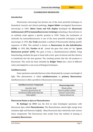
Autofluorescence
Some specimens naturally fluoresce when illuminated by a proper wavelength of
light. This phenomenon is called autofluorescence or primary fluorescence.
Autofluorescence is often a problem in fluorescence microscopy.
Autofluorescence Compound Fluorescing Color
Ascorbic Acid Weak yellow
Carotene (provitamin A) Intense golden yellow
Pyridoxine (B4) hydrochloride Intense violet white
Chlorophyll Intense blood red
Fluorescent Stains or dyes or Fluorochromes
M. Haitinger in 1933 was the first to stain histological specimens with
fluorescent dyes called Fluorochromes. The fluorochromes absorb light energy from
excitation light and fluoresce brightly. Many fluorescent dyes selectively stain various
tissue components. The fluorescence that results from this method of staining is
secondary fluorescence.
General microbiology Unit 2
Arthi Mohan Page 2
Immunofluorescence
Albert H. Coons and N. H. Kaplan were the first to attach a fluorescent dye to an
antibody, and this antibody subsequently used to localize its respective antigen in a
tissue section.
Fluorochrome Excitation max Uses
DAPI (Diamidino-2-phenyl indole) 358/461nm
Stains DNA; fluoresces green; binds to A-T rich
regions of ds DNA
FITC (Fluorescein isothiocyanate) 495/517nm
Often attached to antibodies that bind specific
cellular components; fluoresces green
Rodamine 550/573nm
Often attached to antibodies that bind specific
cellular components; fluoresces red
Stokes’ law.
The color of the emitted light has a longer wavelength than the color of the
exciting light, this relationship is known as Stokes’ law.
Fluorochromes Excitation light
Emmisson
light
FITC Blue light Green light
Rhodamine Green light Red light
Fluorescent substances are excited by a range of wavelengths known as their
absorption spectrum. They also emit a range of wavelengths known as their emission
spectrum. For any fluorescent substance the two spectra will show an absorption
(excitation) maximum and an emission maximum and some portions of the spectra will
usually overlap. The difference between the absorption maximum and emission
maximum is the Stokes’ shift.
The absorption and emission spectra for FITC in aqueous solution.
General microbiology Unit 2
Arthi Mohan Page 3
Types of Fluorescence Microscopes
There are two types of fluorescent microscopy
i. Diascopic Fluorescence
ii. Episcopic or epifluorescence microscope
Epifluorescence microscope
It the most commonly used fluorescence microscopy. In epifluorescence
microscopy, the excitation light comes from above the specimen through the objective
lens. This is the most common form of fluorescence microscopy today. This type of
fluorescence microscopy became feasible with the invention of the dichroic mirror
(chromatic beam-splitter) by E.M. Bromberg in 1953.
Advantages of epifluorescence microscope over Diascopic Fluorescence
1. High NA objectives are used at their full aperture, therefore the expected
resolution is much better, and images are brighter at high magnification.
2. The invention of the epi-illumination filter cube by J. S. Ploem in 1970 has
made it easy to interchange filter combinations using the episcopic apparatus.
3. It is easy to combine the fluorescent image with a transmitted light image of the
specimen.
Components of fluorescent microscope
i) Light source
Light sources for fluorescence microscopy, Field Condenser must produce light
within the absorption region of the fluorochrome(s) being used and the intensity of the
light should be high. Several light sources are available.
 Tungsten halogen lamps can be used for FITC.
 High-pressure mercury lamps are a common source since they produce
radiation in the UV as well as the visible spectrum (it is not suitable for all
fluorochromes).
 High-pressure xenon lamp offers an alternative to the Hg lamp, but it has low
emission in the UV.
 CSI lamp, this is a metal-halide arc lamp.
General microbiology Unit 2
Arthi Mohan Page 4
ii) Filter
Optical filters pass only selected wavelengths of light that is necessary in
fluorescence microscopy. Several types of filters accomplish the excitation and emission
in fluorescence microscopy. Each type is characterized by the wavelengths and intensity
of light that it transmits.
Excitation filter
Transmits only the desired wavelength of excitation light. An excitation filter
must select wavelengths of light from a suitable light source that fall in the maximum
absorption region of a fluorescent dye.
Emission filter or Barrier filter
An emission filter allows the emitted light of longer wavelength to pass through.
It blocks out any residual excitation wavelengths which creates dark background.
iii) Dichroic mirror
A special type of filter is the dichroic mirror or chromatic beam-splitter. This
interference filter will reflect light of shorter wavelengths (i.e. the excitation light) but
allows the light of longer wavelengths to pass through. Dichroic mirrors have very
specific reflection and transmission wavelength characteristics.
The optical set-up of fluorescent microscope
General microbiology Unit 2
Arthi Mohan Page 5
When fluorescent molecules absorb radiant energy, they become excited and
later release much of their trapped energy as light and returns to a more stable form.
Any light emitted by an excited molecule will have a longer wavelength (lower
energy) than the radiation originally absorbed. The fluorescence microscope exposes a
specimen to UV, violet or blue light and forms an image of the object with the resulting
fluorescent light. The objective lens of epifluorescence microscope also acts as a
condenser. A mercury vapour arc or other source produces an intense beam of light
that passes through an exciter filter. The exciter filter transmits only desired
wavelength of excitation light. The excitation light is directed down the microscope by a
special mirror called dichromatic mirror. It reflects the light of shorter wavelengths
(i.e. the excitation light) but allows the light of longer wavelengths to pass through. The
excitation light continues down, passing through the objective lens to specimen, which
is stained with fluorochromes. The fluorochromes absorbs light energy from the
excitation light and fluoresce brightly. The emitted fluorescent light travels up through
the objective lens into the microscope. The emitted florescence light has a longer
wavelength, it passes through the dichromatic mirror to a barrier filter, which blocks
out any residual excitation light. Finally, the emitted light reaches the eyepiece.
Application of Fluorescence Microscope
 The Fluorescence microscope has become an essential tool in medical
microbiology and microbial ecology.
 Bacterial pathogens can be identified after staining them with fluorescent or
specifically labeling them with fluorescent antibodies using
immunofluorescence procedures. eg. Mycobacterium tuberculosis.
 In ecological studies, the Fluorescence microscope is used to observe
microorganisms stained with Fluorochrome-label probes or Fluorochromes that
bind specific cell constituents.
 To visualize photosynthetic microbes, as their pigments naturally fluoresce
when excited by light of specific wavelengths.
 To distinguish live and dead bacteria by the color they fluoresce after
treatment with a mixture of stains.
 Another Important use of the fluorescence microscope is the localization of
specific proteins within the cell.
General microbiology Unit 2
Arthi Mohan Page 6
Advantages of Fluorescence Microscope
 It is used to study the dynamic behaviour exhibited in live-cell imaging.
 It can trace the location of a specific protein in the cell.
 Due to the presence of higher sensitivity, it can detect the 50 molecules per cubic
micrometer.
 It allows multicolor staining of the specimen.
Disadvantages of Fluorescence Microscope
 During the process of photobleaching, the fluorophores lose their ability to
fluoresce.
 Fluorescent molecules can generate reactive chemical species during the
illumination process which enhances the phototoxic effect.
 The specimen must be stained with the fluorescent dyes.
 Specimen preparation is a costly process.

More Related Content

Similar to 5. Fluorescence Microscope.pdf

FLUORESCENCE MICROSCOPY .pptx
FLUORESCENCE MICROSCOPY .pptxFLUORESCENCE MICROSCOPY .pptx
FLUORESCENCE MICROSCOPY .pptx
CHIRANTANMONDAL2
 
Fluorescence Microscopy
Fluorescence MicroscopyFluorescence Microscopy
Fluorescence Microscopy
VIVEK KUMAR SINGH
 
Fluorescence Microscopy
Fluorescence MicroscopyFluorescence Microscopy
Fluorescence Microscopy
University of Allahabad
 
Fluoresence microscope
Fluoresence microscopeFluoresence microscope
Fluoresence microscope
SuganyaPaulraj
 
Fluorescence microscopy presentation
Fluorescence microscopy presentationFluorescence microscopy presentation
Fluorescence microscopy presentation
Mandira bhosale
 
Fluorescence microscopy
Fluorescence microscopy   Fluorescence microscopy
Fluorescence microscopy
Fida Muhammad
 
Microscope ppt, by jitendra kumar pandey,medical micro,2nd yr, mgm medical co...
Microscope ppt, by jitendra kumar pandey,medical micro,2nd yr, mgm medical co...Microscope ppt, by jitendra kumar pandey,medical micro,2nd yr, mgm medical co...
Microscope ppt, by jitendra kumar pandey,medical micro,2nd yr, mgm medical co...
jitendra Pandey
 
fluroscence microscopy final- ppt.pptx
fluroscence microscopy final- ppt.pptxfluroscence microscopy final- ppt.pptx
fluroscence microscopy final- ppt.pptx
MonalisaMathan1
 
Types of microscope
Types of microscopeTypes of microscope
Fluorescence Microscopy.pptx
Fluorescence Microscopy.pptxFluorescence Microscopy.pptx
Fluorescence Microscopy.pptx
CHIRANTANMONDAL2
 
Phase contrast & Flurosence microscope
Phase contrast & Flurosence microscopePhase contrast & Flurosence microscope
Phase contrast & Flurosence microscope
microbiology Notes
 
Flurosence microscopy.pptx
Flurosence  microscopy.pptxFlurosence  microscopy.pptx
Flurosence microscopy.pptx
CHIRANTANMONDAL2
 
Fluorimetry
FluorimetryFluorimetry
Fluorimetry
ushaSanmugaraj
 
maitri218principlesandapplicationoflightphaseconstrastfluorescencemicroscope-...
maitri218principlesandapplicationoflightphaseconstrastfluorescencemicroscope-...maitri218principlesandapplicationoflightphaseconstrastfluorescencemicroscope-...
maitri218principlesandapplicationoflightphaseconstrastfluorescencemicroscope-...
AbhaySharma686520
 
Principles and application of light, phase constrast and fluorescence microscope
Principles and application of light, phase constrast and fluorescence microscopePrinciples and application of light, phase constrast and fluorescence microscope
Principles and application of light, phase constrast and fluorescence microscope
MaitriThakor
 
Phase contrast and Fluorescent microscope
Phase contrast and Fluorescent microscopePhase contrast and Fluorescent microscope
Phase contrast and Fluorescent microscope
Nithiya Pappuraj
 
Fluorescent Microscopy
Fluorescent MicroscopyFluorescent Microscopy
Fluorescent Microscopy
Syed Muhammad Khan
 
Fluorimetry ANALYSIS- SPECTOSCOPY PPT
Fluorimetry ANALYSIS- SPECTOSCOPY PPTFluorimetry ANALYSIS- SPECTOSCOPY PPT
Fluorimetry ANALYSIS- SPECTOSCOPY PPT
Dr Duggirala Mahendra
 

Similar to 5. Fluorescence Microscope.pdf (20)

FLUORESCENCE MICROSCOPY .pptx
FLUORESCENCE MICROSCOPY .pptxFLUORESCENCE MICROSCOPY .pptx
FLUORESCENCE MICROSCOPY .pptx
 
Fluorescence Microscopy
Fluorescence MicroscopyFluorescence Microscopy
Fluorescence Microscopy
 
123456
123456123456
123456
 
Fluorescence Microscopy
Fluorescence MicroscopyFluorescence Microscopy
Fluorescence Microscopy
 
Fluoresence microscope
Fluoresence microscopeFluoresence microscope
Fluoresence microscope
 
Fluorescence microscopy presentation
Fluorescence microscopy presentationFluorescence microscopy presentation
Fluorescence microscopy presentation
 
Fluorescence microscopy
Fluorescence microscopy   Fluorescence microscopy
Fluorescence microscopy
 
Microscope ppt, by jitendra kumar pandey,medical micro,2nd yr, mgm medical co...
Microscope ppt, by jitendra kumar pandey,medical micro,2nd yr, mgm medical co...Microscope ppt, by jitendra kumar pandey,medical micro,2nd yr, mgm medical co...
Microscope ppt, by jitendra kumar pandey,medical micro,2nd yr, mgm medical co...
 
fluroscence microscopy final- ppt.pptx
fluroscence microscopy final- ppt.pptxfluroscence microscopy final- ppt.pptx
fluroscence microscopy final- ppt.pptx
 
Types of microscope
Types of microscopeTypes of microscope
Types of microscope
 
Fluorescence Microscopy.pptx
Fluorescence Microscopy.pptxFluorescence Microscopy.pptx
Fluorescence Microscopy.pptx
 
Phase contrast & Flurosence microscope
Phase contrast & Flurosence microscopePhase contrast & Flurosence microscope
Phase contrast & Flurosence microscope
 
Flurosence microscopy.pptx
Flurosence  microscopy.pptxFlurosence  microscopy.pptx
Flurosence microscopy.pptx
 
Immunofluorescence
ImmunofluorescenceImmunofluorescence
Immunofluorescence
 
Fluorimetry
FluorimetryFluorimetry
Fluorimetry
 
maitri218principlesandapplicationoflightphaseconstrastfluorescencemicroscope-...
maitri218principlesandapplicationoflightphaseconstrastfluorescencemicroscope-...maitri218principlesandapplicationoflightphaseconstrastfluorescencemicroscope-...
maitri218principlesandapplicationoflightphaseconstrastfluorescencemicroscope-...
 
Principles and application of light, phase constrast and fluorescence microscope
Principles and application of light, phase constrast and fluorescence microscopePrinciples and application of light, phase constrast and fluorescence microscope
Principles and application of light, phase constrast and fluorescence microscope
 
Phase contrast and Fluorescent microscope
Phase contrast and Fluorescent microscopePhase contrast and Fluorescent microscope
Phase contrast and Fluorescent microscope
 
Fluorescent Microscopy
Fluorescent MicroscopyFluorescent Microscopy
Fluorescent Microscopy
 
Fluorimetry ANALYSIS- SPECTOSCOPY PPT
Fluorimetry ANALYSIS- SPECTOSCOPY PPTFluorimetry ANALYSIS- SPECTOSCOPY PPT
Fluorimetry ANALYSIS- SPECTOSCOPY PPT
 

More from MicrobiologyMicro

Translation .pptx
Translation                         .pptxTranslation                         .pptx
Translation .pptx
MicrobiologyMicro
 
Structure and types of RNA .pptx
Structure and types of RNA          .pptxStructure and types of RNA          .pptx
Structure and types of RNA .pptx
MicrobiologyMicro
 
Transcription - DNA dependent RNA synthesis.pptx
Transcription - DNA dependent RNA synthesis.pptxTranscription - DNA dependent RNA synthesis.pptx
Transcription - DNA dependent RNA synthesis.pptx
MicrobiologyMicro
 
. DNA Repair Mechanisms.pptx
.             DNA Repair Mechanisms.pptx.             DNA Repair Mechanisms.pptx
. DNA Repair Mechanisms.pptx
MicrobiologyMicro
 
1. Immunology and its history.pptx
1.       Immunology and its history.pptx1.       Immunology and its history.pptx
1. Immunology and its history.pptx
MicrobiologyMicro
 
5. ATOMIC FORCE MICROSCOPE (AFM).pdf
5.     ATOMIC FORCE MICROSCOPE (AFM).pdf5.     ATOMIC FORCE MICROSCOPE (AFM).pdf
5. ATOMIC FORCE MICROSCOPE (AFM).pdf
MicrobiologyMicro
 
4. Phase-contrast microscope.pdf
4.         Phase-contrast microscope.pdf4.         Phase-contrast microscope.pdf
4. Phase-contrast microscope.pdf
MicrobiologyMicro
 
3. Dark-field microscopy.pdf
3.             Dark-field microscopy.pdf3.             Dark-field microscopy.pdf
3. Dark-field microscopy.pdf
MicrobiologyMicro
 
2. Types and Bright field microscopy.pdf
2. Types and Bright field microscopy.pdf2. Types and Bright field microscopy.pdf
2. Types and Bright field microscopy.pdf
MicrobiologyMicro
 
1. Introduction to Microscopy.pdf
1.        Introduction to Microscopy.pdf1.        Introduction to Microscopy.pdf
1. Introduction to Microscopy.pdf
MicrobiologyMicro
 
3. Differences between Prokaryotes and Eukaryotes.pdf
3. Differences between Prokaryotes and Eukaryotes.pdf3. Differences between Prokaryotes and Eukaryotes.pdf
3. Differences between Prokaryotes and Eukaryotes.pdf
MicrobiologyMicro
 
History of Microbiology - General microbiology.pdf
History of Microbiology - General microbiology.pdfHistory of Microbiology - General microbiology.pdf
History of Microbiology - General microbiology.pdf
MicrobiologyMicro
 
Definition and scope of microbiology.pdf
Definition and scope of microbiology.pdfDefinition and scope of microbiology.pdf
Definition and scope of microbiology.pdf
MicrobiologyMicro
 
Transposons, types and transposition.pptx
Transposons, types and transposition.pptxTransposons, types and transposition.pptx
Transposons, types and transposition.pptx
MicrobiologyMicro
 
Prokaryotic & Eukaryotic Replication.pptx
Prokaryotic & Eukaryotic Replication.pptxProkaryotic & Eukaryotic Replication.pptx
Prokaryotic & Eukaryotic Replication.pptx
MicrobiologyMicro
 
Microbial Assessment of Air Quality.pptx
Microbial Assessment of Air Quality.pptxMicrobial Assessment of Air Quality.pptx
Microbial Assessment of Air Quality.pptx
MicrobiologyMicro
 
Air Sanitation.pptx
Air Sanitation.pptxAir Sanitation.pptx
Air Sanitation.pptx
MicrobiologyMicro
 
Composition and Distribution of Air.pptx
Composition and Distribution of Air.pptxComposition and Distribution of Air.pptx
Composition and Distribution of Air.pptx
MicrobiologyMicro
 
Interaction of microorganisms with Insects.pptx
Interaction of microorganisms with Insects.pptxInteraction of microorganisms with Insects.pptx
Interaction of microorganisms with Insects.pptx
MicrobiologyMicro
 
Interaction of microorganisms with Animals.pptx
Interaction of microorganisms with Animals.pptxInteraction of microorganisms with Animals.pptx
Interaction of microorganisms with Animals.pptx
MicrobiologyMicro
 

More from MicrobiologyMicro (20)

Translation .pptx
Translation                         .pptxTranslation                         .pptx
Translation .pptx
 
Structure and types of RNA .pptx
Structure and types of RNA          .pptxStructure and types of RNA          .pptx
Structure and types of RNA .pptx
 
Transcription - DNA dependent RNA synthesis.pptx
Transcription - DNA dependent RNA synthesis.pptxTranscription - DNA dependent RNA synthesis.pptx
Transcription - DNA dependent RNA synthesis.pptx
 
. DNA Repair Mechanisms.pptx
.             DNA Repair Mechanisms.pptx.             DNA Repair Mechanisms.pptx
. DNA Repair Mechanisms.pptx
 
1. Immunology and its history.pptx
1.       Immunology and its history.pptx1.       Immunology and its history.pptx
1. Immunology and its history.pptx
 
5. ATOMIC FORCE MICROSCOPE (AFM).pdf
5.     ATOMIC FORCE MICROSCOPE (AFM).pdf5.     ATOMIC FORCE MICROSCOPE (AFM).pdf
5. ATOMIC FORCE MICROSCOPE (AFM).pdf
 
4. Phase-contrast microscope.pdf
4.         Phase-contrast microscope.pdf4.         Phase-contrast microscope.pdf
4. Phase-contrast microscope.pdf
 
3. Dark-field microscopy.pdf
3.             Dark-field microscopy.pdf3.             Dark-field microscopy.pdf
3. Dark-field microscopy.pdf
 
2. Types and Bright field microscopy.pdf
2. Types and Bright field microscopy.pdf2. Types and Bright field microscopy.pdf
2. Types and Bright field microscopy.pdf
 
1. Introduction to Microscopy.pdf
1.        Introduction to Microscopy.pdf1.        Introduction to Microscopy.pdf
1. Introduction to Microscopy.pdf
 
3. Differences between Prokaryotes and Eukaryotes.pdf
3. Differences between Prokaryotes and Eukaryotes.pdf3. Differences between Prokaryotes and Eukaryotes.pdf
3. Differences between Prokaryotes and Eukaryotes.pdf
 
History of Microbiology - General microbiology.pdf
History of Microbiology - General microbiology.pdfHistory of Microbiology - General microbiology.pdf
History of Microbiology - General microbiology.pdf
 
Definition and scope of microbiology.pdf
Definition and scope of microbiology.pdfDefinition and scope of microbiology.pdf
Definition and scope of microbiology.pdf
 
Transposons, types and transposition.pptx
Transposons, types and transposition.pptxTransposons, types and transposition.pptx
Transposons, types and transposition.pptx
 
Prokaryotic & Eukaryotic Replication.pptx
Prokaryotic & Eukaryotic Replication.pptxProkaryotic & Eukaryotic Replication.pptx
Prokaryotic & Eukaryotic Replication.pptx
 
Microbial Assessment of Air Quality.pptx
Microbial Assessment of Air Quality.pptxMicrobial Assessment of Air Quality.pptx
Microbial Assessment of Air Quality.pptx
 
Air Sanitation.pptx
Air Sanitation.pptxAir Sanitation.pptx
Air Sanitation.pptx
 
Composition and Distribution of Air.pptx
Composition and Distribution of Air.pptxComposition and Distribution of Air.pptx
Composition and Distribution of Air.pptx
 
Interaction of microorganisms with Insects.pptx
Interaction of microorganisms with Insects.pptxInteraction of microorganisms with Insects.pptx
Interaction of microorganisms with Insects.pptx
 
Interaction of microorganisms with Animals.pptx
Interaction of microorganisms with Animals.pptxInteraction of microorganisms with Animals.pptx
Interaction of microorganisms with Animals.pptx
 

Recently uploaded

Earliest Galaxies in the JADES Origins Field: Luminosity Function and Cosmic ...
Earliest Galaxies in the JADES Origins Field: Luminosity Function and Cosmic ...Earliest Galaxies in the JADES Origins Field: Luminosity Function and Cosmic ...
Earliest Galaxies in the JADES Origins Field: Luminosity Function and Cosmic ...
Sérgio Sacani
 
Orion Air Quality Monitoring Systems - CWS
Orion Air Quality Monitoring Systems - CWSOrion Air Quality Monitoring Systems - CWS
Orion Air Quality Monitoring Systems - CWS
Columbia Weather Systems
 
Structures and textures of metamorphic rocks
Structures and textures of metamorphic rocksStructures and textures of metamorphic rocks
Structures and textures of metamorphic rocks
kumarmathi863
 
insect taxonomy importance systematics and classification
insect taxonomy importance systematics and classificationinsect taxonomy importance systematics and classification
insect taxonomy importance systematics and classification
anitaento25
 
general properties of oerganologametal.ppt
general properties of oerganologametal.pptgeneral properties of oerganologametal.ppt
general properties of oerganologametal.ppt
IqrimaNabilatulhusni
 
platelets- lifespan -Clot retraction-disorders.pptx
platelets- lifespan -Clot retraction-disorders.pptxplatelets- lifespan -Clot retraction-disorders.pptx
platelets- lifespan -Clot retraction-disorders.pptx
muralinath2
 
Leaf Initiation, Growth and Differentiation.pdf
Leaf Initiation, Growth and Differentiation.pdfLeaf Initiation, Growth and Differentiation.pdf
Leaf Initiation, Growth and Differentiation.pdf
RenuJangid3
 
PRESENTATION ABOUT PRINCIPLE OF COSMATIC EVALUATION
PRESENTATION ABOUT PRINCIPLE OF COSMATIC EVALUATIONPRESENTATION ABOUT PRINCIPLE OF COSMATIC EVALUATION
PRESENTATION ABOUT PRINCIPLE OF COSMATIC EVALUATION
ChetanK57
 
THE IMPORTANCE OF MARTIAN ATMOSPHERE SAMPLE RETURN.
THE IMPORTANCE OF MARTIAN ATMOSPHERE SAMPLE RETURN.THE IMPORTANCE OF MARTIAN ATMOSPHERE SAMPLE RETURN.
THE IMPORTANCE OF MARTIAN ATMOSPHERE SAMPLE RETURN.
Sérgio Sacani
 
role of pramana in research.pptx in science
role of pramana in research.pptx in sciencerole of pramana in research.pptx in science
role of pramana in research.pptx in science
sonaliswain16
 
GBSN- Microbiology (Lab 3) Gram Staining
GBSN- Microbiology (Lab 3) Gram StainingGBSN- Microbiology (Lab 3) Gram Staining
GBSN- Microbiology (Lab 3) Gram Staining
Areesha Ahmad
 
GBSN - Microbiology (Lab 4) Culture Media
GBSN - Microbiology (Lab 4) Culture MediaGBSN - Microbiology (Lab 4) Culture Media
GBSN - Microbiology (Lab 4) Culture Media
Areesha Ahmad
 
What is greenhouse gasses and how many gasses are there to affect the Earth.
What is greenhouse gasses and how many gasses are there to affect the Earth.What is greenhouse gasses and how many gasses are there to affect the Earth.
What is greenhouse gasses and how many gasses are there to affect the Earth.
moosaasad1975
 
NuGOweek 2024 Ghent - programme - final version
NuGOweek 2024 Ghent - programme - final versionNuGOweek 2024 Ghent - programme - final version
NuGOweek 2024 Ghent - programme - final version
pablovgd
 
Comparative structure of adrenal gland in vertebrates
Comparative structure of adrenal gland in vertebratesComparative structure of adrenal gland in vertebrates
Comparative structure of adrenal gland in vertebrates
sachin783648
 
RNA INTERFERENCE: UNRAVELING GENETIC SILENCING
RNA INTERFERENCE: UNRAVELING GENETIC SILENCINGRNA INTERFERENCE: UNRAVELING GENETIC SILENCING
RNA INTERFERENCE: UNRAVELING GENETIC SILENCING
AADYARAJPANDEY1
 
Lab report on liquid viscosity of glycerin
Lab report on liquid viscosity of glycerinLab report on liquid viscosity of glycerin
Lab report on liquid viscosity of glycerin
ossaicprecious19
 
4. An Overview of Sugarcane White Leaf Disease in Vietnam.pdf
4. An Overview of Sugarcane White Leaf Disease in Vietnam.pdf4. An Overview of Sugarcane White Leaf Disease in Vietnam.pdf
4. An Overview of Sugarcane White Leaf Disease in Vietnam.pdf
ssuserbfdca9
 
erythropoiesis-I_mechanism& clinical significance.pptx
erythropoiesis-I_mechanism& clinical significance.pptxerythropoiesis-I_mechanism& clinical significance.pptx
erythropoiesis-I_mechanism& clinical significance.pptx
muralinath2
 
Nucleic Acid-its structural and functional complexity.
Nucleic Acid-its structural and functional complexity.Nucleic Acid-its structural and functional complexity.
Nucleic Acid-its structural and functional complexity.
Nistarini College, Purulia (W.B) India
 

Recently uploaded (20)

Earliest Galaxies in the JADES Origins Field: Luminosity Function and Cosmic ...
Earliest Galaxies in the JADES Origins Field: Luminosity Function and Cosmic ...Earliest Galaxies in the JADES Origins Field: Luminosity Function and Cosmic ...
Earliest Galaxies in the JADES Origins Field: Luminosity Function and Cosmic ...
 
Orion Air Quality Monitoring Systems - CWS
Orion Air Quality Monitoring Systems - CWSOrion Air Quality Monitoring Systems - CWS
Orion Air Quality Monitoring Systems - CWS
 
Structures and textures of metamorphic rocks
Structures and textures of metamorphic rocksStructures and textures of metamorphic rocks
Structures and textures of metamorphic rocks
 
insect taxonomy importance systematics and classification
insect taxonomy importance systematics and classificationinsect taxonomy importance systematics and classification
insect taxonomy importance systematics and classification
 
general properties of oerganologametal.ppt
general properties of oerganologametal.pptgeneral properties of oerganologametal.ppt
general properties of oerganologametal.ppt
 
platelets- lifespan -Clot retraction-disorders.pptx
platelets- lifespan -Clot retraction-disorders.pptxplatelets- lifespan -Clot retraction-disorders.pptx
platelets- lifespan -Clot retraction-disorders.pptx
 
Leaf Initiation, Growth and Differentiation.pdf
Leaf Initiation, Growth and Differentiation.pdfLeaf Initiation, Growth and Differentiation.pdf
Leaf Initiation, Growth and Differentiation.pdf
 
PRESENTATION ABOUT PRINCIPLE OF COSMATIC EVALUATION
PRESENTATION ABOUT PRINCIPLE OF COSMATIC EVALUATIONPRESENTATION ABOUT PRINCIPLE OF COSMATIC EVALUATION
PRESENTATION ABOUT PRINCIPLE OF COSMATIC EVALUATION
 
THE IMPORTANCE OF MARTIAN ATMOSPHERE SAMPLE RETURN.
THE IMPORTANCE OF MARTIAN ATMOSPHERE SAMPLE RETURN.THE IMPORTANCE OF MARTIAN ATMOSPHERE SAMPLE RETURN.
THE IMPORTANCE OF MARTIAN ATMOSPHERE SAMPLE RETURN.
 
role of pramana in research.pptx in science
role of pramana in research.pptx in sciencerole of pramana in research.pptx in science
role of pramana in research.pptx in science
 
GBSN- Microbiology (Lab 3) Gram Staining
GBSN- Microbiology (Lab 3) Gram StainingGBSN- Microbiology (Lab 3) Gram Staining
GBSN- Microbiology (Lab 3) Gram Staining
 
GBSN - Microbiology (Lab 4) Culture Media
GBSN - Microbiology (Lab 4) Culture MediaGBSN - Microbiology (Lab 4) Culture Media
GBSN - Microbiology (Lab 4) Culture Media
 
What is greenhouse gasses and how many gasses are there to affect the Earth.
What is greenhouse gasses and how many gasses are there to affect the Earth.What is greenhouse gasses and how many gasses are there to affect the Earth.
What is greenhouse gasses and how many gasses are there to affect the Earth.
 
NuGOweek 2024 Ghent - programme - final version
NuGOweek 2024 Ghent - programme - final versionNuGOweek 2024 Ghent - programme - final version
NuGOweek 2024 Ghent - programme - final version
 
Comparative structure of adrenal gland in vertebrates
Comparative structure of adrenal gland in vertebratesComparative structure of adrenal gland in vertebrates
Comparative structure of adrenal gland in vertebrates
 
RNA INTERFERENCE: UNRAVELING GENETIC SILENCING
RNA INTERFERENCE: UNRAVELING GENETIC SILENCINGRNA INTERFERENCE: UNRAVELING GENETIC SILENCING
RNA INTERFERENCE: UNRAVELING GENETIC SILENCING
 
Lab report on liquid viscosity of glycerin
Lab report on liquid viscosity of glycerinLab report on liquid viscosity of glycerin
Lab report on liquid viscosity of glycerin
 
4. An Overview of Sugarcane White Leaf Disease in Vietnam.pdf
4. An Overview of Sugarcane White Leaf Disease in Vietnam.pdf4. An Overview of Sugarcane White Leaf Disease in Vietnam.pdf
4. An Overview of Sugarcane White Leaf Disease in Vietnam.pdf
 
erythropoiesis-I_mechanism& clinical significance.pptx
erythropoiesis-I_mechanism& clinical significance.pptxerythropoiesis-I_mechanism& clinical significance.pptx
erythropoiesis-I_mechanism& clinical significance.pptx
 
Nucleic Acid-its structural and functional complexity.
Nucleic Acid-its structural and functional complexity.Nucleic Acid-its structural and functional complexity.
Nucleic Acid-its structural and functional complexity.
 

5. Fluorescence Microscope.pdf

  • 1. General microbiology Unit 2 Arthi Mohan Page 1 FLUORESCENCE MICROSCOPE Introduction Fluorescence microscopy has become one of the most powerful techniques in biomedical research and clinical pathology. August Köhler investigated fluorescence microscopy in 1904. Albert Coons and N.H. Kaplan developed the fluorescein isothiocyanate (FITC) immunofluorescence technique (attaching a fluorochrome to an antibody made against a specific protein) in 1950. Today, the localization of molecules by immunofluorescence is one of the most powerful techniques in light microscopy. In 1991, B.J. Trask described a method of fluorescently labeling specific sequences of DNA. This method is known as fluorescence in situ hybridization (FISH). In 1992, D.C. Parsher et al. cloned the gene that codes for the “green fluorescent protein” (GFP). The gene is from a chemiluminescent jellyfish. Using biotechnology methods the gene is fused with a host gene of interest and this chimera is transfected into the host genome. The resulting protein that the cell produces is fluorescent. This work has been extended by Rodger Tsien into a host of different colors and adapted to a vast array of biological techniques. Autofluorescence Some specimens naturally fluoresce when illuminated by a proper wavelength of light. This phenomenon is called autofluorescence or primary fluorescence. Autofluorescence is often a problem in fluorescence microscopy. Autofluorescence Compound Fluorescing Color Ascorbic Acid Weak yellow Carotene (provitamin A) Intense golden yellow Pyridoxine (B4) hydrochloride Intense violet white Chlorophyll Intense blood red Fluorescent Stains or dyes or Fluorochromes M. Haitinger in 1933 was the first to stain histological specimens with fluorescent dyes called Fluorochromes. The fluorochromes absorb light energy from excitation light and fluoresce brightly. Many fluorescent dyes selectively stain various tissue components. The fluorescence that results from this method of staining is secondary fluorescence.
  • 2. General microbiology Unit 2 Arthi Mohan Page 2 Immunofluorescence Albert H. Coons and N. H. Kaplan were the first to attach a fluorescent dye to an antibody, and this antibody subsequently used to localize its respective antigen in a tissue section. Fluorochrome Excitation max Uses DAPI (Diamidino-2-phenyl indole) 358/461nm Stains DNA; fluoresces green; binds to A-T rich regions of ds DNA FITC (Fluorescein isothiocyanate) 495/517nm Often attached to antibodies that bind specific cellular components; fluoresces green Rodamine 550/573nm Often attached to antibodies that bind specific cellular components; fluoresces red Stokes’ law. The color of the emitted light has a longer wavelength than the color of the exciting light, this relationship is known as Stokes’ law. Fluorochromes Excitation light Emmisson light FITC Blue light Green light Rhodamine Green light Red light Fluorescent substances are excited by a range of wavelengths known as their absorption spectrum. They also emit a range of wavelengths known as their emission spectrum. For any fluorescent substance the two spectra will show an absorption (excitation) maximum and an emission maximum and some portions of the spectra will usually overlap. The difference between the absorption maximum and emission maximum is the Stokes’ shift. The absorption and emission spectra for FITC in aqueous solution.
  • 3. General microbiology Unit 2 Arthi Mohan Page 3 Types of Fluorescence Microscopes There are two types of fluorescent microscopy i. Diascopic Fluorescence ii. Episcopic or epifluorescence microscope Epifluorescence microscope It the most commonly used fluorescence microscopy. In epifluorescence microscopy, the excitation light comes from above the specimen through the objective lens. This is the most common form of fluorescence microscopy today. This type of fluorescence microscopy became feasible with the invention of the dichroic mirror (chromatic beam-splitter) by E.M. Bromberg in 1953. Advantages of epifluorescence microscope over Diascopic Fluorescence 1. High NA objectives are used at their full aperture, therefore the expected resolution is much better, and images are brighter at high magnification. 2. The invention of the epi-illumination filter cube by J. S. Ploem in 1970 has made it easy to interchange filter combinations using the episcopic apparatus. 3. It is easy to combine the fluorescent image with a transmitted light image of the specimen. Components of fluorescent microscope i) Light source Light sources for fluorescence microscopy, Field Condenser must produce light within the absorption region of the fluorochrome(s) being used and the intensity of the light should be high. Several light sources are available.  Tungsten halogen lamps can be used for FITC.  High-pressure mercury lamps are a common source since they produce radiation in the UV as well as the visible spectrum (it is not suitable for all fluorochromes).  High-pressure xenon lamp offers an alternative to the Hg lamp, but it has low emission in the UV.  CSI lamp, this is a metal-halide arc lamp.
  • 4. General microbiology Unit 2 Arthi Mohan Page 4 ii) Filter Optical filters pass only selected wavelengths of light that is necessary in fluorescence microscopy. Several types of filters accomplish the excitation and emission in fluorescence microscopy. Each type is characterized by the wavelengths and intensity of light that it transmits. Excitation filter Transmits only the desired wavelength of excitation light. An excitation filter must select wavelengths of light from a suitable light source that fall in the maximum absorption region of a fluorescent dye. Emission filter or Barrier filter An emission filter allows the emitted light of longer wavelength to pass through. It blocks out any residual excitation wavelengths which creates dark background. iii) Dichroic mirror A special type of filter is the dichroic mirror or chromatic beam-splitter. This interference filter will reflect light of shorter wavelengths (i.e. the excitation light) but allows the light of longer wavelengths to pass through. Dichroic mirrors have very specific reflection and transmission wavelength characteristics. The optical set-up of fluorescent microscope
  • 5. General microbiology Unit 2 Arthi Mohan Page 5 When fluorescent molecules absorb radiant energy, they become excited and later release much of their trapped energy as light and returns to a more stable form. Any light emitted by an excited molecule will have a longer wavelength (lower energy) than the radiation originally absorbed. The fluorescence microscope exposes a specimen to UV, violet or blue light and forms an image of the object with the resulting fluorescent light. The objective lens of epifluorescence microscope also acts as a condenser. A mercury vapour arc or other source produces an intense beam of light that passes through an exciter filter. The exciter filter transmits only desired wavelength of excitation light. The excitation light is directed down the microscope by a special mirror called dichromatic mirror. It reflects the light of shorter wavelengths (i.e. the excitation light) but allows the light of longer wavelengths to pass through. The excitation light continues down, passing through the objective lens to specimen, which is stained with fluorochromes. The fluorochromes absorbs light energy from the excitation light and fluoresce brightly. The emitted fluorescent light travels up through the objective lens into the microscope. The emitted florescence light has a longer wavelength, it passes through the dichromatic mirror to a barrier filter, which blocks out any residual excitation light. Finally, the emitted light reaches the eyepiece. Application of Fluorescence Microscope  The Fluorescence microscope has become an essential tool in medical microbiology and microbial ecology.  Bacterial pathogens can be identified after staining them with fluorescent or specifically labeling them with fluorescent antibodies using immunofluorescence procedures. eg. Mycobacterium tuberculosis.  In ecological studies, the Fluorescence microscope is used to observe microorganisms stained with Fluorochrome-label probes or Fluorochromes that bind specific cell constituents.  To visualize photosynthetic microbes, as their pigments naturally fluoresce when excited by light of specific wavelengths.  To distinguish live and dead bacteria by the color they fluoresce after treatment with a mixture of stains.  Another Important use of the fluorescence microscope is the localization of specific proteins within the cell.
  • 6. General microbiology Unit 2 Arthi Mohan Page 6 Advantages of Fluorescence Microscope  It is used to study the dynamic behaviour exhibited in live-cell imaging.  It can trace the location of a specific protein in the cell.  Due to the presence of higher sensitivity, it can detect the 50 molecules per cubic micrometer.  It allows multicolor staining of the specimen. Disadvantages of Fluorescence Microscope  During the process of photobleaching, the fluorophores lose their ability to fluoresce.  Fluorescent molecules can generate reactive chemical species during the illumination process which enhances the phototoxic effect.  The specimen must be stained with the fluorescent dyes.  Specimen preparation is a costly process.