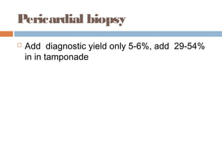
PERICARDIUM
Dr Ramachandra
Enclosed lubricated space forthe beating
heart/fix it in chest
Rule of Two’s
 Double-layered: visceral=epicardium & parietal
 Thickness=2 mm
 Fibroblastic Parietal =Meshwork of 2 fibres
 collagen (types I and III) and elastic.
 Pericardial fluid volume=2x10 ml=20-30ml
 Two sinuses=Oblique(4PV AND IVC-SVC)
 & Transverse=AO and PA
 Pressure in pericardium 2mmHg lower than
intrathoracic pressure.
 Innervation :2=ANS(parasympathetic
( vagus)/sympathetic ( stellate ganglia) and somatic=
the phrenic nerve.
Embryology
From mesoderm (the pleuropericardial membranes)
that divide the primitive thoracic cavity of the
intraembryonic coelom into pericardial and pleural
compartments. The endocardial heart tube
invaginates into the developing pericardial cavity,
whereby the innermost serosal layer adheres to the
myocardium and then folds back onto itself to create
visceral and parietal layers.This process of
invagination eventually results in suspending the adult
heart within the developing pericardial cavity by blood
vessels at its cranial and caudal attachments.The
pericardium receives its blood supply via the internal
mammary artery. Phrenic nerves are enveloped in the
pleuropericardial membranes, and in the adult, reach
the diaphragm by traveling through the fibrous
pericardium .
Physiology
Pericardial fluid act as lubricant between visceral and parietal
layers to facilitate frictionless beating of the heart. The
intrapericardial fluid function as a reservoir of paracrinemodulators,
notably compounds such as prostanoids, natriuretic peptides, and
endothelins, that may regulate sympathetic tone, coronary
vasomotor tone, heart rate, and bloodpressure, but also
immunologically active moieties, such as complement factors.
Pericardial fluid is in equilibrium with pleural fluid, as there is
hydraulic conductance across the parietal pericardium when
intrapericardial pressures exceed pleural pressures.Numerous
attachments from the pericardium to other thoracic structures
secure the heart in place within the chest cavity. Cranially, the
reflections on great vessels interdigitate with vascular adventitia to
fasten the superior borders of the pericardium; a diaphragmatic
tendon anchors the caudal end.Ventrally the pericardium connects
with the interior of the sternum (superior and inferior
pericaridosternal ligaments), and dorsally it affixes to the
esophagus and spine.
Contd
The fibrous nature of the pericardium likely also serves as a
barrier to insulate the heart from exogenous tumors and spread
of infections.Despite its fibrous character, the pericardium rests
at slightly lower pressures (_3 to _6 mm Hg) compared to the
rest of the intrathoracic cavity and is able to transmit hanges in
intrathoracic pressures with respiration.Intact pericardium is
rather nondistensible and provides a strong mechanical
constraint on overall heart volume and chamber enlargement.
Important consequences of nondistensibility are equalizing
compliance of right and left ventricles as well as
interdependence of the cardiac chambers.Interdependence
occurs because total intrapercardial volume (namely pericardial
cavity and cardiac chamber volume) is fixed in the short term,
and expansion of 1 chamber necessarily impedes expansion of
another.
Contd.......
Especially as regards ventricular interdependence,acute
expansion of either right or left ventricular volume will impair
filling of the other chamber, at the limit resulting in signs such
as the pulsus paradoxus. The same principles apply in acute
pericardial effusion and tamponade physiology, because
there is very little reserve volume for expansion in the
pericardial space before intrapericardial pressure rises
Sharply. In situations where pericardial effusion develops
gradually,pericardial compliance can increase to
accommodate significant fluid volumes, often a liter or more.
Efferent painful stimuli (eg, inflammation, distention) are
carried via phrenic nerves and stellate ganglion; however, not
every surface of the pericardium is innervated. The
pericardium appears insensate to temperature and vibration.
Contd
Interdependence occurs because total intrapercardial volume
(namely pericardial cavity and cardiac chamber volume) is
fixed in the short term, and expansion of 1 chamber
necessarily impedes expansion of another. Especially as
regards ventricular interdependence,acute expansion of
either right or left ventricular volume will impair filling of the
other chamber, at the limit resulting in signs such as the
pulsus paradoxus. The same principles apply in acute
pericardial effusion and tamponade physiology, because
there is very little reserve volume for expansion in the
pericardial space before intrapericardial pressure rises
Sharply. In situations where pericardial effusion develops
gradually,pericardial compliance can increase to
accommodate significant fluid volumes, often a liter or more.
Efferent painful stimuli (eg, inflammation, distention) are
carried via phrenic nerves and stellate ganglion; however, not
every surface of the pericardium is innervated. The
pericardium appears insensate to temperature and vibration.
Congenital defects
 Abse nce : “stabbing” chest pains with “heart
shifting
RBBB/Clo ckwise lo o p, levoposition and posterior
rotation,of the cardiac apex, as well as
pathognomonic interdigitation of lung tissue
between the inferior heart surface and diaphragm
as well as between the aorta and pulmonary
artery. “teardrop” shaped, because of atrial
elongation and ventricle widening in the absence
of typical pericardial tethering.
 Pe ricardialMasse s(e m bryo nic co e lo m ic cavity. )
EpicardialFat
 The closest D/D of pericardial effusion
Pericardial tumors
Pericardial collection
Pe ricardialEffusio ns
 Mild:<5mm
 Moerate:5-10mm
 Severe(Tamponade)>10mm
 In acute situation even mild may cause
tamponade
Pe ricarditis
 Infiltration is the sine qua non of pericarditis
 self-limited(mostly)
 80-90% of cases are idiopathic,
 “bread-and-butter pericardium”=supporation
 Peri-infarction - 5%(TLT+) vs 10-20%(TLT-
Ve), Dressler’s < 1%
 Dx:At least 2 of 4 cardinal features
Contd.......
Unique pain radiation to the trapezius ridge
The pericardial friction rub
A triphasic(50%) cadence with a discrete
“sandpaper” sound occurring with atrial
systole, ventricular systole, and early
ventricular diastole. 100% specific but
evanescent ,less sensitive.
EKG changes in acute
pericarditis
STAGE-I
Recurrent orRelapsing
Pericarditis
 Latent periods of up to 6 weeks
 12 weeks of therapy of NSAID in tappering
 CORE trial:Colchicine (1-2 mg followed by 0.5 mg daily or twice
daily for 6 months; addition to high-dose aspirin therapy for first
recurrence of idiopathic,viral, or autoimmune pericarditis,24%
recurrence at 18 months as compared to 51% in the control, a two-
thirds reduction (10% vs 31%) in symptoms at 72 hours(CLASS I-
EHA)
 Steroid for auto immuno causes-0.5mg-1mg/Kg
 Intrapericardial corticosteroids(A single instillation of triamcinolone
(300
mg/m2 to 600 mg/m2 in 100 mL isotonic saline) over 24 hours on the
background of maintenance colchicine therapy prevented 84%
recurrences
at 1 year
 Immunosuppresants:Cyclosporine /Azathioprine, remains
investigational.
Hemodynamic Consequences
Claude Schaeffer Beck (1894 – 1971) was
a pioneer American cardiac surgeon,
famous for innovating various cardiac
surgery techniques, and performing the
first defibrillation in 1947.[ He was the first
American professor of cardiovascular
surgery, from 1952 through 1965.
1930
Tamponade physiology
 Resting intrapericardial pressure is 3 to 6 mm Hg
less than the diastolic pressures in the cardiac
chambers and thus does not affect intracardiac
blood flows with 2-4 mmHg respiratory variation.
 Pericardial pressure at which the point where
cardiac filling is compromised.
 Pericardial fluid becomes like a fifth cardiac
chamber, occupying space within the pericardium
and thus limiting the ability of atria and ventricles
to expand and fill appropriately ( impaired diastolic
compliance).
May be slow or fast
Contd........
 Chamber-filling pressure is the ∆ between
intracardiac and intrapericardial pressures
 Mean diastolic pressures equalize across cardiac
chambers as they need to exceed the increased
pressures in the intrapericardial space
 Physiologicalventricular interdependence
becomes magnified due to isolation of the heart
from normal respiratory ∆
 Inspiration will increase right-sided flows at the
expense of the left-sided chambers, thus reducing
left-sided flow
Contd.......
Contd....
Left- and right-ventricular filling pressures are usually elevated at 15-
20 mm Hg in tamponade. However, so-called low-pressure
tamponade with much lower ventricular filling pressures can occur in
patients with reduced blood volume, eg, dehydration, overdiuresis, or
hemorrhage.
 A pulsus paradoxus ≥10 mm Hg a likelihood
ratio of 3.3 for the presence of tamponade
 Value ≤10 confers a negative likelihood ratio
of
0.03
 PP= 20 mm Hg ≥75% Tamponade
 Palpable pulsus (or “total paradox”) in almost
20%
DX of pulsus paradoxus
Pulsus suppression
RV or LV are markedly noncompliant= severe
left ventricular (LV) failure, right ventricular
hypertrophy, left ventricular hypertrophy, acute
myocardial infarction,severe aortic
insufficiency (elevated left ventricular end
diastolicpressure and damping of respiratory
variation), or in uremia, atrial septal defect (as
venous return is offset by right to left
shunting), and pericardial adhesions
QRS alternans
 QRS alternans is present in 20% and total
alternans (of P, QRS, and T waves) in 8% of
patients.
 Low voltage complex
Echocardiographic signs of
tamponade
ROVT collapse is quite
consistent
Inflow and out flow doppler
Mitral 35% and aortic 25% variation
and TV=80%
Pe ricardialCo nstrictio n
 ≥ 5mm echo and ≥4mm CT(N=1-2mm,<4mm)
 CP Physiology with non thickened 20%
 Kussmaul’s sign(JVP distension in inspiration)
 pericardial knock≥frequency than S3
 Friedrich’s sign=Rapid Y descent of early
diastolic suction
 “square-root”=sign of no further filling occurs
after early diastole
 Equalized diastolic in all 4 chambers
Hemodynamic=CATH/ECHO
diastolic functionareload-dependent
Echo Doppler
CT Scan
 CT is the best imaging
 ≥4 mm diagnostic
 sigmoid-shaped ventricular septum
 Persistent concordance of tagged signals
between the pericardium and the myocardium
during both systolic and the diastolic phases
is a sign of pericardial adhesions and is a
reliable sign of pericardial constriction.
CMRI+ CE T1 We ig hte d
More soft tissue clarification at more cost
Pericardial biopsy
 Add diagnostic yield only 5-6%, add 29-54%
in in tamponade
Thanks, you came so long with
me.....

Pericardium

  • 1.
  • 2.
    Enclosed lubricated spaceforthe beating heart/fix it in chest
  • 3.
    Rule of Two’s Double-layered: visceral=epicardium & parietal  Thickness=2 mm  Fibroblastic Parietal =Meshwork of 2 fibres  collagen (types I and III) and elastic.  Pericardial fluid volume=2x10 ml=20-30ml  Two sinuses=Oblique(4PV AND IVC-SVC)  & Transverse=AO and PA  Pressure in pericardium 2mmHg lower than intrathoracic pressure.  Innervation :2=ANS(parasympathetic ( vagus)/sympathetic ( stellate ganglia) and somatic= the phrenic nerve.
  • 4.
    Embryology From mesoderm (thepleuropericardial membranes) that divide the primitive thoracic cavity of the intraembryonic coelom into pericardial and pleural compartments. The endocardial heart tube invaginates into the developing pericardial cavity, whereby the innermost serosal layer adheres to the myocardium and then folds back onto itself to create visceral and parietal layers.This process of invagination eventually results in suspending the adult heart within the developing pericardial cavity by blood vessels at its cranial and caudal attachments.The pericardium receives its blood supply via the internal mammary artery. Phrenic nerves are enveloped in the pleuropericardial membranes, and in the adult, reach the diaphragm by traveling through the fibrous pericardium .
  • 5.
    Physiology Pericardial fluid actas lubricant between visceral and parietal layers to facilitate frictionless beating of the heart. The intrapericardial fluid function as a reservoir of paracrinemodulators, notably compounds such as prostanoids, natriuretic peptides, and endothelins, that may regulate sympathetic tone, coronary vasomotor tone, heart rate, and bloodpressure, but also immunologically active moieties, such as complement factors. Pericardial fluid is in equilibrium with pleural fluid, as there is hydraulic conductance across the parietal pericardium when intrapericardial pressures exceed pleural pressures.Numerous attachments from the pericardium to other thoracic structures secure the heart in place within the chest cavity. Cranially, the reflections on great vessels interdigitate with vascular adventitia to fasten the superior borders of the pericardium; a diaphragmatic tendon anchors the caudal end.Ventrally the pericardium connects with the interior of the sternum (superior and inferior pericaridosternal ligaments), and dorsally it affixes to the esophagus and spine.
  • 6.
    Contd The fibrous natureof the pericardium likely also serves as a barrier to insulate the heart from exogenous tumors and spread of infections.Despite its fibrous character, the pericardium rests at slightly lower pressures (_3 to _6 mm Hg) compared to the rest of the intrathoracic cavity and is able to transmit hanges in intrathoracic pressures with respiration.Intact pericardium is rather nondistensible and provides a strong mechanical constraint on overall heart volume and chamber enlargement. Important consequences of nondistensibility are equalizing compliance of right and left ventricles as well as interdependence of the cardiac chambers.Interdependence occurs because total intrapercardial volume (namely pericardial cavity and cardiac chamber volume) is fixed in the short term, and expansion of 1 chamber necessarily impedes expansion of another.
  • 7.
    Contd....... Especially as regardsventricular interdependence,acute expansion of either right or left ventricular volume will impair filling of the other chamber, at the limit resulting in signs such as the pulsus paradoxus. The same principles apply in acute pericardial effusion and tamponade physiology, because there is very little reserve volume for expansion in the pericardial space before intrapericardial pressure rises Sharply. In situations where pericardial effusion develops gradually,pericardial compliance can increase to accommodate significant fluid volumes, often a liter or more. Efferent painful stimuli (eg, inflammation, distention) are carried via phrenic nerves and stellate ganglion; however, not every surface of the pericardium is innervated. The pericardium appears insensate to temperature and vibration.
  • 8.
    Contd Interdependence occurs becausetotal intrapercardial volume (namely pericardial cavity and cardiac chamber volume) is fixed in the short term, and expansion of 1 chamber necessarily impedes expansion of another. Especially as regards ventricular interdependence,acute expansion of either right or left ventricular volume will impair filling of the other chamber, at the limit resulting in signs such as the pulsus paradoxus. The same principles apply in acute pericardial effusion and tamponade physiology, because there is very little reserve volume for expansion in the pericardial space before intrapericardial pressure rises Sharply. In situations where pericardial effusion develops gradually,pericardial compliance can increase to accommodate significant fluid volumes, often a liter or more. Efferent painful stimuli (eg, inflammation, distention) are carried via phrenic nerves and stellate ganglion; however, not every surface of the pericardium is innervated. The pericardium appears insensate to temperature and vibration.
  • 9.
    Congenital defects  Absence : “stabbing” chest pains with “heart shifting RBBB/Clo ckwise lo o p, levoposition and posterior rotation,of the cardiac apex, as well as pathognomonic interdigitation of lung tissue between the inferior heart surface and diaphragm as well as between the aorta and pulmonary artery. “teardrop” shaped, because of atrial elongation and ventricle widening in the absence of typical pericardial tethering.  Pe ricardialMasse s(e m bryo nic co e lo m ic cavity. )
  • 10.
    EpicardialFat  The closestD/D of pericardial effusion
  • 11.
  • 12.
  • 13.
    Pe ricardialEffusio ns Mild:<5mm  Moerate:5-10mm  Severe(Tamponade)>10mm  In acute situation even mild may cause tamponade
  • 14.
    Pe ricarditis  Infiltrationis the sine qua non of pericarditis  self-limited(mostly)  80-90% of cases are idiopathic,  “bread-and-butter pericardium”=supporation  Peri-infarction - 5%(TLT+) vs 10-20%(TLT- Ve), Dressler’s < 1%  Dx:At least 2 of 4 cardinal features
  • 15.
    Contd....... Unique pain radiationto the trapezius ridge
  • 16.
    The pericardial frictionrub A triphasic(50%) cadence with a discrete “sandpaper” sound occurring with atrial systole, ventricular systole, and early ventricular diastole. 100% specific but evanescent ,less sensitive.
  • 17.
    EKG changes inacute pericarditis
  • 18.
  • 19.
    Recurrent orRelapsing Pericarditis  Latentperiods of up to 6 weeks  12 weeks of therapy of NSAID in tappering  CORE trial:Colchicine (1-2 mg followed by 0.5 mg daily or twice daily for 6 months; addition to high-dose aspirin therapy for first recurrence of idiopathic,viral, or autoimmune pericarditis,24% recurrence at 18 months as compared to 51% in the control, a two- thirds reduction (10% vs 31%) in symptoms at 72 hours(CLASS I- EHA)  Steroid for auto immuno causes-0.5mg-1mg/Kg  Intrapericardial corticosteroids(A single instillation of triamcinolone (300 mg/m2 to 600 mg/m2 in 100 mL isotonic saline) over 24 hours on the background of maintenance colchicine therapy prevented 84% recurrences at 1 year  Immunosuppresants:Cyclosporine /Azathioprine, remains investigational.
  • 20.
    Hemodynamic Consequences Claude SchaefferBeck (1894 – 1971) was a pioneer American cardiac surgeon, famous for innovating various cardiac surgery techniques, and performing the first defibrillation in 1947.[ He was the first American professor of cardiovascular surgery, from 1952 through 1965. 1930
  • 21.
    Tamponade physiology  Restingintrapericardial pressure is 3 to 6 mm Hg less than the diastolic pressures in the cardiac chambers and thus does not affect intracardiac blood flows with 2-4 mmHg respiratory variation.  Pericardial pressure at which the point where cardiac filling is compromised.  Pericardial fluid becomes like a fifth cardiac chamber, occupying space within the pericardium and thus limiting the ability of atria and ventricles to expand and fill appropriately ( impaired diastolic compliance).
  • 22.
    May be slowor fast
  • 23.
    Contd........  Chamber-filling pressureis the ∆ between intracardiac and intrapericardial pressures  Mean diastolic pressures equalize across cardiac chambers as they need to exceed the increased pressures in the intrapericardial space  Physiologicalventricular interdependence becomes magnified due to isolation of the heart from normal respiratory ∆  Inspiration will increase right-sided flows at the expense of the left-sided chambers, thus reducing left-sided flow
  • 24.
  • 25.
    Contd.... Left- and right-ventricularfilling pressures are usually elevated at 15- 20 mm Hg in tamponade. However, so-called low-pressure tamponade with much lower ventricular filling pressures can occur in patients with reduced blood volume, eg, dehydration, overdiuresis, or hemorrhage.
  • 26.
     A pulsusparadoxus ≥10 mm Hg a likelihood ratio of 3.3 for the presence of tamponade  Value ≤10 confers a negative likelihood ratio of 0.03  PP= 20 mm Hg ≥75% Tamponade  Palpable pulsus (or “total paradox”) in almost 20%
  • 27.
    DX of pulsusparadoxus
  • 28.
    Pulsus suppression RV orLV are markedly noncompliant= severe left ventricular (LV) failure, right ventricular hypertrophy, left ventricular hypertrophy, acute myocardial infarction,severe aortic insufficiency (elevated left ventricular end diastolicpressure and damping of respiratory variation), or in uremia, atrial septal defect (as venous return is offset by right to left shunting), and pericardial adhesions
  • 29.
    QRS alternans  QRSalternans is present in 20% and total alternans (of P, QRS, and T waves) in 8% of patients.  Low voltage complex
  • 30.
  • 31.
    ROVT collapse isquite consistent
  • 32.
    Inflow and outflow doppler Mitral 35% and aortic 25% variation and TV=80%
  • 33.
    Pe ricardialCo nstriction  ≥ 5mm echo and ≥4mm CT(N=1-2mm,<4mm)  CP Physiology with non thickened 20%  Kussmaul’s sign(JVP distension in inspiration)  pericardial knock≥frequency than S3  Friedrich’s sign=Rapid Y descent of early diastolic suction  “square-root”=sign of no further filling occurs after early diastole  Equalized diastolic in all 4 chambers
  • 34.
  • 35.
  • 36.
    CT Scan  CTis the best imaging  ≥4 mm diagnostic  sigmoid-shaped ventricular septum  Persistent concordance of tagged signals between the pericardium and the myocardium during both systolic and the diastolic phases is a sign of pericardial adhesions and is a reliable sign of pericardial constriction.
  • 37.
    CMRI+ CE T1We ig hte d More soft tissue clarification at more cost
  • 38.
    Pericardial biopsy  Adddiagnostic yield only 5-6%, add 29-54% in in tamponade
  • 39.
    Thanks, you cameso long with me.....