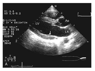
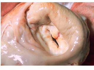
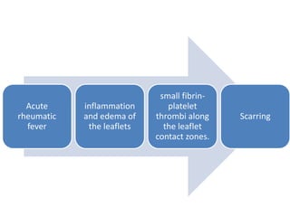
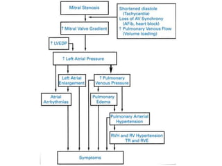
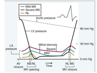
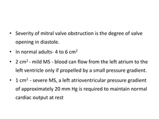
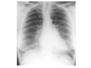
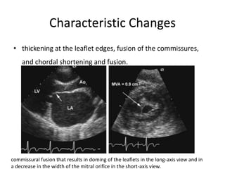
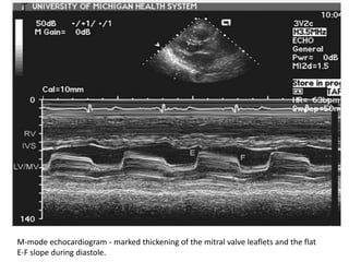
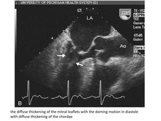
SlideShare a Scribd company logo
Discuss mitral valve apparatus
Clinical features, investigation and
management of Mitral Stenosis
-Dr. Uphar Gupta
Moderator – Dr. Prabhakar K
The mitral valve
• AKA bicuspid valve or left
atrioventricular valve
• dual-flap valve
• lies between the left atrium
(LA) and the left ventricle
(LV)
The mitral apparatus
• composed of the left atrial wall, the annulus,
the leaflets, the chordae tendineae, the
papillary muscles, and the left ventricular wall.
Left atrial wall
• The left atrial myocardium extends over the
proximal portion of the posterior leaflet.
• Left atrial enlargement can result in mitral
regurgitation by affecting the posterior leaflet.
• The anterior leaflet is not affected, because of
its attachment to the root of the aorta
Mitral annulus
• fibrous ring that connects with the leaflets.
• not a continuous ring around the mitral orifice
• D-shaped
• The straight border of the annulus is posterior to the
aortic valve.
• The aortic valve is located between the ventricular
septum and the mitral valve.
• The annulus functions as a sphincter that
contracts and reduces the surface area of the
valve during systole to ensure complete closure
of the leaflets.
• Annular dilatation of the mitral valve causes poor
leaflet apposition - results in mitral regurgitation.
Mitral valve leaflets
• continuous veil inserted around the circumference of the mitral
orifice.
• The free edges of the leaflets have several indentations.
• Two of these indentations, the anterolateral and posteromedial
commissures, divide the leaflets into anterior and posterior.
• These commissures can be accurately identified by the insertions of
the commissural chordae tendineae into the leaflets.
• Normally, the leaflets are thin, pliable,
translucent, and soft.
• Each leaflet has an atrial and a ventricular
surface.
Anterior leaflet
• located posterior to the aortic root and is also anchored to the
aortic root, unlike the posterior leaflet.
• also known as the aortic, septal, greater, or anteromedial leaflet.
• The anterior leaflet is large and semicircular in shape.
• The 2 zones on the anterior leaflet are referred to as rough and
clear zones, according to the chordae tendineae insertion.
Posterior leaflet
• ventricular, mural, smaller, or posterolateral leaflet.
• The posterior leaflet is the section of the mitral valve
that is located posterior to the 2 commissural areas.
• It has a wider attachment to the annulus than the
anterior leaflet.
• It is divided into 3 scallops by 2 indentations or clefts.
Chordae tendineae
• Small fibrous strings that originate either from
the apical portion of the papillary muscles or
directly from the ventricular wall and insert
into the valve leaflets or the muscle.
Commissural chordae
• Commissural chordae are the chordae that insert into
the interleaflet or commissural areas located at the
junction of the anterior and posterior leaflets.
• Two types of commissural chordae exist.
• Posteromedial commissural chordae and anterolateral
commissural
Leaflet chordae
• insert into the anterior or posterior leaflets.
• Two types of chordae tendineae are connected to the
anterior leaflet.
• The first is rough zone chordae, which insert into the distal
portion of the anterior leaflet known as the rough zone.
• The second is strut chordae, which are the chordae that
branch before inserting into the anterior leaflet.
Papillary muscles and left
ventricular wall
• represent the muscular components of the mitral apparatus.
• The papillary muscles normally arise from the apex and middle third
of the left ventricular wall.
• The anterolateral papillary muscle is normally larger than the
posteromedial papillary muscle and is supplied by the left anterior
descending artery or the left circumflex artery.
• The posteromedial papillary muscle is supplied by the right
coronary artery.
• Extreme fusion of papillary muscle can result
into mitral stenosis.
• On the other hand, rupture of a papillary
muscle, usually the complication of acute
myocardial infarction, will result in acute
mitral regurgitation.
Microscopic Anatomy
• The 3 layers of the ventricular wall are the
endocardium, the myocardium, and the epicardium.
• The endocardium consists of a simple squamous
endothelium and a thin subendothelial tissue.
• The myocardium consists of cardiac muscle fibers.
• The epicardium consists of a simple squamous
mesothelium and subepicardial tissue.
• There is a layer of dense fibrous connective tissue, called
the annulus fibrosus, located between the atrium and
ventricle.
• The mitral valve connects the left atrium (LA) and the left
ventricle (LV).
• The mitral valve leaflets are composed of an outer layer of
endocardium and a dense connective tissue core, which is
continuous with the annulus fibrosus.
2- to 3-mm zone of overlap (the zona coapta).
Apical four-chamber view recorded in systole in a normal patient
MITRAL STENOSIS
• Obstruction to blood flow between the left atrium and the left ventricle
• ETIOLOGY:-
– rheumatic carditis.
– Congenital MS is uncommon.
– MS, usually rheumatic, in association with atrial septal defect is called
Lutembacher syndrome.
– massive mitral valve annular calcification. This process occurs most frequently
in elderly patients and produces MS by limiting leaflet motion.
• Rheumatic changes present in 99% of stenotic mitral valves excised
at the time of mitral valve (MV) replacement.
• 25% - isolated MS
• 40% - combined MS and MR.
• Multivalve involvement - 38% of MS patients,
– Aortic valve 35%
– tricuspid valve 6%.
– pulmonic valve is rarely affected.
• Two thirds of all patients with rheumatic MS are female.
Acute
rheumatic
fever
inflammation
and edema of
the leaflets
small fibrin-
platelet
thrombi along
the leaflet
contact zones.
Scarring
Earlier Stages
• relatively flexible leaflets snap open in diastole into a curved shape
because of restriction of motion at the leaflet tips.
• This diastolic doming is most evident in the motion of the anterior
leaflet and becomes less prominent as the leaflets become more
fibrotic and calcified.
• The symmetrical fusion of the commissures results in a small
central oval orifice in diastole that on pathologic specimens is
shaped like a fish mouth or buttonhole because the anterior leaflet
is not in the physiological open position .
End-stage Disease
• thickened leaflets -adherent and rigid that they cannot open or
shut, reducing or, rarely, even abolishing the first heart sound and
leading to combined MS and MR.
• When rheumatic fever results exclusively or predominantly in
contraction and fusion of the chordae tendineae, with little fusion
of the valvular commissures, dominant MR results.
• Aschoff bodies, the pathologic hallmark of rheumatic disease, are
most frequently seen in the myocardium, not the valve tissue.
OTHER ETIOLOGIES
• rare complication of :-
– malignant carcinoid disease
– systemic lupus erythematosus
– rheumatoid arthritis
– mucopolysaccharidoses of the Hunter-Hurler phenotype,
Fabry disease, and Whipple disease
– Methysergide therapy is an unusual but documented
cause of MS.
Other causes of LA – LV Flow
obstruction
– a left atrial tumor, particularly myxoma
– ball valve thrombus in the left atrium
– infective endocarditis with large vegetations
– congenital membrane in the left atrium (cor
triatriatum)
• Severity of mitral valve obstruction is the degree of valve
opening in diastole.
• In normal adults- 4 to 6 cm2
• 2 cm2 - mild MS - blood can flow from the left atrium to the
left ventricle only if propelled by a small pressure gradient.
• 1 cm2 - severe MS, a left atrioventricular pressure gradient
of approximately 20 mm Hg is required to maintain normal
cardiac output at rest
Pulmonary Hypertension
• mean left atrial pressure is elevated - prominent atrial contraction
(a wave), with a gradual pressure decline after mitral valve opening
(y descent).
• mild to moderate MS - pulmonary arterial pressure may be normal
or only minimally elevated at rest but rises during exercise.
• severe MS and those in whom the pulmonary vascular resistance is
significantly increased - pulmonary arterial pressure is elevated
when the patient is at rest.
(1) passive backward transmission of the elevated left atrial
pressure
(2) pulmonary arteriolar constriction, which presumably is
triggered by left atrial and pulmonary venous hypertension
(reactive pulmonary hypertension)
(3) organic obliterative changes in the pulmonary vascular
bed, which may be considered to be a complication of long-
standing and severe MS
• also exert a protective effect
• the elevated precapillary resistance makes the
development of symptoms of pulmonary congestion less
likely to occur by tending to prevent blood from surging
into the pulmonary capillary bed and damming up behind
the stenotic mitral valve.
• However, this protection occurs at the expense of a
reduced cardiac output.
• In severe MS - pulmonary vein–bronchial vein shunts occur.
• Their rupture may cause hemoptysis.
• manifest a reduction in pulmonary compliance, increase in the work
of breathing, and redistribution of pulmonary blood flow from the
base to the apex.
• In time, severe pulmonary hypertension results in right-sided heart
failure, with dilation of the right ventricle and its annulus,
secondary tricuspid regurgitation (TR), and sometimes pulmonic
regurgitation.
Left Ventricular Function
• The LV chamber typically is normal or small, with
normal systolic function and normal LV end-
diastolic pressure.
• However, coexisting MR, aortic valve lesions,
systemic hypertension, ischemic heart disease,
and cardiomyopathy may all be responsible for
elevations of LV diastolic pressure
Left Atrial Changes
• The combination of mitral valve disease and
atrial inflammation secondary to rheumatic
carditis causes the following:
– (1) left atrial dilation;
– (2) fibrosis of the atrial wall;
– (3) disorganization of the atrial muscle bundles.
• Premature atrial activation, caused by an automatic focus or
reentry, may stimulate the left atrium during the vulnerable period
and thereby precipitate AF.
• AF is often episodic at first but then becomes more persistent.
• AF per se causes diffuse atrophy of atrial muscle, further atrial
enlargement, and further inhomogeneity of refractoriness and
conduction.
• These changes, in turn, lead to irreversible AF.
Symptoms:- Dyspnea
• The most common presenting symptoms - dyspnea, fatigue, and
decreased exercise tolerance.
• Symptoms are caused by a reduced ability to increase cardiac
output normally with exercise or elevated pulmonary venous
pressures and reduced pulmonary compliance.
• Dyspnea may be accompanied by cough and wheezing.
• Vital capacity is reduced, presumably because of the presence of
engorged pulmonary vessels and interstitial edema.
• critical obstruction to left atrial emptying and dyspnea with
ordinary activity (NYHA functional Class III) - orthopnea as well and
frank pulmonary edema.
• The latter may be precipitated by effort, emotional stress,
respiratory infection, fever pregnancy, or AF with a rapid ventricular
rate or other tachyarrhythmia.
• In patients with a markedly elevated pulmonary vascular resistance,
RV function is often impaired and the presentation may also include
symptoms and signs of right heart failure.
Hemoptysis
• sudden and severe - caused by rupture of thin-walled, dilated
bronchial veins, usually as a consequence of a sudden rise in left
atrial pressure.
• milder, with only blood-stained sputum associated with attacks of
paroxysmal nocturnal dyspnea.
• pink frothy sputum characteristic of acute pulmonary edema with
rupture of alveolar capillaries.
• also may be caused by pulmonary infarction, a late complication of
MS associated with heart failure.
Chest Pain
• not a typical symptom
• 15%
• indistinguishable from that of angina pectoris
• caused by :-
– severe RV hypertension secondary to the pulmonary vascular disease
– concomitant coronary atherosclerosis
– coronary obstruction caused by coronary embolization
Palpitations and Embolic Events
• Patients with AF often are initially diagnosed
when they present with AF or an embolic
event.
Other Symptoms
• Compression of the left recurrent laryngeal nerve by a greatly
dilated left atrium, enlarged tracheobronchial lymph nodes, and
dilated pulmonary artery may cause hoarseness (Ortner syndrome).
• repeated hemoptysis with pulmonary hemosiderosis.
• Systemic venous hypertension, hepatomegaly, edema, ascites, and
hydrothorax are all signs of severe MS with elevated pulmonary
vascular resistance and right-sided heart failure.
Physical Examination
• irregular pulse caused by AF and signs of left and right
heart failure
• systemic vasoconstriction may exhibit the so-called mitral
facies, characterized by pinkish-purple patches on the
cheeks
• The arterial pulse is usually normal, but in patients with a
reduced stroke volume, the pulse may be low in volume.
• The jugular venous pulse usually exhibits a
prominent a wave in patients with sinus
rhythm and elevated pulmonary vascular
resistance.
• In patients with AF, the x descent of the
jugular venous pulse disappears, and there is
only one crest, a prominent v or c-v wave, per
cardiac cycle.
• Palpation of the cardiac apex usually reveals an inconspicuous left
ventricle; the presence of a palpable presystolic expansion wave or
an early diastolic rapid filling wave speaks strongly against serious
MS.
• A readily palpable, tapping S1 suggests that the anterior mitral valve
leaflet is pliable.
• When the patient is in the left lateral recumbent position, a
diastolic thrill of MS may be palpable at the apex.
• RV lift is felt in the left parasternal region in patients with
pulmonary hypertension.
• A markedly enlarged right ventricle may displace the left
ventricle posteriorly and produce a prominent RV apex beat
that can be confused with a LV lift.
• A loud P2 may be palpable in the second left intercostal
space in patients with MS and pulmonary hypertension.
Auscultation
• accentuated S1
• As pulmonary arterial pressure rises, P2 at first becomes
accentuated and widely transmitted and can often be readily heard
at both the mitral and the aortic areas.
• With further elevation of pulmonary arterial pressure, splitting of S2
narrows because of reduced compliance of the pulmonary vascular
bed, with earlier pulmonic valve closure.
• Finally, S2 becomes single and accentuated.
• Other signs of severe pulmonary hypertension
– nonvalvular pulmonic ejection sound that diminishes during
inspiration, because of dilation of the pulmonary artery,
– systolic murmur of TR,
– Graham Steell murmur of pulmonic regurgitation,
– S4 originating from the right ventricle.
• An S3 gallop originating from the left ventricle is absent in patients
with MS unless significant MR or AR coexists
• The opening snap (OS) of the mitral valve is caused by a sudden tensing of
the valve leaflets after the valve cusps have completed their opening
excursion.
• The OS occurs when the movement of the mitral dome into the left
ventricle suddenly stops.
• The OS can usually be differentiated from P2 because the OS occurs later,
unless right bundle branch block is present.
• In addition, the OS usually is loudest at the apex, whereas S2 is best heard
at the cardiac base.
• The mitral valve cannot be totally rigid if it produces an OS, so an
OS is usually accompanied by an accentuated S1.
• Calcification confined to the tip of the mitral valve leaflets does not
preclude an OS, although calcification of the body and tip does.
• The mitral OS follows A2 by 0.04 to 0.12 second; this interval varies
inversely with the left atrial pressure.
• A short A2-OS interval is a reliable indicator of severe MS
• The diastolic, low-pitched, rumbling murmur of MS is best heard at the
apex, with the bell of the stethoscope (low-frequency mode on electronic
stethoscopes) and with the patient in the left lateral recumbent position.
• When this murmur is soft, it is limited to the apex but, when louder, it may
radiate to the left axilla or the lower left sternal area.
• Although the intensity of the diastolic murmur is not closely related to the
severity of stenosis, the duration of the murmur is a guide to the severity
of mitral valve narrowing.
• The murmur persists for as long as the left atrioventricular
pressure gradient exceeds approximately 3 mm Hg.
• The murmur usually commences immediately after the OS.
• In mild MS, the early diastolic murmur is brief but, in the
presence of sinus rhythm, it resumes in presystole.
• In severe MS, the murmur persists until end-diastole, with
presystolic accentuation while sinus rhythm is maintained.
Differential Diagnosis
• rare diagnosis in developed countries
• In older patients - mitral annular calcification
• Left atrial myxoma
• HOCM
Radiography
• left atrial enlargement
• Extreme left atrial enlargement rarely occurs in isolated
MS - when present, MR is usually severe
• fluoroscopy is required to detect valvular calcification.
• Radiologic changes in the lung fields indirectly reflect
the severity of MS.
• Interstitial edema, an indication of severe obstruction, is manifested as
Kerley B lines (dense, short, horizontal lines most commonly seen in the
costophrenic angles).
• This finding is present in 30% of patients with resting pulmonary arterial
wedge pressures less than 20 mm Hg and in 70% of patients with pressures
greater than 20 mm Hg.
• Severe long-standing mitral obstruction often results in Kerley A lines
(straight, dense lines up to 4 cm in length, running toward the hilum), as
well as the findings of pulmonary hemosiderosis and rarely of parenchymal
ossification.
• Kerley B lines
• subpleural
perpendicular lines
1-3 cm in length
Calcification in the mitral ring(lateral view )
Electrocardiography
• Relatively insensitive for detecting mild MS
• shows characteristic changes in moderate or severe obstruction
• Left atrial enlargement (P wave duration in lead II >0.12 second
and/or a P wave axis between +45 and −30 degrees) is a principal
electrocardiographic feature of MS and is found in 90% of patients
with significant MS and sinus rhythm.
• AF is common with long-standing MS.
• Electrocardiographic evidence of RV hypertrophy correlates with RV
systolic pressure.
• When RV systolic pressure is 70 to 100 mm Hg, approximately 50% of
patients manifest ECG criteria for RV hypertrophy, including a mean QRS
axis greater than 80 degrees in the frontal plane and an R:S ratio greater
than 1 in lead V1.
• When RV systolic pressure is greater than 100 mm Hg in patients with
isolated or predominant MS, electrocardiographic evidence of RV
hypertrophy is consistently found.
Echocardiography
• characteristic anatomy with leaflet thickening and restriction of
opening caused by symmetric fusion of the commissures, resulting in
“doming” of the leaflets in diastole
• As disease becomes more severe, thickening extends from the
leaflet tips toward the base with further restriction of motion and
less curvature of the leaflet in diastole.
• The mitral chords are variably thickening, fused, and shortened and
there may be superimposed calcification of the valve apparatus
• A score of 0 to 4+ is given for leaflet thickness, mobility,
calcification, and chordal involvement to provide an overall
score that is favorable (low) or unfavorable (high) for
valvuloplasty
• degree of anterior leaflet doming, symmetry of commissural
fusion, and distribution of leaflet calcification.
• left atrial size, pulmonary artery pressures, LV size and systolic
function, and RV size and systolic function.
• When pulmonary hypertension is present, the right
ventricle is frequently dilated, with reduced systolic
function.
• TR may be secondary to RV dysfunction and annular
dilation or may be caused by rheumatic involvement of the
tricuspid valve.
• Complete evaluation of aortic valve anatomy and function
is also important because the aortic valve is affected in
approximately one third of patients with MS.
• When transthoracic images are suboptimal, TEE is
appropriate.
• TEE is also necessary to exclude left atrial thrombus and
evaluate MR severity when percutaneous BMV is considered.
Characteristic Changes
• thickening at the leaflet edges, fusion of the commissures,
and chordal shortening and fusion.
commissural fusion that results in doming of the leaflets in the long-axis view and in
a decrease in the width of the mitral orifice in the short-axis view.
M-mode echocardiogram - marked thickening of the mitral valve leaflets and the flat
E-F slope during diastole.
the diffuse thickening of the mitral leaflets with the doming motion in diastole
with diffuse thickening of the chordae
Exercise Testing with Doppler
Echocardiography
• Exercise testing is useful for many patients with MS to ascertain the
level of physical conditioning and elicit covert cardiac symptoms.
• The exercise test can be combined with Doppler echocardiography
to assess exercise pulmonary pressure.
• Useful parameters on exercise testing include the following: (1)
exercise duration; (2) blood pressure and heart rate response; and
(3) increase in pulmonary pressures with exercise, compared with
the expected normal changes.
Cardiac Catheterization
• Catheter-based measurement of left atrial and LV pressures
• allows measurement of the mean transmitral pressure
gradient and, in conjunction with measurement of
transmitral volume flow rate, calculation of the valve area
using the Gorlin formula
• Routine diagnostic cardiac catheterization is not
recommended for the evaluation of MS.
Complications :- Atrial Fibrillation
• The most common complication of MS is AF
• AF may precipitate or worsen symptoms caused by loss of the atrial
contribution to filling and to a short diastolic filling period when the
ventricular rate is not well controlled.
• predisposes to left atrial thrombus formation and systemic embolic
events.
• conveys a worse overall prognosis
Systemic Embolism
• caused by left atrial thrombus formation.
• most often occurs in patients with AF
• When embolization occurs in patients in sinus rhythm, the
possibility of transient AF or underlying infective endocarditis
should be considered.
• loss of atrial appendage contractile function, despite electrical
evidence of sinus rhythm, leads to blood flow stasis and thrombus
formation.
Infective Endocarditis
• MS is a predisposing factor for endocarditis in less than 1% of
cases in clinical series of bacterial endocarditis.
• The estimated risk of endocarditis in patients with MS is
0.17/1000 patient-years, which is much lower than the risk in
patients with MR or aortic valve disease.
Medical Treatment
• The medical management of MS is primarily directed
toward the following:
– (1) prevention of recurrent rheumatic fever;
– (2) prevention and treatment of complications of MS; and
– (3) monitoring disease progression to allow intervention at
the optimal time point.
• Patients with MS caused by rheumatic heart disease
should receive penicillin prophylaxis for beta-hemolytic
streptococcal infections to prevent recurrent rheumatic
fever
• Prophylaxis for infective endocarditis is no longer
recommended
• Anemia and infections should be treated promptly and
aggressively in patients with valvular heart disease.
• Anticoagulant therapy is indicated for prevention of systemic embolism in
MS patients with AF (persistent or paroxysmal), any prior embolic events
(even if in sinus rhythm), and documented left atrial thrombus.
• Anticoagulation also may be considered for patients with severe MS and
sinus rhythm when there is severe left atrial enlargement (diameter
>55 mm) or spontaneous contrast on echocardiography.
• Treatment with warfarin is used to maintain the international normalized
ratio (INR) between 2 and 3.
• Asymptomatic patients with mild to moderate rheumatic mitral
valve disease should have a history and physical examination
annually, with echocardiography every 3 to 5 years for mild
stenosis, every 1 to 2 years for moderate stenosis, and annually for
severe stenosis.
• More frequent evaluation is appropriate for any change in signs or
symptoms.
• All patients with significant MS should be advised to avoid
occupations requiring strenuous exertion.
• In patients with severe MS, with persistent symptoms after
intervention or when intervention is not possible, medical
therapy with oral diuretics and the restriction of sodium
intake may improve symptoms.
• Digitalis glycosides - slowing the ventricular rate in patients
with AF and in treating patients with right-sided heart failure.
• Hemoptysis is managed by measures designed to reduce
pulmonary venous pressure, including sedation, assumption
of the upright position, and aggressive diuresis.
Treatment of Arrhythmias
• Immediate treatment of AF includes administration of intravenous heparin
followed by oral warfarin.
• The ventricular rate should be slowed, initially with an intravenous beta
blocker or calcium channel antagonist, followed by long-term rate control
with oral doses of these agents.
• When these medications are ineffective or when additional rate control is
necessary, digoxin or amiodarone may be considered.
• Digoxin alone for long-term management of AF may be considered in
patients with concurrent LV dysfunction or a sedentary lifestyle.
• Paroxysmal AF and repeated conversions, spontaneous or induced, carry
the risk of embolization.
• In patients who cannot be converted or maintained in sinus rhythm,
digitalis should be used to maintain the ventricular rate at rest at
approximately 60 beats/min.
• If this is not possible, small doses of a beta-blocking agent, such as
atenolol (25 mg daily) or metoprolol (50 to 100 mg daily), may be added.
• Beta blockers are particularly helpful in preventing rapid ventricular
responses that develop during exertion.
• Multiple repeat cardioversions are not indicated if the patient fails
to sustain sinus rhythm while on adequate doses of an
antiarrhythmic.
• Patients with chronic AF who undergo surgical MV repair or MV
replacement may undergo the maze procedure (atrial compartment
operation).
• Early intervention with percutaneous valvotomy may prevent the
development of AF.
Percutaneous Balloon Mitral Valvotomy
• Patients with mild to moderate MS who are asymptomatic frequently
remain so for years, and clinical outcomes are similar to age-matched
normal patients.
• However, severe or symptomatic MS is associated with poor long-term
outcomes if the stenosis is not relieved mechanically
• Percutaneous BMV is the procedure of choice for the treatment of MS so
that surgical intervention is now reserved for patients who require
intervention and are not candidates for a percutaneous procedure.
• BMV is recommended for symptomatic patients with
– moderate to severe MS
– with favorable valve morphology
– no or mild MR
– no evidence of left atrial thrombus.
• recommended for asymptomatic patients with
– moderate to severe MS
– when mitral valve obstruction has resulted in pulmonary hypertension
with a pulmonary systolic pressure greater than 50 mm Hg at rest or
60 mm Hg with exercise.
• BMV also is reasonable for symptomatic patients who are at
high risk for surgery, even when valve morphology is not ideal,
including patients with restenosis after a previous BMV or
previous commissurotomy who are unsuitable for surgery
because of very high risk.
• BMV may be considered for patients with moderate to severe
MS and new-onset AF and those with mild MS when
significant pulmonary hypertension is present .
• This percutaneous technique consists of advancing a small balloon
flotation catheter across the interatrial septum (after transseptal
puncture), enlarging the opening, advancing a large (23- to 25-mm)
hourglass-shaped balloon (the Inoue balloon), and inflating it within
the orifice
• Alternatively, two smaller (15- to 20-mm) side by side balloons
across the mitral orifice may be used.
• A third technique involves retrograde, nontransseptal dilation of
the mitral valve, in which the balloon is positioned across the mitral
valve using a steerable guidewire.
Mitral valve
Mitral valve
Mitral valve

More Related Content

What's hot

Congenitally corrected transposition of great arteries
Congenitally corrected transposition of great arteriesCongenitally corrected transposition of great arteries
Congenitally corrected transposition of great arteriesDheeraj Sharma
 
Echocardiographic Evaluation of Hypertrophic Cardiomyopathy
Echocardiographic Evaluation of Hypertrophic CardiomyopathyEchocardiographic Evaluation of Hypertrophic Cardiomyopathy
Echocardiographic Evaluation of Hypertrophic Cardiomyopathy
Hatem Soliman Aboumarie
 
Mitral valve disease
Mitral valve diseaseMitral valve disease
Mitral valve disease
DR NIKUNJ SHEKHADA
 
Mitral stenosis - Echocardiography
Mitral stenosis - EchocardiographyMitral stenosis - Echocardiography
Mitral stenosis - Echocardiography
Ankur Gupta
 
Right ventricle (RV) anatomy and functions
Right ventricle (RV) anatomy and functionsRight ventricle (RV) anatomy and functions
Right ventricle (RV) anatomy and functions
Malleswara rao Dangeti
 
Asd echo assessment
Asd echo assessmentAsd echo assessment
Asd echo assessment
Mashiul Alam
 
MVP Mitral Valve Prolapse - Echocardiographic Evaluation
 MVP Mitral Valve  Prolapse - Echocardiographic Evaluation MVP Mitral Valve  Prolapse - Echocardiographic Evaluation
MVP Mitral Valve Prolapse - Echocardiographic Evaluation
Praveen Nagula
 
Coronary artery anatomy
Coronary artery anatomyCoronary artery anatomy
Coronary artery anatomy
Dr. Yash Kumar Achantani
 
CONTRAST ECHOCARDIOGRAPHY
CONTRAST ECHOCARDIOGRAPHYCONTRAST ECHOCARDIOGRAPHY
CONTRAST ECHOCARDIOGRAPHY
soumenprasad
 
Mitral Regurgitation
Mitral RegurgitationMitral Regurgitation
Mitral Regurgitation
Sujay Iyer
 
Echo assessment of cardiomyopathy pdf
Echo assessment of cardiomyopathy pdfEcho assessment of cardiomyopathy pdf
Echo assessment of cardiomyopathy pdf
Nizam Uddin
 
Tricuspid valve
Tricuspid valveTricuspid valve
Tricuspid valve
Ramachandra Barik
 
segment approach to congenital heart diseases
segment approach to congenital heart diseasessegment approach to congenital heart diseases
segment approach to congenital heart diseases
Sumiya Arshad
 
Cath hemodynamics vir
Cath hemodynamics virCath hemodynamics vir
Cath hemodynamics vir
Dr Virbhan Balai
 
Diastolic Dysfunction 2016
Diastolic Dysfunction 2016Diastolic Dysfunction 2016
Diastolic Dysfunction 2016
drabhishekbabbu
 
SEGMENTAL ANALYSIS OF CONGENITAL HEART DISEASE
SEGMENTAL ANALYSIS OF CONGENITAL HEART DISEASE SEGMENTAL ANALYSIS OF CONGENITAL HEART DISEASE
SEGMENTAL ANALYSIS OF CONGENITAL HEART DISEASE
Dr. Murtaza Kamal MD,DNB,DrNB Ped Cardiology
 
Echocardiography in mitral stenosis
Echocardiography in mitral stenosisEchocardiography in mitral stenosis
Echocardiography in mitral stenosis
Malleswara rao Dangeti
 
Aortic stenosis Echo
Aortic stenosis Echo Aortic stenosis Echo
Aortic stenosis Echo
madhusiva03
 
Constrictive pericarditis
Constrictive pericarditis Constrictive pericarditis
Constrictive pericarditis
Ramachandra Barik
 
Echocardiography in ischemic heart disease
Echocardiography in ischemic heart diseaseEchocardiography in ischemic heart disease
Echocardiography in ischemic heart disease
Bhargav Kiran
 

What's hot (20)

Congenitally corrected transposition of great arteries
Congenitally corrected transposition of great arteriesCongenitally corrected transposition of great arteries
Congenitally corrected transposition of great arteries
 
Echocardiographic Evaluation of Hypertrophic Cardiomyopathy
Echocardiographic Evaluation of Hypertrophic CardiomyopathyEchocardiographic Evaluation of Hypertrophic Cardiomyopathy
Echocardiographic Evaluation of Hypertrophic Cardiomyopathy
 
Mitral valve disease
Mitral valve diseaseMitral valve disease
Mitral valve disease
 
Mitral stenosis - Echocardiography
Mitral stenosis - EchocardiographyMitral stenosis - Echocardiography
Mitral stenosis - Echocardiography
 
Right ventricle (RV) anatomy and functions
Right ventricle (RV) anatomy and functionsRight ventricle (RV) anatomy and functions
Right ventricle (RV) anatomy and functions
 
Asd echo assessment
Asd echo assessmentAsd echo assessment
Asd echo assessment
 
MVP Mitral Valve Prolapse - Echocardiographic Evaluation
 MVP Mitral Valve  Prolapse - Echocardiographic Evaluation MVP Mitral Valve  Prolapse - Echocardiographic Evaluation
MVP Mitral Valve Prolapse - Echocardiographic Evaluation
 
Coronary artery anatomy
Coronary artery anatomyCoronary artery anatomy
Coronary artery anatomy
 
CONTRAST ECHOCARDIOGRAPHY
CONTRAST ECHOCARDIOGRAPHYCONTRAST ECHOCARDIOGRAPHY
CONTRAST ECHOCARDIOGRAPHY
 
Mitral Regurgitation
Mitral RegurgitationMitral Regurgitation
Mitral Regurgitation
 
Echo assessment of cardiomyopathy pdf
Echo assessment of cardiomyopathy pdfEcho assessment of cardiomyopathy pdf
Echo assessment of cardiomyopathy pdf
 
Tricuspid valve
Tricuspid valveTricuspid valve
Tricuspid valve
 
segment approach to congenital heart diseases
segment approach to congenital heart diseasessegment approach to congenital heart diseases
segment approach to congenital heart diseases
 
Cath hemodynamics vir
Cath hemodynamics virCath hemodynamics vir
Cath hemodynamics vir
 
Diastolic Dysfunction 2016
Diastolic Dysfunction 2016Diastolic Dysfunction 2016
Diastolic Dysfunction 2016
 
SEGMENTAL ANALYSIS OF CONGENITAL HEART DISEASE
SEGMENTAL ANALYSIS OF CONGENITAL HEART DISEASE SEGMENTAL ANALYSIS OF CONGENITAL HEART DISEASE
SEGMENTAL ANALYSIS OF CONGENITAL HEART DISEASE
 
Echocardiography in mitral stenosis
Echocardiography in mitral stenosisEchocardiography in mitral stenosis
Echocardiography in mitral stenosis
 
Aortic stenosis Echo
Aortic stenosis Echo Aortic stenosis Echo
Aortic stenosis Echo
 
Constrictive pericarditis
Constrictive pericarditis Constrictive pericarditis
Constrictive pericarditis
 
Echocardiography in ischemic heart disease
Echocardiography in ischemic heart diseaseEchocardiography in ischemic heart disease
Echocardiography in ischemic heart disease
 

Viewers also liked

Central vertigo
Central vertigoCentral vertigo
Central vertigo
Uphar Gupta
 
The hypothalamus
The hypothalamusThe hypothalamus
The hypothalamus
Uphar Gupta
 
Craniovertebral JUNCTION ANOMALIES
Craniovertebral JUNCTION ANOMALIESCraniovertebral JUNCTION ANOMALIES
Craniovertebral JUNCTION ANOMALIES
Uphar Gupta
 
Pericardial deseases
Pericardial deseasesPericardial deseases
Pericardial deseasesairwave12
 
Pulmonary circulation
Pulmonary circulationPulmonary circulation
Pulmonary circulation
Uphar Gupta
 
Mitral regurgitation
Mitral regurgitationMitral regurgitation
Mitral regurgitation
Uphar Gupta
 
Coronarystents phpapp02
Coronarystents phpapp02Coronarystents phpapp02
Coronarystents phpapp02
Saurabh Gupta
 
Pericarditis, Pericardial Effusion, & Cardiac Tamponade - BMH/Tele
Pericarditis, Pericardial Effusion, & Cardiac Tamponade - BMH/TelePericarditis, Pericardial Effusion, & Cardiac Tamponade - BMH/Tele
Pericarditis, Pericardial Effusion, & Cardiac Tamponade - BMH/TeleTeleClinEd
 
Cardiac tamponade
Cardiac tamponadeCardiac tamponade
Cardiac tamponade
Satyam Rajvanshi
 
17 pericardial disease
17 pericardial disease17 pericardial disease
17 pericardial diseaseinternalmed
 

Viewers also liked (10)

Central vertigo
Central vertigoCentral vertigo
Central vertigo
 
The hypothalamus
The hypothalamusThe hypothalamus
The hypothalamus
 
Craniovertebral JUNCTION ANOMALIES
Craniovertebral JUNCTION ANOMALIESCraniovertebral JUNCTION ANOMALIES
Craniovertebral JUNCTION ANOMALIES
 
Pericardial deseases
Pericardial deseasesPericardial deseases
Pericardial deseases
 
Pulmonary circulation
Pulmonary circulationPulmonary circulation
Pulmonary circulation
 
Mitral regurgitation
Mitral regurgitationMitral regurgitation
Mitral regurgitation
 
Coronarystents phpapp02
Coronarystents phpapp02Coronarystents phpapp02
Coronarystents phpapp02
 
Pericarditis, Pericardial Effusion, & Cardiac Tamponade - BMH/Tele
Pericarditis, Pericardial Effusion, & Cardiac Tamponade - BMH/TelePericarditis, Pericardial Effusion, & Cardiac Tamponade - BMH/Tele
Pericarditis, Pericardial Effusion, & Cardiac Tamponade - BMH/Tele
 
Cardiac tamponade
Cardiac tamponadeCardiac tamponade
Cardiac tamponade
 
17 pericardial disease
17 pericardial disease17 pericardial disease
17 pericardial disease
 

Similar to Mitral valve

Pathophysiology of the Mitral Valve .pptx
Pathophysiology of the Mitral Valve .pptxPathophysiology of the Mitral Valve .pptx
Pathophysiology of the Mitral Valve .pptx
Nora Albogami
 
Valvular Heart Disease, Esther
Valvular Heart Disease, EstherValvular Heart Disease, Esther
Valvular Heart Disease, Esther
Esther Mary Mathew
 
mitral prolpse and regutition.pptx
mitral prolpse and regutition.pptxmitral prolpse and regutition.pptx
mitral prolpse and regutition.pptx
AbdifatahSalahOmar1
 
Pathophysiology aortic valve disease
Pathophysiology aortic valve diseasePathophysiology aortic valve disease
Pathophysiology aortic valve disease
India CTVS
 
Ventricular Septal Defects - A Review
Ventricular Septal Defects - A ReviewVentricular Septal Defects - A Review
Ventricular Septal Defects - A Review
Vivek Rana
 
Mitral stenosis
Mitral stenosis Mitral stenosis
Mitral stenosis
Kamaldeep Sidhu
 
Echo and CAD-2.pptx
Echo and CAD-2.pptxEcho and CAD-2.pptx
Echo and CAD-2.pptx
AnayaAnaya14
 
mitral s.pdf
mitral s.pdfmitral s.pdf
mitral s.pdf
HibaP5
 
Mitral regurgitation
Mitral regurgitationMitral regurgitation
Mitral regurgitation
Diaa Srahin
 
Mitral valve prolapse
Mitral valve prolapseMitral valve prolapse
Mitral valve prolapse
Radhapriya Yalamanchi
 
Aortic valve disease
Aortic valve diseaseAortic valve disease
Aortic valve disease
DR NIKUNJ SHEKHADA
 
Aortic valve disease
Aortic valve diseaseAortic valve disease
Aortic valve disease
DR NIKUNJ SHEKHADA
 
10- cardiovascular diseases - Part 1.ppt
10- cardiovascular diseases - Part 1.ppt10- cardiovascular diseases - Part 1.ppt
10- cardiovascular diseases - Part 1.ppt
AhmedFaisal59561
 
10- cardiovascular diseases - Part 1.ppt
10- cardiovascular diseases - Part 1.ppt10- cardiovascular diseases - Part 1.ppt
10- cardiovascular diseases - Part 1.ppt
AhmedFaisal59561
 
The anatomy of the cardiac valves and their abnormalities
The anatomy of the cardiac valves and their abnormalitiesThe anatomy of the cardiac valves and their abnormalities
The anatomy of the cardiac valves and their abnormalities
Ibrahim Tawfeeq
 
Acquired valvular heart disease
Acquired valvular heart diseaseAcquired valvular heart disease
Acquired valvular heart disease
KamalAdhikari13
 
Mitral regurgitation
Mitral regurgitationMitral regurgitation
Mitral regurgitation
Parth Shanishwara
 
Valvular heart disease
Valvular heart diseaseValvular heart disease
Valvular heart disease
MR. JAGDISH SAMBAD
 
valvularheartdisease-180524125726 (1).pdf
valvularheartdisease-180524125726 (1).pdfvalvularheartdisease-180524125726 (1).pdf
valvularheartdisease-180524125726 (1).pdf
jiregnaetichadako
 
valvularheartdisease-180524125726 (1).pptx
valvularheartdisease-180524125726 (1).pptxvalvularheartdisease-180524125726 (1).pptx
valvularheartdisease-180524125726 (1).pptx
ArpitaHalder8
 

Similar to Mitral valve (20)

Pathophysiology of the Mitral Valve .pptx
Pathophysiology of the Mitral Valve .pptxPathophysiology of the Mitral Valve .pptx
Pathophysiology of the Mitral Valve .pptx
 
Valvular Heart Disease, Esther
Valvular Heart Disease, EstherValvular Heart Disease, Esther
Valvular Heart Disease, Esther
 
mitral prolpse and regutition.pptx
mitral prolpse and regutition.pptxmitral prolpse and regutition.pptx
mitral prolpse and regutition.pptx
 
Pathophysiology aortic valve disease
Pathophysiology aortic valve diseasePathophysiology aortic valve disease
Pathophysiology aortic valve disease
 
Ventricular Septal Defects - A Review
Ventricular Septal Defects - A ReviewVentricular Septal Defects - A Review
Ventricular Septal Defects - A Review
 
Mitral stenosis
Mitral stenosis Mitral stenosis
Mitral stenosis
 
Echo and CAD-2.pptx
Echo and CAD-2.pptxEcho and CAD-2.pptx
Echo and CAD-2.pptx
 
mitral s.pdf
mitral s.pdfmitral s.pdf
mitral s.pdf
 
Mitral regurgitation
Mitral regurgitationMitral regurgitation
Mitral regurgitation
 
Mitral valve prolapse
Mitral valve prolapseMitral valve prolapse
Mitral valve prolapse
 
Aortic valve disease
Aortic valve diseaseAortic valve disease
Aortic valve disease
 
Aortic valve disease
Aortic valve diseaseAortic valve disease
Aortic valve disease
 
10- cardiovascular diseases - Part 1.ppt
10- cardiovascular diseases - Part 1.ppt10- cardiovascular diseases - Part 1.ppt
10- cardiovascular diseases - Part 1.ppt
 
10- cardiovascular diseases - Part 1.ppt
10- cardiovascular diseases - Part 1.ppt10- cardiovascular diseases - Part 1.ppt
10- cardiovascular diseases - Part 1.ppt
 
The anatomy of the cardiac valves and their abnormalities
The anatomy of the cardiac valves and their abnormalitiesThe anatomy of the cardiac valves and their abnormalities
The anatomy of the cardiac valves and their abnormalities
 
Acquired valvular heart disease
Acquired valvular heart diseaseAcquired valvular heart disease
Acquired valvular heart disease
 
Mitral regurgitation
Mitral regurgitationMitral regurgitation
Mitral regurgitation
 
Valvular heart disease
Valvular heart diseaseValvular heart disease
Valvular heart disease
 
valvularheartdisease-180524125726 (1).pdf
valvularheartdisease-180524125726 (1).pdfvalvularheartdisease-180524125726 (1).pdf
valvularheartdisease-180524125726 (1).pdf
 
valvularheartdisease-180524125726 (1).pptx
valvularheartdisease-180524125726 (1).pptxvalvularheartdisease-180524125726 (1).pptx
valvularheartdisease-180524125726 (1).pptx
 

Recently uploaded

Pulmonary Thromboembolism - etilogy, types, medical- Surgical and nursing man...
Pulmonary Thromboembolism - etilogy, types, medical- Surgical and nursing man...Pulmonary Thromboembolism - etilogy, types, medical- Surgical and nursing man...
Pulmonary Thromboembolism - etilogy, types, medical- Surgical and nursing man...
VarunMahajani
 
Physiology of Special Chemical Sensation of Taste
Physiology of Special Chemical Sensation of TastePhysiology of Special Chemical Sensation of Taste
Physiology of Special Chemical Sensation of Taste
MedicoseAcademics
 
Ocular injury ppt Upendra pal optometrist upums saifai etawah
Ocular injury  ppt  Upendra pal  optometrist upums saifai etawahOcular injury  ppt  Upendra pal  optometrist upums saifai etawah
Ocular injury ppt Upendra pal optometrist upums saifai etawah
pal078100
 
POST OPERATIVE OLIGURIA and its management
POST OPERATIVE OLIGURIA and its managementPOST OPERATIVE OLIGURIA and its management
POST OPERATIVE OLIGURIA and its management
touseefaziz1
 
KDIGO 2024 guidelines for diabetologists
KDIGO 2024 guidelines for diabetologistsKDIGO 2024 guidelines for diabetologists
KDIGO 2024 guidelines for diabetologists
د.محمود نجيب
 
Maxilla, Mandible & Hyoid Bone & Clinical Correlations by Dr. RIG.pptx
Maxilla, Mandible & Hyoid Bone & Clinical Correlations by Dr. RIG.pptxMaxilla, Mandible & Hyoid Bone & Clinical Correlations by Dr. RIG.pptx
Maxilla, Mandible & Hyoid Bone & Clinical Correlations by Dr. RIG.pptx
Dr. Rabia Inam Gandapore
 
ARTHROLOGY PPT NCISM SYLLABUS AYURVEDA STUDENTS
ARTHROLOGY PPT NCISM SYLLABUS AYURVEDA STUDENTSARTHROLOGY PPT NCISM SYLLABUS AYURVEDA STUDENTS
ARTHROLOGY PPT NCISM SYLLABUS AYURVEDA STUDENTS
Dr. Vinay Pareek
 
BRACHYTHERAPY OVERVIEW AND APPLICATORS
BRACHYTHERAPY OVERVIEW  AND  APPLICATORSBRACHYTHERAPY OVERVIEW  AND  APPLICATORS
BRACHYTHERAPY OVERVIEW AND APPLICATORS
Krishan Murari
 
Surgical Site Infections, pathophysiology, and prevention.pptx
Surgical Site Infections, pathophysiology, and prevention.pptxSurgical Site Infections, pathophysiology, and prevention.pptx
Surgical Site Infections, pathophysiology, and prevention.pptx
jval Landero
 
Flu Vaccine Alert in Bangalore Karnataka
Flu Vaccine Alert in Bangalore KarnatakaFlu Vaccine Alert in Bangalore Karnataka
Flu Vaccine Alert in Bangalore Karnataka
addon Scans
 
Are There Any Natural Remedies To Treat Syphilis.pdf
Are There Any Natural Remedies To Treat Syphilis.pdfAre There Any Natural Remedies To Treat Syphilis.pdf
Are There Any Natural Remedies To Treat Syphilis.pdf
Little Cross Family Clinic
 
Physiology of Chemical Sensation of smell.pdf
Physiology of Chemical Sensation of smell.pdfPhysiology of Chemical Sensation of smell.pdf
Physiology of Chemical Sensation of smell.pdf
MedicoseAcademics
 
The Normal Electrocardiogram - Part I of II
The Normal Electrocardiogram - Part I of IIThe Normal Electrocardiogram - Part I of II
The Normal Electrocardiogram - Part I of II
MedicoseAcademics
 
Evaluation of antidepressant activity of clitoris ternatea in animals
Evaluation of antidepressant activity of clitoris ternatea in animalsEvaluation of antidepressant activity of clitoris ternatea in animals
Evaluation of antidepressant activity of clitoris ternatea in animals
Shweta
 
BENIGN PROSTATIC HYPERPLASIA.BPH. BPHpdf
BENIGN PROSTATIC HYPERPLASIA.BPH. BPHpdfBENIGN PROSTATIC HYPERPLASIA.BPH. BPHpdf
BENIGN PROSTATIC HYPERPLASIA.BPH. BPHpdf
DR SETH JOTHAM
 
THOA 2.ppt Human Organ Transplantation Act
THOA 2.ppt Human Organ Transplantation ActTHOA 2.ppt Human Organ Transplantation Act
THOA 2.ppt Human Organ Transplantation Act
DrSathishMS1
 
The hemodynamic and autonomic determinants of elevated blood pressure in obes...
The hemodynamic and autonomic determinants of elevated blood pressure in obes...The hemodynamic and autonomic determinants of elevated blood pressure in obes...
The hemodynamic and autonomic determinants of elevated blood pressure in obes...
Catherine Liao
 
24 Upakrama.pptx class ppt useful in all
24 Upakrama.pptx class ppt useful in all24 Upakrama.pptx class ppt useful in all
24 Upakrama.pptx class ppt useful in all
DrSathishMS1
 
Couples presenting to the infertility clinic- Do they really have infertility...
Couples presenting to the infertility clinic- Do they really have infertility...Couples presenting to the infertility clinic- Do they really have infertility...
Couples presenting to the infertility clinic- Do they really have infertility...
Sujoy Dasgupta
 
Charaka Samhita Sutra sthana Chapter 15 Upakalpaniyaadhyaya
Charaka Samhita Sutra sthana Chapter 15 UpakalpaniyaadhyayaCharaka Samhita Sutra sthana Chapter 15 Upakalpaniyaadhyaya
Charaka Samhita Sutra sthana Chapter 15 Upakalpaniyaadhyaya
Dr KHALID B.M
 

Recently uploaded (20)

Pulmonary Thromboembolism - etilogy, types, medical- Surgical and nursing man...
Pulmonary Thromboembolism - etilogy, types, medical- Surgical and nursing man...Pulmonary Thromboembolism - etilogy, types, medical- Surgical and nursing man...
Pulmonary Thromboembolism - etilogy, types, medical- Surgical and nursing man...
 
Physiology of Special Chemical Sensation of Taste
Physiology of Special Chemical Sensation of TastePhysiology of Special Chemical Sensation of Taste
Physiology of Special Chemical Sensation of Taste
 
Ocular injury ppt Upendra pal optometrist upums saifai etawah
Ocular injury  ppt  Upendra pal  optometrist upums saifai etawahOcular injury  ppt  Upendra pal  optometrist upums saifai etawah
Ocular injury ppt Upendra pal optometrist upums saifai etawah
 
POST OPERATIVE OLIGURIA and its management
POST OPERATIVE OLIGURIA and its managementPOST OPERATIVE OLIGURIA and its management
POST OPERATIVE OLIGURIA and its management
 
KDIGO 2024 guidelines for diabetologists
KDIGO 2024 guidelines for diabetologistsKDIGO 2024 guidelines for diabetologists
KDIGO 2024 guidelines for diabetologists
 
Maxilla, Mandible & Hyoid Bone & Clinical Correlations by Dr. RIG.pptx
Maxilla, Mandible & Hyoid Bone & Clinical Correlations by Dr. RIG.pptxMaxilla, Mandible & Hyoid Bone & Clinical Correlations by Dr. RIG.pptx
Maxilla, Mandible & Hyoid Bone & Clinical Correlations by Dr. RIG.pptx
 
ARTHROLOGY PPT NCISM SYLLABUS AYURVEDA STUDENTS
ARTHROLOGY PPT NCISM SYLLABUS AYURVEDA STUDENTSARTHROLOGY PPT NCISM SYLLABUS AYURVEDA STUDENTS
ARTHROLOGY PPT NCISM SYLLABUS AYURVEDA STUDENTS
 
BRACHYTHERAPY OVERVIEW AND APPLICATORS
BRACHYTHERAPY OVERVIEW  AND  APPLICATORSBRACHYTHERAPY OVERVIEW  AND  APPLICATORS
BRACHYTHERAPY OVERVIEW AND APPLICATORS
 
Surgical Site Infections, pathophysiology, and prevention.pptx
Surgical Site Infections, pathophysiology, and prevention.pptxSurgical Site Infections, pathophysiology, and prevention.pptx
Surgical Site Infections, pathophysiology, and prevention.pptx
 
Flu Vaccine Alert in Bangalore Karnataka
Flu Vaccine Alert in Bangalore KarnatakaFlu Vaccine Alert in Bangalore Karnataka
Flu Vaccine Alert in Bangalore Karnataka
 
Are There Any Natural Remedies To Treat Syphilis.pdf
Are There Any Natural Remedies To Treat Syphilis.pdfAre There Any Natural Remedies To Treat Syphilis.pdf
Are There Any Natural Remedies To Treat Syphilis.pdf
 
Physiology of Chemical Sensation of smell.pdf
Physiology of Chemical Sensation of smell.pdfPhysiology of Chemical Sensation of smell.pdf
Physiology of Chemical Sensation of smell.pdf
 
The Normal Electrocardiogram - Part I of II
The Normal Electrocardiogram - Part I of IIThe Normal Electrocardiogram - Part I of II
The Normal Electrocardiogram - Part I of II
 
Evaluation of antidepressant activity of clitoris ternatea in animals
Evaluation of antidepressant activity of clitoris ternatea in animalsEvaluation of antidepressant activity of clitoris ternatea in animals
Evaluation of antidepressant activity of clitoris ternatea in animals
 
BENIGN PROSTATIC HYPERPLASIA.BPH. BPHpdf
BENIGN PROSTATIC HYPERPLASIA.BPH. BPHpdfBENIGN PROSTATIC HYPERPLASIA.BPH. BPHpdf
BENIGN PROSTATIC HYPERPLASIA.BPH. BPHpdf
 
THOA 2.ppt Human Organ Transplantation Act
THOA 2.ppt Human Organ Transplantation ActTHOA 2.ppt Human Organ Transplantation Act
THOA 2.ppt Human Organ Transplantation Act
 
The hemodynamic and autonomic determinants of elevated blood pressure in obes...
The hemodynamic and autonomic determinants of elevated blood pressure in obes...The hemodynamic and autonomic determinants of elevated blood pressure in obes...
The hemodynamic and autonomic determinants of elevated blood pressure in obes...
 
24 Upakrama.pptx class ppt useful in all
24 Upakrama.pptx class ppt useful in all24 Upakrama.pptx class ppt useful in all
24 Upakrama.pptx class ppt useful in all
 
Couples presenting to the infertility clinic- Do they really have infertility...
Couples presenting to the infertility clinic- Do they really have infertility...Couples presenting to the infertility clinic- Do they really have infertility...
Couples presenting to the infertility clinic- Do they really have infertility...
 
Charaka Samhita Sutra sthana Chapter 15 Upakalpaniyaadhyaya
Charaka Samhita Sutra sthana Chapter 15 UpakalpaniyaadhyayaCharaka Samhita Sutra sthana Chapter 15 Upakalpaniyaadhyaya
Charaka Samhita Sutra sthana Chapter 15 Upakalpaniyaadhyaya
 

Mitral valve

  • 1. Discuss mitral valve apparatus Clinical features, investigation and management of Mitral Stenosis -Dr. Uphar Gupta Moderator – Dr. Prabhakar K
  • 2. The mitral valve • AKA bicuspid valve or left atrioventricular valve • dual-flap valve • lies between the left atrium (LA) and the left ventricle (LV)
  • 3.
  • 4.
  • 5. The mitral apparatus • composed of the left atrial wall, the annulus, the leaflets, the chordae tendineae, the papillary muscles, and the left ventricular wall.
  • 6.
  • 7. Left atrial wall • The left atrial myocardium extends over the proximal portion of the posterior leaflet. • Left atrial enlargement can result in mitral regurgitation by affecting the posterior leaflet. • The anterior leaflet is not affected, because of its attachment to the root of the aorta
  • 8. Mitral annulus • fibrous ring that connects with the leaflets. • not a continuous ring around the mitral orifice • D-shaped • The straight border of the annulus is posterior to the aortic valve. • The aortic valve is located between the ventricular septum and the mitral valve.
  • 9. • The annulus functions as a sphincter that contracts and reduces the surface area of the valve during systole to ensure complete closure of the leaflets. • Annular dilatation of the mitral valve causes poor leaflet apposition - results in mitral regurgitation.
  • 10. Mitral valve leaflets • continuous veil inserted around the circumference of the mitral orifice. • The free edges of the leaflets have several indentations. • Two of these indentations, the anterolateral and posteromedial commissures, divide the leaflets into anterior and posterior. • These commissures can be accurately identified by the insertions of the commissural chordae tendineae into the leaflets.
  • 11. • Normally, the leaflets are thin, pliable, translucent, and soft. • Each leaflet has an atrial and a ventricular surface.
  • 12.
  • 13.
  • 14. Anterior leaflet • located posterior to the aortic root and is also anchored to the aortic root, unlike the posterior leaflet. • also known as the aortic, septal, greater, or anteromedial leaflet. • The anterior leaflet is large and semicircular in shape. • The 2 zones on the anterior leaflet are referred to as rough and clear zones, according to the chordae tendineae insertion.
  • 15. Posterior leaflet • ventricular, mural, smaller, or posterolateral leaflet. • The posterior leaflet is the section of the mitral valve that is located posterior to the 2 commissural areas. • It has a wider attachment to the annulus than the anterior leaflet. • It is divided into 3 scallops by 2 indentations or clefts.
  • 16. Chordae tendineae • Small fibrous strings that originate either from the apical portion of the papillary muscles or directly from the ventricular wall and insert into the valve leaflets or the muscle.
  • 17. Commissural chordae • Commissural chordae are the chordae that insert into the interleaflet or commissural areas located at the junction of the anterior and posterior leaflets. • Two types of commissural chordae exist. • Posteromedial commissural chordae and anterolateral commissural
  • 18. Leaflet chordae • insert into the anterior or posterior leaflets. • Two types of chordae tendineae are connected to the anterior leaflet. • The first is rough zone chordae, which insert into the distal portion of the anterior leaflet known as the rough zone. • The second is strut chordae, which are the chordae that branch before inserting into the anterior leaflet.
  • 19. Papillary muscles and left ventricular wall • represent the muscular components of the mitral apparatus. • The papillary muscles normally arise from the apex and middle third of the left ventricular wall. • The anterolateral papillary muscle is normally larger than the posteromedial papillary muscle and is supplied by the left anterior descending artery or the left circumflex artery. • The posteromedial papillary muscle is supplied by the right coronary artery.
  • 20. • Extreme fusion of papillary muscle can result into mitral stenosis. • On the other hand, rupture of a papillary muscle, usually the complication of acute myocardial infarction, will result in acute mitral regurgitation.
  • 21.
  • 22. Microscopic Anatomy • The 3 layers of the ventricular wall are the endocardium, the myocardium, and the epicardium. • The endocardium consists of a simple squamous endothelium and a thin subendothelial tissue. • The myocardium consists of cardiac muscle fibers. • The epicardium consists of a simple squamous mesothelium and subepicardial tissue.
  • 23. • There is a layer of dense fibrous connective tissue, called the annulus fibrosus, located between the atrium and ventricle. • The mitral valve connects the left atrium (LA) and the left ventricle (LV). • The mitral valve leaflets are composed of an outer layer of endocardium and a dense connective tissue core, which is continuous with the annulus fibrosus.
  • 24.
  • 25. 2- to 3-mm zone of overlap (the zona coapta).
  • 26. Apical four-chamber view recorded in systole in a normal patient
  • 27. MITRAL STENOSIS • Obstruction to blood flow between the left atrium and the left ventricle • ETIOLOGY:- – rheumatic carditis. – Congenital MS is uncommon. – MS, usually rheumatic, in association with atrial septal defect is called Lutembacher syndrome. – massive mitral valve annular calcification. This process occurs most frequently in elderly patients and produces MS by limiting leaflet motion.
  • 28. • Rheumatic changes present in 99% of stenotic mitral valves excised at the time of mitral valve (MV) replacement. • 25% - isolated MS • 40% - combined MS and MR. • Multivalve involvement - 38% of MS patients, – Aortic valve 35% – tricuspid valve 6%. – pulmonic valve is rarely affected. • Two thirds of all patients with rheumatic MS are female.
  • 29.
  • 30. Acute rheumatic fever inflammation and edema of the leaflets small fibrin- platelet thrombi along the leaflet contact zones. Scarring
  • 31. Earlier Stages • relatively flexible leaflets snap open in diastole into a curved shape because of restriction of motion at the leaflet tips. • This diastolic doming is most evident in the motion of the anterior leaflet and becomes less prominent as the leaflets become more fibrotic and calcified. • The symmetrical fusion of the commissures results in a small central oval orifice in diastole that on pathologic specimens is shaped like a fish mouth or buttonhole because the anterior leaflet is not in the physiological open position .
  • 32. End-stage Disease • thickened leaflets -adherent and rigid that they cannot open or shut, reducing or, rarely, even abolishing the first heart sound and leading to combined MS and MR. • When rheumatic fever results exclusively or predominantly in contraction and fusion of the chordae tendineae, with little fusion of the valvular commissures, dominant MR results. • Aschoff bodies, the pathologic hallmark of rheumatic disease, are most frequently seen in the myocardium, not the valve tissue.
  • 33. OTHER ETIOLOGIES • rare complication of :- – malignant carcinoid disease – systemic lupus erythematosus – rheumatoid arthritis – mucopolysaccharidoses of the Hunter-Hurler phenotype, Fabry disease, and Whipple disease – Methysergide therapy is an unusual but documented cause of MS.
  • 34. Other causes of LA – LV Flow obstruction – a left atrial tumor, particularly myxoma – ball valve thrombus in the left atrium – infective endocarditis with large vegetations – congenital membrane in the left atrium (cor triatriatum)
  • 35.
  • 36.
  • 37. • Severity of mitral valve obstruction is the degree of valve opening in diastole. • In normal adults- 4 to 6 cm2 • 2 cm2 - mild MS - blood can flow from the left atrium to the left ventricle only if propelled by a small pressure gradient. • 1 cm2 - severe MS, a left atrioventricular pressure gradient of approximately 20 mm Hg is required to maintain normal cardiac output at rest
  • 38. Pulmonary Hypertension • mean left atrial pressure is elevated - prominent atrial contraction (a wave), with a gradual pressure decline after mitral valve opening (y descent). • mild to moderate MS - pulmonary arterial pressure may be normal or only minimally elevated at rest but rises during exercise. • severe MS and those in whom the pulmonary vascular resistance is significantly increased - pulmonary arterial pressure is elevated when the patient is at rest.
  • 39. (1) passive backward transmission of the elevated left atrial pressure (2) pulmonary arteriolar constriction, which presumably is triggered by left atrial and pulmonary venous hypertension (reactive pulmonary hypertension) (3) organic obliterative changes in the pulmonary vascular bed, which may be considered to be a complication of long- standing and severe MS
  • 40. • also exert a protective effect • the elevated precapillary resistance makes the development of symptoms of pulmonary congestion less likely to occur by tending to prevent blood from surging into the pulmonary capillary bed and damming up behind the stenotic mitral valve. • However, this protection occurs at the expense of a reduced cardiac output.
  • 41. • In severe MS - pulmonary vein–bronchial vein shunts occur. • Their rupture may cause hemoptysis. • manifest a reduction in pulmonary compliance, increase in the work of breathing, and redistribution of pulmonary blood flow from the base to the apex. • In time, severe pulmonary hypertension results in right-sided heart failure, with dilation of the right ventricle and its annulus, secondary tricuspid regurgitation (TR), and sometimes pulmonic regurgitation.
  • 42. Left Ventricular Function • The LV chamber typically is normal or small, with normal systolic function and normal LV end- diastolic pressure. • However, coexisting MR, aortic valve lesions, systemic hypertension, ischemic heart disease, and cardiomyopathy may all be responsible for elevations of LV diastolic pressure
  • 43. Left Atrial Changes • The combination of mitral valve disease and atrial inflammation secondary to rheumatic carditis causes the following: – (1) left atrial dilation; – (2) fibrosis of the atrial wall; – (3) disorganization of the atrial muscle bundles.
  • 44. • Premature atrial activation, caused by an automatic focus or reentry, may stimulate the left atrium during the vulnerable period and thereby precipitate AF. • AF is often episodic at first but then becomes more persistent. • AF per se causes diffuse atrophy of atrial muscle, further atrial enlargement, and further inhomogeneity of refractoriness and conduction. • These changes, in turn, lead to irreversible AF.
  • 45. Symptoms:- Dyspnea • The most common presenting symptoms - dyspnea, fatigue, and decreased exercise tolerance. • Symptoms are caused by a reduced ability to increase cardiac output normally with exercise or elevated pulmonary venous pressures and reduced pulmonary compliance. • Dyspnea may be accompanied by cough and wheezing. • Vital capacity is reduced, presumably because of the presence of engorged pulmonary vessels and interstitial edema.
  • 46. • critical obstruction to left atrial emptying and dyspnea with ordinary activity (NYHA functional Class III) - orthopnea as well and frank pulmonary edema. • The latter may be precipitated by effort, emotional stress, respiratory infection, fever pregnancy, or AF with a rapid ventricular rate or other tachyarrhythmia. • In patients with a markedly elevated pulmonary vascular resistance, RV function is often impaired and the presentation may also include symptoms and signs of right heart failure.
  • 47. Hemoptysis • sudden and severe - caused by rupture of thin-walled, dilated bronchial veins, usually as a consequence of a sudden rise in left atrial pressure. • milder, with only blood-stained sputum associated with attacks of paroxysmal nocturnal dyspnea. • pink frothy sputum characteristic of acute pulmonary edema with rupture of alveolar capillaries. • also may be caused by pulmonary infarction, a late complication of MS associated with heart failure.
  • 48. Chest Pain • not a typical symptom • 15% • indistinguishable from that of angina pectoris • caused by :- – severe RV hypertension secondary to the pulmonary vascular disease – concomitant coronary atherosclerosis – coronary obstruction caused by coronary embolization
  • 49. Palpitations and Embolic Events • Patients with AF often are initially diagnosed when they present with AF or an embolic event.
  • 50. Other Symptoms • Compression of the left recurrent laryngeal nerve by a greatly dilated left atrium, enlarged tracheobronchial lymph nodes, and dilated pulmonary artery may cause hoarseness (Ortner syndrome). • repeated hemoptysis with pulmonary hemosiderosis. • Systemic venous hypertension, hepatomegaly, edema, ascites, and hydrothorax are all signs of severe MS with elevated pulmonary vascular resistance and right-sided heart failure.
  • 51. Physical Examination • irregular pulse caused by AF and signs of left and right heart failure • systemic vasoconstriction may exhibit the so-called mitral facies, characterized by pinkish-purple patches on the cheeks • The arterial pulse is usually normal, but in patients with a reduced stroke volume, the pulse may be low in volume.
  • 52. • The jugular venous pulse usually exhibits a prominent a wave in patients with sinus rhythm and elevated pulmonary vascular resistance. • In patients with AF, the x descent of the jugular venous pulse disappears, and there is only one crest, a prominent v or c-v wave, per cardiac cycle.
  • 53.
  • 54. • Palpation of the cardiac apex usually reveals an inconspicuous left ventricle; the presence of a palpable presystolic expansion wave or an early diastolic rapid filling wave speaks strongly against serious MS. • A readily palpable, tapping S1 suggests that the anterior mitral valve leaflet is pliable. • When the patient is in the left lateral recumbent position, a diastolic thrill of MS may be palpable at the apex.
  • 55. • RV lift is felt in the left parasternal region in patients with pulmonary hypertension. • A markedly enlarged right ventricle may displace the left ventricle posteriorly and produce a prominent RV apex beat that can be confused with a LV lift. • A loud P2 may be palpable in the second left intercostal space in patients with MS and pulmonary hypertension.
  • 56. Auscultation • accentuated S1 • As pulmonary arterial pressure rises, P2 at first becomes accentuated and widely transmitted and can often be readily heard at both the mitral and the aortic areas. • With further elevation of pulmonary arterial pressure, splitting of S2 narrows because of reduced compliance of the pulmonary vascular bed, with earlier pulmonic valve closure. • Finally, S2 becomes single and accentuated.
  • 57.
  • 58. • Other signs of severe pulmonary hypertension – nonvalvular pulmonic ejection sound that diminishes during inspiration, because of dilation of the pulmonary artery, – systolic murmur of TR, – Graham Steell murmur of pulmonic regurgitation, – S4 originating from the right ventricle. • An S3 gallop originating from the left ventricle is absent in patients with MS unless significant MR or AR coexists
  • 59. • The opening snap (OS) of the mitral valve is caused by a sudden tensing of the valve leaflets after the valve cusps have completed their opening excursion. • The OS occurs when the movement of the mitral dome into the left ventricle suddenly stops. • The OS can usually be differentiated from P2 because the OS occurs later, unless right bundle branch block is present. • In addition, the OS usually is loudest at the apex, whereas S2 is best heard at the cardiac base.
  • 60. • The mitral valve cannot be totally rigid if it produces an OS, so an OS is usually accompanied by an accentuated S1. • Calcification confined to the tip of the mitral valve leaflets does not preclude an OS, although calcification of the body and tip does. • The mitral OS follows A2 by 0.04 to 0.12 second; this interval varies inversely with the left atrial pressure. • A short A2-OS interval is a reliable indicator of severe MS
  • 61. • The diastolic, low-pitched, rumbling murmur of MS is best heard at the apex, with the bell of the stethoscope (low-frequency mode on electronic stethoscopes) and with the patient in the left lateral recumbent position. • When this murmur is soft, it is limited to the apex but, when louder, it may radiate to the left axilla or the lower left sternal area. • Although the intensity of the diastolic murmur is not closely related to the severity of stenosis, the duration of the murmur is a guide to the severity of mitral valve narrowing.
  • 62. • The murmur persists for as long as the left atrioventricular pressure gradient exceeds approximately 3 mm Hg. • The murmur usually commences immediately after the OS. • In mild MS, the early diastolic murmur is brief but, in the presence of sinus rhythm, it resumes in presystole. • In severe MS, the murmur persists until end-diastole, with presystolic accentuation while sinus rhythm is maintained.
  • 63.
  • 64. Differential Diagnosis • rare diagnosis in developed countries • In older patients - mitral annular calcification • Left atrial myxoma • HOCM
  • 65. Radiography • left atrial enlargement • Extreme left atrial enlargement rarely occurs in isolated MS - when present, MR is usually severe • fluoroscopy is required to detect valvular calcification. • Radiologic changes in the lung fields indirectly reflect the severity of MS.
  • 66. • Interstitial edema, an indication of severe obstruction, is manifested as Kerley B lines (dense, short, horizontal lines most commonly seen in the costophrenic angles). • This finding is present in 30% of patients with resting pulmonary arterial wedge pressures less than 20 mm Hg and in 70% of patients with pressures greater than 20 mm Hg. • Severe long-standing mitral obstruction often results in Kerley A lines (straight, dense lines up to 4 cm in length, running toward the hilum), as well as the findings of pulmonary hemosiderosis and rarely of parenchymal ossification.
  • 67. • Kerley B lines • subpleural perpendicular lines 1-3 cm in length
  • 68.
  • 69. Calcification in the mitral ring(lateral view )
  • 70.
  • 71. Electrocardiography • Relatively insensitive for detecting mild MS • shows characteristic changes in moderate or severe obstruction • Left atrial enlargement (P wave duration in lead II >0.12 second and/or a P wave axis between +45 and −30 degrees) is a principal electrocardiographic feature of MS and is found in 90% of patients with significant MS and sinus rhythm. • AF is common with long-standing MS.
  • 72. • Electrocardiographic evidence of RV hypertrophy correlates with RV systolic pressure. • When RV systolic pressure is 70 to 100 mm Hg, approximately 50% of patients manifest ECG criteria for RV hypertrophy, including a mean QRS axis greater than 80 degrees in the frontal plane and an R:S ratio greater than 1 in lead V1. • When RV systolic pressure is greater than 100 mm Hg in patients with isolated or predominant MS, electrocardiographic evidence of RV hypertrophy is consistently found.
  • 73.
  • 74. Echocardiography • characteristic anatomy with leaflet thickening and restriction of opening caused by symmetric fusion of the commissures, resulting in “doming” of the leaflets in diastole • As disease becomes more severe, thickening extends from the leaflet tips toward the base with further restriction of motion and less curvature of the leaflet in diastole. • The mitral chords are variably thickening, fused, and shortened and there may be superimposed calcification of the valve apparatus
  • 75. • A score of 0 to 4+ is given for leaflet thickness, mobility, calcification, and chordal involvement to provide an overall score that is favorable (low) or unfavorable (high) for valvuloplasty • degree of anterior leaflet doming, symmetry of commissural fusion, and distribution of leaflet calcification. • left atrial size, pulmonary artery pressures, LV size and systolic function, and RV size and systolic function.
  • 76. • When pulmonary hypertension is present, the right ventricle is frequently dilated, with reduced systolic function. • TR may be secondary to RV dysfunction and annular dilation or may be caused by rheumatic involvement of the tricuspid valve. • Complete evaluation of aortic valve anatomy and function is also important because the aortic valve is affected in approximately one third of patients with MS.
  • 77. • When transthoracic images are suboptimal, TEE is appropriate. • TEE is also necessary to exclude left atrial thrombus and evaluate MR severity when percutaneous BMV is considered.
  • 78. Characteristic Changes • thickening at the leaflet edges, fusion of the commissures, and chordal shortening and fusion. commissural fusion that results in doming of the leaflets in the long-axis view and in a decrease in the width of the mitral orifice in the short-axis view.
  • 79. M-mode echocardiogram - marked thickening of the mitral valve leaflets and the flat E-F slope during diastole.
  • 80. the diffuse thickening of the mitral leaflets with the doming motion in diastole with diffuse thickening of the chordae
  • 81. Exercise Testing with Doppler Echocardiography • Exercise testing is useful for many patients with MS to ascertain the level of physical conditioning and elicit covert cardiac symptoms. • The exercise test can be combined with Doppler echocardiography to assess exercise pulmonary pressure. • Useful parameters on exercise testing include the following: (1) exercise duration; (2) blood pressure and heart rate response; and (3) increase in pulmonary pressures with exercise, compared with the expected normal changes.
  • 82.
  • 83. Cardiac Catheterization • Catheter-based measurement of left atrial and LV pressures • allows measurement of the mean transmitral pressure gradient and, in conjunction with measurement of transmitral volume flow rate, calculation of the valve area using the Gorlin formula • Routine diagnostic cardiac catheterization is not recommended for the evaluation of MS.
  • 84. Complications :- Atrial Fibrillation • The most common complication of MS is AF • AF may precipitate or worsen symptoms caused by loss of the atrial contribution to filling and to a short diastolic filling period when the ventricular rate is not well controlled. • predisposes to left atrial thrombus formation and systemic embolic events. • conveys a worse overall prognosis
  • 85. Systemic Embolism • caused by left atrial thrombus formation. • most often occurs in patients with AF • When embolization occurs in patients in sinus rhythm, the possibility of transient AF or underlying infective endocarditis should be considered. • loss of atrial appendage contractile function, despite electrical evidence of sinus rhythm, leads to blood flow stasis and thrombus formation.
  • 86. Infective Endocarditis • MS is a predisposing factor for endocarditis in less than 1% of cases in clinical series of bacterial endocarditis. • The estimated risk of endocarditis in patients with MS is 0.17/1000 patient-years, which is much lower than the risk in patients with MR or aortic valve disease.
  • 87. Medical Treatment • The medical management of MS is primarily directed toward the following: – (1) prevention of recurrent rheumatic fever; – (2) prevention and treatment of complications of MS; and – (3) monitoring disease progression to allow intervention at the optimal time point.
  • 88. • Patients with MS caused by rheumatic heart disease should receive penicillin prophylaxis for beta-hemolytic streptococcal infections to prevent recurrent rheumatic fever • Prophylaxis for infective endocarditis is no longer recommended • Anemia and infections should be treated promptly and aggressively in patients with valvular heart disease.
  • 89. • Anticoagulant therapy is indicated for prevention of systemic embolism in MS patients with AF (persistent or paroxysmal), any prior embolic events (even if in sinus rhythm), and documented left atrial thrombus. • Anticoagulation also may be considered for patients with severe MS and sinus rhythm when there is severe left atrial enlargement (diameter >55 mm) or spontaneous contrast on echocardiography. • Treatment with warfarin is used to maintain the international normalized ratio (INR) between 2 and 3.
  • 90. • Asymptomatic patients with mild to moderate rheumatic mitral valve disease should have a history and physical examination annually, with echocardiography every 3 to 5 years for mild stenosis, every 1 to 2 years for moderate stenosis, and annually for severe stenosis. • More frequent evaluation is appropriate for any change in signs or symptoms. • All patients with significant MS should be advised to avoid occupations requiring strenuous exertion.
  • 91. • In patients with severe MS, with persistent symptoms after intervention or when intervention is not possible, medical therapy with oral diuretics and the restriction of sodium intake may improve symptoms. • Digitalis glycosides - slowing the ventricular rate in patients with AF and in treating patients with right-sided heart failure. • Hemoptysis is managed by measures designed to reduce pulmonary venous pressure, including sedation, assumption of the upright position, and aggressive diuresis.
  • 92. Treatment of Arrhythmias • Immediate treatment of AF includes administration of intravenous heparin followed by oral warfarin. • The ventricular rate should be slowed, initially with an intravenous beta blocker or calcium channel antagonist, followed by long-term rate control with oral doses of these agents. • When these medications are ineffective or when additional rate control is necessary, digoxin or amiodarone may be considered. • Digoxin alone for long-term management of AF may be considered in patients with concurrent LV dysfunction or a sedentary lifestyle.
  • 93. • Paroxysmal AF and repeated conversions, spontaneous or induced, carry the risk of embolization. • In patients who cannot be converted or maintained in sinus rhythm, digitalis should be used to maintain the ventricular rate at rest at approximately 60 beats/min. • If this is not possible, small doses of a beta-blocking agent, such as atenolol (25 mg daily) or metoprolol (50 to 100 mg daily), may be added. • Beta blockers are particularly helpful in preventing rapid ventricular responses that develop during exertion.
  • 94. • Multiple repeat cardioversions are not indicated if the patient fails to sustain sinus rhythm while on adequate doses of an antiarrhythmic. • Patients with chronic AF who undergo surgical MV repair or MV replacement may undergo the maze procedure (atrial compartment operation). • Early intervention with percutaneous valvotomy may prevent the development of AF.
  • 95. Percutaneous Balloon Mitral Valvotomy • Patients with mild to moderate MS who are asymptomatic frequently remain so for years, and clinical outcomes are similar to age-matched normal patients. • However, severe or symptomatic MS is associated with poor long-term outcomes if the stenosis is not relieved mechanically • Percutaneous BMV is the procedure of choice for the treatment of MS so that surgical intervention is now reserved for patients who require intervention and are not candidates for a percutaneous procedure.
  • 96. • BMV is recommended for symptomatic patients with – moderate to severe MS – with favorable valve morphology – no or mild MR – no evidence of left atrial thrombus. • recommended for asymptomatic patients with – moderate to severe MS – when mitral valve obstruction has resulted in pulmonary hypertension with a pulmonary systolic pressure greater than 50 mm Hg at rest or 60 mm Hg with exercise.
  • 97. • BMV also is reasonable for symptomatic patients who are at high risk for surgery, even when valve morphology is not ideal, including patients with restenosis after a previous BMV or previous commissurotomy who are unsuitable for surgery because of very high risk. • BMV may be considered for patients with moderate to severe MS and new-onset AF and those with mild MS when significant pulmonary hypertension is present .
  • 98. • This percutaneous technique consists of advancing a small balloon flotation catheter across the interatrial septum (after transseptal puncture), enlarging the opening, advancing a large (23- to 25-mm) hourglass-shaped balloon (the Inoue balloon), and inflating it within the orifice • Alternatively, two smaller (15- to 20-mm) side by side balloons across the mitral orifice may be used. • A third technique involves retrograde, nontransseptal dilation of the mitral valve, in which the balloon is positioned across the mitral valve using a steerable guidewire.

Editor's Notes

  1. During systole or mitral valve closure, the rough zone of the anterior leaflet will appose to the rough zone of the posterior leaflet.
  2. Para sternal long axis view . Note the anterior and posterior mitral valve leaflets. The posterior leaflet lies against the inferoposterior wall of the left ventricle (LV) (arrow) and may not be clearly seen when fully open.
  3. Both leaflets have moved toward the center of the left ventricular cavity and have closed with a
  4. Apical four-chamber view recorded in systole in a normal patient. In this image, the normal closure pattern of the anterior and posterior leaflets of the mitral valve is clearly demonstrated. At the upper right, the closure pattern has been expanded. Note that the anterior and posterior mitral valve leaflets do not close tip to tip but rather along a 4-mm length [the zona coapta (ZC)].
  5. Acute rheumatic carditis is a pancarditis involving the pericardium, myocardium, and endocardium. MS is usually the result of repeated episodes of carditis alternating with healing and is characterized by the deposition of fibrous tissue. MS may result from fusion of the commissures, cusps, or chordae, or a combination of these.2 Ultimately, the deformed valve is subject to nonspecific fibrosis and calcification. Lesions along the line of closure result in fusion of the commissures and contracture and thickening of the valve leaflets. The chordal lesions manifest as shortening and fusion of these structures. The combination of commissural fusion, valve leaflet contracture, and fusion of the chordae tendineae results in a narrow, funnel-shaped orifice, which restricts the flow of blood from the LA to the LV. The rapidity with which patients become symptomatic may depend on the number and severity of repeated bouts of rheumatic valvulitis. Frequently, the rheumatic episodes are not clinically apparent.
  6. Two thirds of all patients with rheumatic MS are female. The interval between the initial episode of rheumatic fever (see Chap. 88) and clinical evidence of MV obstruction is variable, ranging from a few years to more than 20 years.
  7. leads to the characteristic valve deformity, with obliteration of the normal leaflet architecture by fibrosis, neovascularization and increased collagen and tissue cellularity.
  8. in which a congenital membrane is present in the LA.
  9. (and therefore, in the presence of a normal LV diastolic pressure, a mean left atrial pressure >25 mm Hg)
  10. Rarely, in patients with extremely elevated pulmonary vascular resistance, pulmonary arterial pressure may exceed systemic arterial pressure.
  11. Pulmonary hypertension in patients with MS results from the following:
  12. Accentuation of S1 occurs when the mitral valve leaflets are flexible. It is caused, in part, by the rapidity with which LV pressure rises at the time of mitral valve closure, as well as by the wide closing excursion of the leaflets. Marked calcification and/or thickening of the mitral valve leaflets reduce the amplitude of S1, probably because of diminished motion of the leaflets.
  13. , caused by early diastolic flow into the hypertrophied, nondistensible left ventricle (see Chap. 69).
  14. on the lateral and left anterior oblique views (see Chap. 16 and Figs. 16-10, 16-17, and 16-18Fig. 16-10 Fig. 15-17 Fig. 15-18), although the cardiac silhouette may be normal in the frontal projection.
  15. Mitral stenosis with a normal size heart. In the early years of this chronic disease there is often a normal heart size with only subtle signs of left atrial enlargement being evident. The left atrial appendage is prominent and there is pulmonary venous hypertension.
  16. Left atrial dilatation in mitral stenosis. The grossly enlarged left atrium (arrows) extends beyond the right heart border. Note that the border of the right atrium can be identified where it is joined by the IVC coming up through the diaphragm.
  17. ECG from a 45-year-old woman with severe mitral stenosis showing multiple abnormalities. The rhythm is sinus tachycardia. Right axis deviation and a tall R wave in lead V1 are consistent with right ventricular hypertrophy. The very prominent biphasic P wave in lead V1 indicates left atrial abnormality and enlargement. The tall P waves in lead II suggest concomitant right abnormality. Nonspecific ST-T changes and incomplete right bundle branch block are also present. The combination of right ventricular hypertrophy and marked left or biatrial abnormality is highly suggestive of mitral stenosis
  18. Parasternal long-axis (left) and short-axis (right) two-dimensional echocardiographic views showing the characteristic findings in rheumatic mitral stenosis. Note the
  19. Transmitral pressure gradient
  20. Percutaneous balloon mitral valvotomy (BMV) for mitral stenosis using the Inoue technique (see Chap. 59). A, The catheter is advanced into the left atrium via the transseptal technique and guided antegrade across the mitral orifice. As the balloon is inflated, its distal portion expands first and is pulled back so that it fits snugly against the orifice. With further inflation, the proximal portion of the balloon expands to center the balloon within the stenotic orifice (left). Further inflation expands the central “waist” portion of the balloon (right), resulting in commissural splitting and enlargement of the orifice. B, Successful BMV results in significant increase in mitral valve area, as reflected by a reduction in the diastolic pressure gradient between left ventricle (magenta) and pulmonary capillary wedge (blue) pressure, as indicated by the shaded area