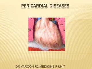
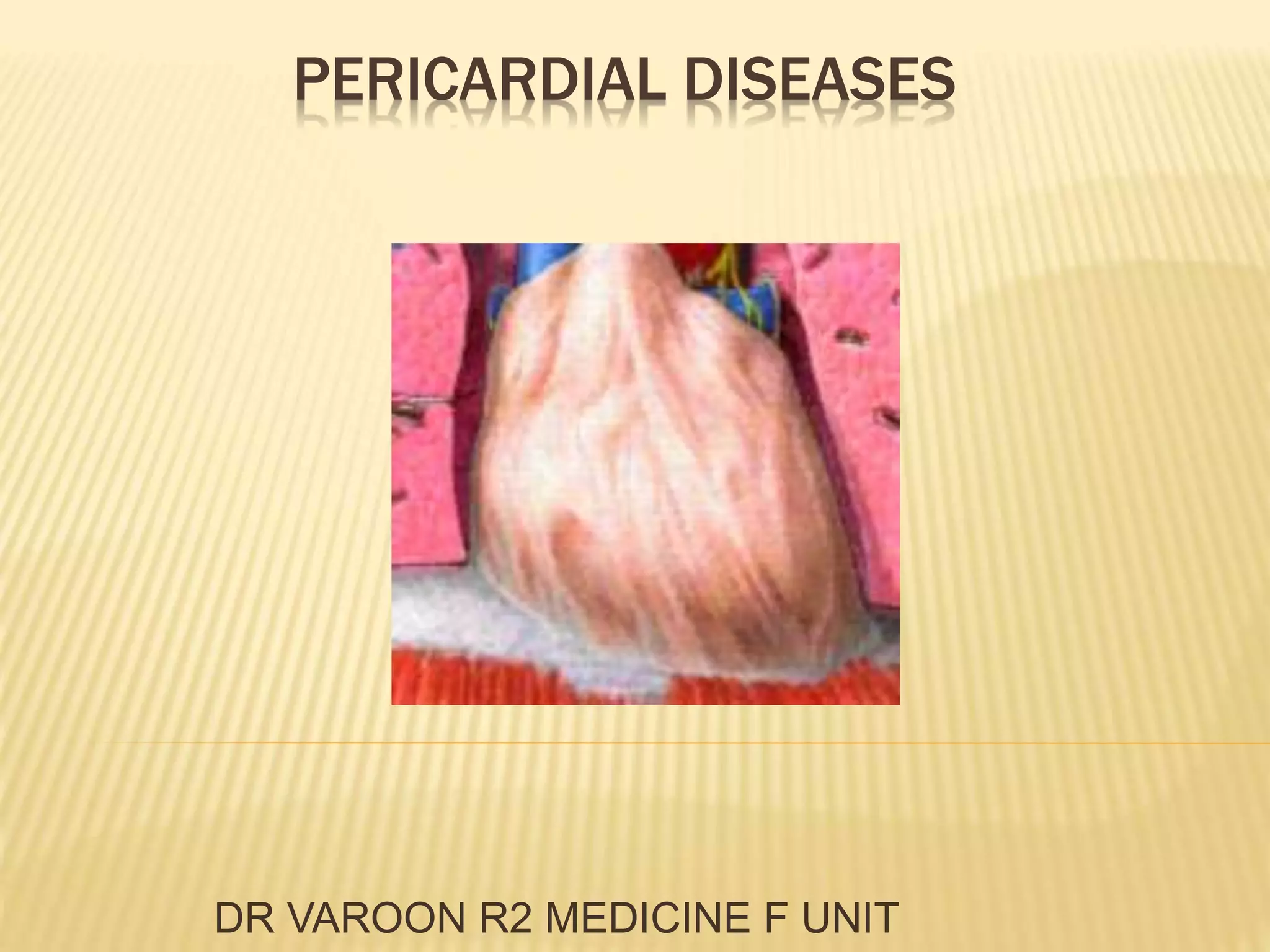
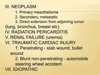
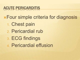
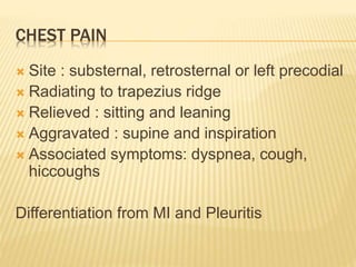
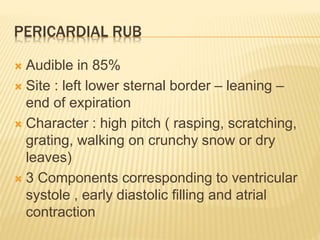
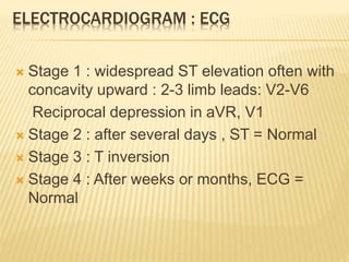
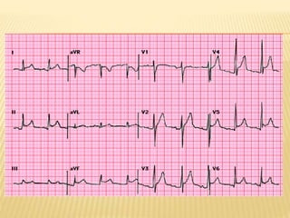
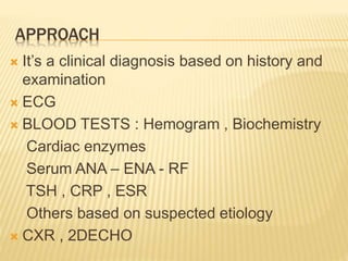
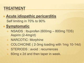
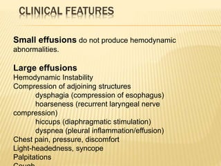
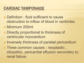
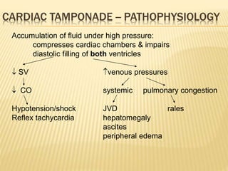
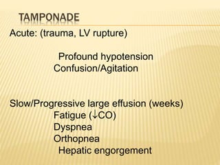
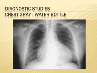
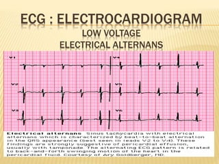
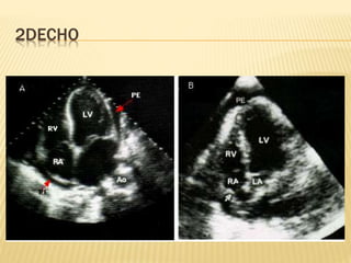
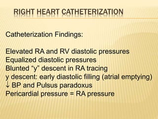
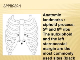
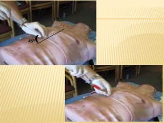
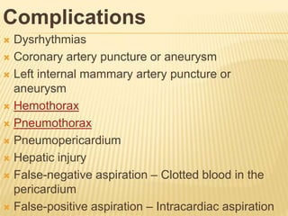
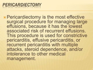
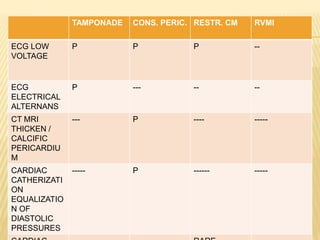
This document discusses pericardial diseases, focusing on pericarditis, pericardial effusion, and cardiac tamponade. It defines the pericardium and its functions. It describes the symptoms, signs, and diagnostic criteria for acute pericarditis. Causes of pericarditis include infections, autoimmune disorders, neoplasms, radiation, renal failure, and trauma. Treatment involves NSAIDs, colchicine, or steroids depending on severity and recurrence risk. Pericardial effusion and tamponade can develop as complications, requiring drainage procedures or surgery.
The document discusses various pericardial diseases including normal anatomy, pericarditis, pericardial effusions, cardiac tamponade, and constrictive pericarditis. Key points include:
- Pericardial effusions can lead to cardiac tamponade by exerting pressure on the heart and restricting filling.
- Cardiac tamponade is diagnosed using echocardiogram, chest x-ray and equalized diastolic pressures on catheterization.
- Constrictive pericarditis involves thickened pericardium constraining all chambers and is diagnosed using characteristic hemodynamic tracings on catheterization.
The document discusses the anatomy, functions, pathophysiology, etiologies, clinical features, diagnosis and treatment of pericardial diseases including pericarditis, pericardial effusion, cardiac tamponade, and constrictive pericarditis. Key points include: the pericardium has visceral and parietal layers separated by pericardial fluid; pericarditis can be caused by infection, autoimmune disorders, neoplasms or trauma; tamponade occurs when fluid rapidly accumulates under pressure, compressing the heart; constrictive pericarditis involves fibrosis and scarring that restricts diastolic filling.
This document discusses pericardial disease and provides information on the anatomy, physiology, and diseases of the pericardium. It covers the following key points in 3 sentences:
The pericardium has inner serous and outer fibrous layers and normally contains 15-35mL of fluid. Diseases of the pericardium include congenital defects, infections, malignancies, and acquired conditions like acute pericarditis which can lead to cardiac tamponade physiology from excess fluid accumulation. Constrictive pericarditis occurs when the pericardium thickens and restricts heart chamber filling, showing signs on echocardiogram like equalized diastolic pressures between chambers.
The document summarizes pericardial diseases. It discusses the anatomy and physiology of the pericardium, acute pericarditis including symptoms, diagnosis and treatment, and pericardial effusion and tamponade. Acute pericarditis is usually self-limited and treated with NSAIDs. Larger effusions may require hospitalization. Pericardial effusion can progress to tamponade, where fluid accumulation compresses the heart and impairs filling.
The document discusses the anatomy and physiology of the pericardium and various pericardial diseases. It describes the pericardium's functions in maintaining cardiac structure and output. It then covers acute pericarditis and its symptoms of chest pain, pericardial friction rub, and ECG changes. Pericardial effusion and its causes are discussed along with cardiac tamponade, which results from excessive fluid accumulation compressing the heart. Diagnosis involves echocardiography and treatment involves drainage of excessive fluid in tamponade.
1. Pericardial effusion is an abnormal accumulation of fluid in the pericardial cavity, while cardiac tamponade is a clinical syndrome caused by excess fluid in the pericardial space, reducing heart filling and function.
2. Causes of pericardial effusion include infections, autoimmune diseases, cancer, trauma, and uremia. Symptoms vary depending on the rate and amount of fluid accumulation but can include chest pain, dyspnea, and hypotension in tamponade.
3. Diagnosis involves echocardiography, which can detect fluid and signs of tamponade like heart chamber collapse. Treatment of tamponade requires pericardiocentesis
This document discusses pericardial diseases and various conditions that affect the pericardium. It begins by describing normal pericardial fluid volume and ventricular interdependence under normal conditions. It then discusses the history of using ultrasound to image the pericardium. Various pathological conditions are covered, including increased pericardial thickness in constrictive pericarditis, how intrapericardial pressure changes with fluid volume and pericardial stiffness, and signs of cardiac tamponade seen on echocardiogram like right atrial and ventricular collapse and IVC plethora. Finally, it describes the presentation of effusive-constrictive pericarditis.
This document provides an overview of echocardiography in pericardial diseases. It begins with an introduction to pericardial anatomy and pathophysiology. It then discusses various pericardial diseases that can be evaluated by echocardiography, including acute pericarditis, recurrent pericarditis, pericardial effusions, cardiac tamponade, and constrictive pericarditis. For each condition, it describes the echocardiographic findings and techniques used to evaluate the condition. It emphasizes that echocardiography is usually the initial imaging test of choice but that CT or CMR may be needed in some complex cases.
The document discusses constrictive pericarditis, providing details on:
1) The pathology of constrictive pericarditis which involves thickening and scarring of the pericardium leading to loss of elasticity.
2) The pathophysiology of constrictive pericarditis where the inelastic pericardium constrains cardiac filling and prevents adaptation to volume changes.
3) Key diagnostic features of constrictive pericarditis seen on echocardiogram include septal bounce, rapid early diastolic mitral inflow, and increased mitral annular velocities that rise with inspiration.
1. The document discusses the classification, diagnosis, and treatment of acute coronary syndromes including unstable angina and myocardial infarction.
2. Key points include definitions of unstable angina, NSTEMI, and STEMI; causes of acute coronary syndromes including plaque rupture and vasospasm; the importance of history, ECG, biomarkers in diagnosis; and the use of antiplatelet agents, beta blockers, nitroglycerin, and anticoagulants in treatment.
3. Primary percutaneous coronary intervention is recommended over thrombolysis when certain criteria are met for STEMI patients.
The document discusses arrhythmias and their management. It begins by describing the normal electrical conduction system of the heart. It then defines arrhythmias as disorders of heart rhythm or rate caused by issues with electrical impulse formation or conduction. Various types of arrhythmias are classified based on the site of abnormal impulse formation or conduction, including sinus node arrhythmias, atrial arrhythmias, junctional arrhythmias, and ventricular arrhythmias. Treatment depends on restoring normal rhythm and addressing any underlying causes.
This document discusses aortic stenosis (AS), including its causes, effects on the heart, symptoms, diagnosis, and management. It causes left ventricular outflow tract obstruction, leading to pressure overload hypertrophy and eventual heart failure if left untreated. Calcific aortic valve disease is the most common cause in adults. Symptoms typically manifest in the 6th or 7th decade of life and include chest pain, syncope, and heart failure. Echocardiography is the main diagnostic test used to evaluate severity based on aortic valve area and transvalvular pressure gradient. Treatment involves managing symptoms medically or replacing the stenotic valve surgically with either bioprosthetic or mechanical valves.
This document discusses cardiac tamponade, which occurs when fluid rapidly accumulates in the pericardial sac, putting pressure on the heart and reducing cardiac function. Key points include:
- Pericardial effusion puts pressure on the heart, causing symptoms like chest pain and shortness of breath.
- Cardiac tamponade occurs when a rapid accumulation of fluid in the pericardial sac severely compresses the heart.
- Echocardiography is useful for diagnosing tamponade by showing findings like pericardial effusion, right ventricular collapse, and reduced respiratory variation in blood flow velocities.
- Tamponade is a medical emergency treated initially with medications and peric
Tissue Doppler Imaging (TDI) provides low velocity, high amplitude signals from the myocardium that can be used to assess systolic and diastolic function. TDI utilizes pulsed wave and color Doppler techniques to measure peak myocardial velocities. The E/E' ratio, where E is transmitral early diastolic velocity and E' is early diastolic mitral annular velocity, correlates well with left ventricular filling pressures and can help distinguish normal from elevated pressures. TDI parameters are useful for evaluating global and regional systolic function, diastolic function, ischemia, and viability as well as distinguishing between restrictive cardiomyopathy and constrictive pericarditis.
This document summarizes the echocardiographic assessment of mitral stenosis (MS). It describes the anatomy of the mitral valve and causes of MS. Methods for assessing MS severity include measuring the pressure gradient, mitral valve area using planimetry and pressure half-time, and pulmonary artery pressure. Suitability for percutaneous transvenous mitral commissurotomy is evaluated. Concomitant valve lesions are also identified. Stress echocardiography may be used when symptoms are equivocal. Transesophageal echocardiography is recommended in some cases.
Chronic constrictive pericarditis is a condition where the pericardium thickens and scar tissue forms, restricting the heart's ability to fill with blood. It results from various causes like infections, surgery, radiation, or autoimmune disorders. On examination, elevated jugular venous pressure and equalization of cardiac filling pressures are seen. Imaging like echocardiograms and CT scans show thickened pericardium. Definitive treatment is surgical removal of the pericardium (pericardiectomy), which improves symptoms in most patients.
This document discusses pericardial diseases seen on echocardiograms. It notes that pericardial effusion is seen when there is over 50ml of fluid between the two layers of the pericardium. Tuberculosis, malignancy, cardiac tamponade, and constrictive pericarditis are pericardial diseases mentioned. Constrictive pericarditis is characterized by the pericardium appearing as a dark, thick line and abrupt anterior motion of the interventricular septum.
This document defines heart failure and discusses its key characteristics. It describes how heart failure occurs when the heart is unable to pump enough blood to meet the body's needs due to problems like abnormal heart muscle function or excessive loads on the heart. The document outlines the pathophysiology and progression of heart failure, including ventricular dilation and hypertrophy as compensatory mechanisms that ultimately fail. It also covers the clinical features, diagnostic tests, medical management, and nursing care considerations for patients with heart failure.
This document summarizes the evaluation of aortic valve stenosis using echocardiography. It describes the normal aortic valve anatomy and various types of aortic valve stenosis including calcific, bicuspid, rheumatic, and supravalvular or subvalvular stenosis. Doppler echocardiography is used to evaluate aortic valve stenosis severity based on valve area, mean gradient, and peak jet velocity. Stress echocardiography with dobutamine can help distinguish true severe from pseudo-severe low-flow, low-gradient aortic stenosis.
Echocardiography is the test of choice for evaluating pericardial effusions and cardiac tamponade. Characteristic findings include identifying the size and location of a pericardial effusion, end-diastolic chamber collapse, and respiratory variations in transvalvular flow velocities. Right atrial and ventricular collapse on expiration as well as dilatation of the IVC that fails to collapse with inspiration also suggest tamponade. Echocardiography allows for rapid assessment but findings may be absent in patients with pulmonary hypertension or RV hypertrophy.
The document summarizes key aspects of cardiac catheterization and hemodynamic data collection. It describes the normal cardiac cycle, pressure measurement systems, normal pressure waveforms, methods to measure cardiac output like thermodilution and Fick, how to evaluate valvular stenosis and regurgitation, determine vascular resistance and shunts. Specific details are provided on assessing aortic stenosis, mitral stenosis, right-sided valves and quantifying regurgitant fractions. Oxygen saturation analysis and Fick principles are outlined for shunt determinations.
The document provides an overview of electrocardiography (ECG). It defines an ECG as a tracing of the heart's electrical activity. It describes how to perform and record an ECG on a patient, including electrode placement and the conduction pathway of the heart. It also lists the objectives of interpreting ECGs, such as identifying abnormalities like myocardial infarction, atrial fibrillation, and various heart blocks.
This document presents information on various types of atrial arrhythmias. It discusses premature atrial complexes, atrial tachycardia, multifocal atrial tachycardia, atrial flutter, atrial fibrillation, and wandering atrial pacemaker. For each type, it covers etiology, characteristics, and treatment approaches. The document is presented by Baby Haokip from the College of Nursing, NEIGRIHMS.
This document discusses various tachyarrhythmias, including:
- Supraventricular tachycardias like atrial flutter, AV nodal reentrant tachycardia, and AV reentrant tachycardia.
- Ventricular arrhythmias including ventricular tachycardia and ventricular flutter.
- Irregular rhythms such as atrial fibrillation.
It provides details on characteristics like rate, morphology, underlying causes, and treatment approaches for each type of tachycardia. Emphasis is placed on distinguishing ventricular tachycardia from supraventricular tachycardia with aberrancy in clinical evaluation.
A 58-year-old man presented with shortness of breath and chest pain. An ECG showed ST segment elevation consistent with pericarditis. Pericarditis is inflammation of the pericardium and can be caused by uremia in patients with chronic kidney disease. The ECG changes in acute pericarditis include diffuse concave ST elevation and upright T waves, except in leads aVR and V1 which are usually depressed. This differs from a myocardial infarction which shows more convex ST elevation and the presence of Q waves.
mitral regurgitation american guidlines 2014Basem Enany
This document discusses mitral regurgitation (MR), including its etiology, clinical manifestations, physical exam findings, diagnostic testing, and management according to American Heart Association guidelines. The most common cause of primary MR in developed countries is mitral valve prolapse. Secondary MR is usually caused by ischemic heart disease, left ventricular dysfunction, or hypertrophic cardiomyopathy. Diagnosis involves echocardiography to determine the severity and mechanism of MR. Management is generally medical for mild MR but may involve surgery for severe primary MR.
Cardiovascular and hemolynphatic diseases word printableDevon Avis
This document summarizes various diseases of the cardiovascular and hemolymphatic systems in horses. It describes the pathogenesis, clinical signs, lesions, diagnosis, treatment, and prevention of conditions like congestive heart failure, acute heart failure, peripheral circulatory failure, valvular diseases, endocarditis, pericarditis, septal defects, and hemolymphatic diseases like anemia and hemorrhage. Key points include that congestive heart failure develops when the heart can no longer maintain circulatory equilibrium at rest, valvular diseases cause murmurs from turbulent blood flow, endocarditis can be caused by bacterial infection of heart valves, and anemia results from a decrease in circulating blood volume.
The document discusses postpericardiotomy syndrome, which is an inflammatory condition that can occur after surgery involving opening of the pericardium. It has three key characteristics:
1) It presents with symptoms like fever, chest pain, and pericardial friction rub within 1-6 weeks after cardiac surgery.
2) It is characterized by pericardial and sometimes pleural effusions seen on imaging like echocardiography.
3) It is typically treated with anti-inflammatory drugs like NSAIDs or corticosteroids, and pericardiocentesis if cardiac tamponade develops.
This document discusses the diagnostic criteria for rheumatic fever. It lists the major criteria as polyarthritis, carditis, subcutaneous nodules, erythema marginatum, and Sydenham's chorea. The minor criteria include fever, arthralgia, elevated inflammatory markers, leukocytosis, and ECG abnormalities. A diagnosis of rheumatic fever requires supporting evidence of a preceding streptococcal infection and presence of either two major criteria or one major criterion with two minor criteria or three minor criteria.
This document defines cardiac failure and heart failure, describes the types and causes, and discusses the pathophysiology, clinical features, investigations, and treatment. Heart failure is a clinical syndrome where the heart cannot pump enough blood to meet the body's needs, or can only do so with elevated filling pressures. It can be systolic or diastolic in nature. Common causes include ischemic heart disease, cardiomyopathy, valvular disease, and hypertension. Symptoms include breathlessness, fatigue, and fluid retention. Echocardiography, biomarkers like BNP, and cardiac imaging are used in diagnosis and assessment. Treatment aims to relieve symptoms, improve quality of life, and reduce mortality through medications, device therapies, and lifestyle changes.
1. Shock is defined as inadequate tissue perfusion to meet metabolic needs due to issues with cardiac performance, vascular performance, or cellular function.
2. The main types of shock are hypovolemic, cardiogenic, obstructive, and distributive. Clinical signs include low blood pressure, fast heart rate, pale skin, confusion and loss of consciousness.
3. Treatment of shock focuses on identifying the type, treating the underlying cause, restoring circulating volume with fluids, and supporting vital organ function with vasopressors or inotropes as needed. The goal is to restore adequate perfusion to prevent multiple organ dysfunction syndrome.
This document discusses the anaesthetic management of closed mitral valvotomy. It begins with the anatomy and pathophysiology of mitral stenosis. It then discusses the indications for closed mitral valvotomy and the pre-anaesthetic assessment. The key aspects of anaesthetic management are maintaining haemodynamic stability, avoiding tachycardia and hypotension, and careful fluid management. Etomidate is recommended for induction due to hemodynamic stability. Post-operatively, risks of pulmonary edema and right heart failure must be assessed and managed.
This document discusses pericarditis, which is inflammation of the pericardium. It describes the different types of pericarditis and various etiologies in cattle, horses, small animals, sheep, goats and pigs. Clinical findings include lethargy, respiratory difficulty and muffled heart sounds. Diagnosis involves thoracic radiography, echocardiography and electrocardiography. Treatment depends on the underlying cause but may include pericardiocentesis, antibiotics, drainage or pericardiectomy. Pericardiocentesis provides immediate relief for cardiac tamponade and allows diagnostic sampling. Prognosis varies based on the causative agent but is generally poor for septic pericard
This document discusses cardiac tamponade, which is the compression of the heart caused by fluid accumulation in the pericardial sac. It defines cardiac tamponade and describes its causes, signs, symptoms, diagnostic tests, and treatments. Key causes include infection, cancer, surgery, and medication side effects. Diagnosis involves echocardiography and other imaging tests. Treatment involves draining excess fluid via pericardiocentesis, a procedure where a needle is used to drain the fluid for diagnostic and therapeutic purposes. Nursing care focuses on monitoring vital signs, administering oxygen, IV fluids, and medications to support heart function.
The document discusses diseases of the pericardium, including acute pericarditis, constrictive pericarditis, pericardial effusion, and cardiac tamponade. It describes the anatomy and functions of the pericardium, symptoms and signs of different pericardial diseases, diagnostic tests including ECG, echo, CT and treatment approaches.
The document discusses tricuspid valve stenosis and insufficiency. It defines the conditions as a narrowing or stiffening of the tricuspid valve opening (stenosis), or the valve not closing tightly enough to prevent leakage (insufficiency). Causes can include rheumatic fever, infections, congenital malformations, and tumors. Risks include right heart failure and liver congestion. Diagnosis involves physical exams, echocardiograms, and cardiac catheterization. Treatment options include medications, valve repairs such as annuloplasty or valvuloplasty, and valve replacements. Complications include heart failure, endocarditis, and liver cirrhosis.
Cardiac tamponade occurs when fluid accumulates in the pericardial space, reducing cardiac filling and output. It can develop acutely or subacutely. Echocardiography is key for diagnosis, showing pericardial effusion, chamber collapse, and respiratory variations in flow velocities. Treatment involves drainage of fluid, usually by pericardiocentesis under ultrasound guidance. Subxiphoid approach carries liver/vessel injury risk but is safest in emergencies, while apical is easiest but risks heart wall puncture. Drainage resolves tamponade, and catheters are typically removed within 2 days if drainage is low.
Cardiac tamponade is caused by fluid accumulation in the pericardial space, reducing heart filling and function. It requires urgent treatment to remove fluid via pericardiocentesis or surgery. Symptoms include low blood pressure, increased heart rate, and difficulty breathing due to restricted heart movement. A diagnosis is made through echocardiography identifying fluid and heart compression.
The document discusses pericarditis, pericardial effusion, cardiac tamponade, and chronic constrictive pericarditis. It describes the functions of the pericardium, signs and symptoms, diagnostic tests including EKG changes and imaging, and treatments for the different conditions including medications, pericardiocentesis, and pericardial stripping. Chronic constrictive pericarditis results from scarring and thickening of the pericardium limiting ventricular filling, with symptoms of exertional dyspnea and elevated jugular venous pressure.
Anaesthetic management of a patient with mitral stenosis put for non-cardiac ...Ankur Khandelwal
Mitral stenosis is a narrowing of the mitral valve that causes obstruction of blood flow from the left atrium to the left ventricle. Rheumatic fever is the most common cause. Symptoms range from none in mild cases, to shortness of breath with exertion in moderate cases, to shortness of breath at rest in severe cases. Diagnosis is made through echocardiogram which can assess the severity based on metrics like mitral valve area and pressure gradients. Treatment depends on symptoms and severity, ranging from medications and lifestyle changes in mild cases, to balloon valvuloplasty or surgical commissurotomy in moderate to severe cases. Anesthetic management aims to avoid tachycardia and
This document provides an overview of pericardial diseases. It begins with the anatomy and functions of the pericardium. It then discusses various pericardial diseases like acute pericarditis, pericardial effusion, and cardiac tamponade. For acute pericarditis, it describes the key symptoms of chest pain, pericardial friction rub, and ECG changes. It also outlines the diagnostic criteria and treatment approaches for pericardial effusion and cardiac tamponade, including the use of echocardiography, medications, and pericardiocentesis.
Cardiogenic shock is a life-threatening condition where the heart is unable to pump enough blood to meet the body's needs. It is usually caused by heart muscle damage from a myocardial infarction, cardiomyopathy, or other acute cardiac conditions. Key features include low blood pressure, rapid heart rate, impaired thinking, and poor peripheral circulation. Treatment focuses on supporting heart function through inotropic drugs or devices, reducing workload on the heart, and treating any underlying causes.
This document discusses cardiac tamponade, including its anatomy, physiology, causes, clinical presentation, diagnosis and treatment. Cardiac tamponade is caused by an accumulation of fluid in the pericardial space that compresses the heart and impairs diastolic filling. It presents with symptoms like chest pain, dyspnea and pulsus paradoxus. Diagnosis is made through echocardiography, ECG changes and chest x-ray. Treatment involves drainage of pericardial fluid, usually through pericardiocentesis, along with medical management including oxygen, medications and ventilation.
The document discusses several diseases of the pericardium, including acute pericarditis, pericardial effusion, cardiac tamponade, constrictive pericarditis. Acute pericarditis is often caused by infections, immunological mechanisms, or after myocardial infarction. It presents with chest pain, pericardial friction, and fever. Pericardial effusion occurs when fluid in the pericardium exceeds normal amounts and can be caused by acute pericarditis. Cardiac tamponade is when excess fluid compresses the heart and hinders its filling. Constrictive pericarditis involves thickening and scarring of the pericardium that limits ventricular filling
Headache Attributed to Nonvascular, Noninfectious
Intracranial Disorders
Headache Attributed to Trauma or Injury to the Head
and/or Neck
Headache Attributed to Infection
Headache Attributed to Cranial or Cervical Vascular
Disorders
Headache Associated with Disorders of Homeostasis
Headache Caused by Disorders of the Cranium, Neck,
Eyes, Ears, Nose, Sinuses, Teeth, Mouth, or Other
Facial or Cranial Structures
Headaches and the Cervical Spine
Migraine
Chronic Daily Headache
Cluster Headache
Other Trigeminal Autonomic Cephalalgias
Other Primary Headaches
1. Neuroplasticity refers to the brain's ability to change and reorganize itself in response to experience or injury. It allows the brain to compensate for damage and to adjust its activity in response to new situations or information.
2. Several mechanisms underlie neuroplasticity including neuronal regeneration, synaptic plasticity, neurogenesis, gliogenesis, dendritic remodeling, and functional reorganization through processes like vicariation.
3. Neuroplasticity can be measured at the cellular level through changes in synapses and at the neural network level through reorganization of maps. Imaging techniques like MRI can also measure plastic changes in gray and white matter.
This document provides information on various brain tumors including their appearance on different MRI sequences and key imaging findings. It discusses tumors such as pineal region tumors, germ cell tumors, embryonal tumors, meningiomas, sarcomas, nerve sheath tumors, sellar region tumors, lymphomas, and hematopoietic tumors. For each tumor type, it presents representative imaging examples with descriptions of imaging features and differential diagnoses.
- Gliomas are the most common malignant primary brain tumors, accounting for 80% of malignant tumors and 28% of all brain tumors.
- Prognosis and survival rates vary significantly by histological subtype, with oligodendroglioma having the best prognosis and glioblastoma having the worst.
- Young age and lower tumor grade are favorable prognostic factors, while radiation exposure and certain genetic mutations can increase risk.
TUBEROUS SCLEROSIS
Cutaneous Features
Neurological Features
Retinal Features
Systemic Features
NEUROFIBROMATOSIS
Cutaneous Features of Neurofibromatosis Type 1
Systemic Features of Neurofibromatosis Type 1
Neurological Features in Neurofibromatosis Type 1
Clinical Features of Neurofibromatosis Type 2
STURGE-WEBER SYNDROME
Cutaneous Features
Ocular Features
Neurological Features
Diagnostic Studies
Treatment
VON HIPPEL-LINDAU SYNDROME
Neurological Features
Ocular Features
Systemic Features
Molecular Genetics
Treatment
HEREDITARY HEMORRHAGIC TELANGIECTASIA
Neurological Features
Treatment
HYPOMELANOSIS OF ITO
Cutaneous Features
Neurological Features
Systemic Features
INCONTINENTIA PIGMENTI
Cutaneous Features
Neurological Features
Genetics
ATAXIA-TELANGIECTASIA
Cutaneous Features
Neurological Features
Immunodeficiency and Cancer Risk
Laboratory Diagnosis
EPIDERMAL NEVUS SYNDROME
Cutaneous Features
Neurological Features
Other Features
Neuroimaging
NEUROCUTANEOUS MELANOSIS
Cutaneous Features
Neurological Features
Laboratory Findings
Neuroimaging
EHLERS-DANLOS SYNDROME
Neurovascular Features
CEREBROTENDINOUS XANTHOMATOSIS
Neurological Features
Xanthomas
Other Clinical Features
Treatment
PROGRESSIVE FACIAL HEMIATROPHY
Clinical Features
KINKY HAIR SYNDROME (MENKES DISEASE)
Cutaneous Features
Other Clinical Features
Neurological Features
Neuroimaging
Genetic Studies
Diagnosis and Treatment
XERODERMA PIGMENTOSUM
Complementation Groups
Related Syndromes
Cutaneous and Ocular Features
Treatment
OTHER NEUROLOGICAL CONDITIONS WITH CUTANEOUS
MANIFESTATIONS
This document discusses migraine, including its classification, pathophysiology, triggers, symptoms, and treatment. It classifies migraines into categories including migraine without aura, migraine with aura, hemiplegic migraine, retinal migraine, and chronic migraine. It describes the pathophysiology involving the trigeminovascular system and brain networks. Common triggers include stress, hormones, missed meals, and certain stimuli. Symptoms involve headache, nausea, sensitivity to light and sound. Treatment includes lifestyle changes, medications like triptans and NSAIDs, and preventative medications. New therapies targeting CGRP are also discussed.
This document discusses new and existing antiepileptic drugs (AEDs), including their mechanisms of action, pharmacokinetic properties, drug interactions, efficacy, side effects, and considerations for use. It introduces several new AEDs approved in recent years for various seizure types including rufinamide, perampanel, cannabidiol, and stiripentol. It also summarizes the mechanisms, spectra of efficacy, drug interactions, common and rare side effects of numerous established AEDs. Finally, it provides guidance on choosing AED therapy, including preferences for monotherapy versus combination treatment based on factors like efficacy, tolerability, and pharmacokinetic profiles.
This document discusses occupational lung diseases including their definitions, etiology, epidemiology, clinical presentation, diagnosis, and treatment. Some key occupational lung diseases covered include coal worker's pneumoconiosis, asbestosis, silicosis, byssinosis, and hypersensitivity pneumonitis caused by exposure to various occupational dusts and chemicals. Diagnostic tools like chest x-rays and CT scans are discussed. Management involves removal from exposure, supportive care, and medications like steroids in some cases.
Can coffee help me lose weight? Yes, 25,422 users in the USA use it for that ...nirahealhty
The South Beach Coffee Java Diet is a variation of the popular South Beach Diet, which was developed by cardiologist Dr. Arthur Agatston. The original South Beach Diet focuses on consuming lean proteins, healthy fats, and low-glycemic index carbohydrates. The South Beach Coffee Java Diet adds the element of coffee, specifically caffeine, to enhance weight loss and improve energy levels.
Hypertension and it's role of physiotherapy in it.Vishal kr Thakur
This particular slides consist of- what is hypertension,what are it's causes and it's effect on body, risk factors, symptoms,complications, diagnosis and role of physiotherapy in it.
This slide is very helpful for physiotherapy students and also for other medical and healthcare students.
Here is summary of hypertension -
Hypertension, also known as high blood pressure, is a serious medical condition that occurs when blood pressure in the body's arteries is consistently too high. Blood pressure is the force of blood pushing against the walls of blood vessels as the heart pumps it. Hypertension can increase the risk of heart disease, brain disease, kidney disease, and premature death.
TEST BANK FOR Health Assessment in Nursing 7th Edition by Weber Chapters 1 - ...rightmanforbloodline
TEST BANK FOR Health Assessment in Nursing 7th Edition by Weber Chapters 1 - 34.
Letter to MREC - application to conduct studyAzreen Aj
Application to conduct study on research title 'Awareness and knowledge of oral cancer and precancer among dental outpatient in Klinik Pergigian Merlimau, Melaka'
We are one of the top Massage Spa Ajman Our highly skilled, experienced, and certified massage therapists from different corners of the world are committed to serving you with a soothing and relaxing experience. Luxuriate yourself at our spas in Sharjah and Ajman, which are indeed enriched with an ambiance of relaxation and tranquility. We could confidently claim that we are one of the most affordable Spa Ajman and Sharjah as well, where you can book the massage session of your choice for just 99 AED at any time as we are open 24 hours a day, 7 days a week.
Visit : https://massagespaajman.com/
Call : 052 987 1315
MBC Support Group for Black Women – Insights in Genetic Testing.pdfbkling
Christina Spears, breast cancer genetic counselor at the Ohio State University Comprehensive Cancer Center, joined us for the MBC Support Group for Black Women to discuss the importance of genetic testing in communities of color and answer pressing questions.
Let's Talk About It: Breast Cancer (What is Mindset and Does it Really Matter?)bkling
Your mindset is the way you make sense of the world around you. This lens influences the way you think, the way you feel, and how you might behave in certain situations. Let's talk about mindset myths that can get us into trouble and ways to cultivate a mindset to support your cancer survivorship in authentic ways. Let’s Talk About It!
This particular slides consist of- what is Pneumothorax,what are it's causes and it's effect on body, risk factors, symptoms,complications, diagnosis and role of physiotherapy in it.
This slide is very helpful for physiotherapy students and also for other medical and healthcare students.
Here is a summary of Pneumothorax:
Pneumothorax, also known as a collapsed lung, is a condition that occurs when air leaks into the space between the lung and chest wall. This air buildup puts pressure on the lung, preventing it from expanding fully when you breathe. A pneumothorax can cause a complete or partial collapse of the lung.
The facial nerve, also known as cranial nerve VII, is one of the 12 cranial nerves originating from the brain. It's a mixed nerve, meaning it contains both sensory and motor fibres, and it plays a crucial role in controlling various facial muscles, as well as conveying sensory information from the taste buds on the anterior two-thirds of the tongue.
3. FUNCTION OF THE PERICARDIUM
1. Stabilization of the heart within the thoracic cavity -
ligamentous attachments -- limiting the heart’s motion.
2. Protection of the heart from mechanical trauma and
infection from adjoining structures.
3. The pericardial fluid functions as a lubricant and
decreases friction of cardiac surface during systole
and diastole.
4. Prevention of excessive dilation of heart especially
during sudden rise in intra-cardiac volume
10. CHEST PAIN
Site : substernal, retrosternal or left precodial
Radiating to trapezius ridge
Relieved : sitting and leaning
Aggravated : supine and inspiration
Associated symptoms: dyspnea, cough,
hiccoughs
Differentiation from MI and Pleuritis
11. PERICARDIAL RUB
Audible in 85%
Site : left lower sternal border – leaning –
end of expiration
Character : high pitch ( rasping, scratching,
grating, walking on crunchy snow or dry
leaves)
3 Components corresponding to ventricular
systole , early diastolic filling and atrial
contraction
12. ELECTROCARDIOGRAM : ECG
Stage 1 : widespread ST elevation often with
concavity upward : 2-3 limb leads: V2-V6
Reciprocal depression in aVR, V1
Stage 2 : after several days , ST = Normal
Stage 3 : T inversion
Stage 4 : After weeks or months, ECG =
Normal
13.
14. APPROACH
It’s a clinical diagnosis based on history and
examination
ECG
BLOOD TESTS : Hemogram , Biochemistry
Cardiac enzymes
Serum ANA – ENA - RF
TSH , CRP , ESR
Others based on suspected etiology
CXR , 2DECHO
15. TREATMENT
Acute idiopathic pericarditis
Self limiting in 70% to 90%
Symptomatic
NSAIDS : Ibuprofen (600mg – 800mg TDS)
Aspirin (2-4mg/d)
NARCOTIC: Morphine
COLCHICINE ( 2-3mg loading with 1mg 10-14d)
STERIODS : avoid : recurrences
60mg x 2d and then taper in week.
18. PERICARDIAL EFFUSION
Normal 15-50 ml of fluid
ETIOLOGY
1. Inflammation : infection, immunologic process.
2. Trauma : bleeding in pericardial space.
3. Noninfectious :
a. increase in pulmonary hydrostatic pressure e.g.
congestive heart failure.
b. increase in capillary permeability e.g.
hypothyroidism
c. decrease in plasma oncotic pressure e.g.
cirrhosis.
4. Decreased drainage : obstruction of thoracic
duct : malignancy or damage during surgery.
20. CLINICAL FEATURES
Small effusions do not produce hemodynamic
abnormalities.
Large effusions
Hemodynamic Instability
Compression of adjoining structures
dysphagia (compression of esophagus)
hoarseness (recurrent laryngeal nerve
compression)
hiccups (diaphragmatic stimulation)
dyspnea (pleural inflammation/effusion)
Chest pain, pressure, discomfort
Light-headedness, syncope
Palpitations
21. CARDIAC TAMPONADE
Definition : fluid sufficient to cause
obstruction to inflow of blood in ventricles
Minimum 200ml
Directly proportional to thickness of
ventricular myocardium
Inversely thickness of parietal pericardium
Three common causes : neoplastic ,
idiopathic, pericardial effusion secondary to
renal failure
22. CARDIAC TAMPONADE -- PATHOPHYSIOLOGY
Accumulation of fluid under high pressure:
compresses cardiac chambers & impairs
diastolic filling of both ventricles
SV venous pressures
CO systemic pulmonary congestion
Hypotension/shock JVD rales
Reflex tachycardia hepatomegaly
ascites
peripheral edema
24. EXAMINATION AND FINDINGS
BECK’S TRIAD
HYPOTENSION
SOFT OR ABSENT HEART SOUNDS
JUGULAR VENOUS DISTENSION WITH
PROMINENT X DESCENT BUT AN ABSENT
Y DESCENT
25. PULSUS PARADOXUS
Inspiration: negative intrathoracic pressure is
transmitted to the pericardial space
IPP
blood return to the right ventricle
jugular venous and right atrial pressures
right ventricular volume interventricular
septum shifts towards the left ventricle
left ventricular volume
LV stroke volume
blood pressure (<10mmHg is normal) during
inspiration
27. SYSTEMIC EXAMINATION
MUFFLED HEART SOUNDS
PERICARDIAL RUB
TUBULAR BREATH SOUNDS : LEFT
AXILLA OR LEF BASE : BRONCHIAL
COMPRESSION
EWART SIGN: DULLNESS TO
PERCUSSION BENEATH THE ANGLE OF
LEFT SCAPULA and INCREASED
FREMITUS
HEPATOSPLENOMEGALY
31. RIGHT HEART CATHETERIZATION
Catheterization Findings:
Elevated RA and RV diastolic pressures
Equalized diastolic pressures
Blunted “y” descent in RA tracing
y descent: early diastolic filling (atrial emptying)
BP and Pulsus paradoxus
Pericardial pressure = RA pressure
34. CONTRAINDICATIONS
Absolute contraindications
In the hemodynamically unstable patient, no
absolute contraindications
Relative contraindications
Uncorrected bleeding disorder
Traumatic cardiac tamponade. Some authors
argue that traumatic cardiac tamponade
should be treated by emergent thoracotomy.
35. POSITION
Position the patient in a semirecumbent
position at a 30- to 45-degree angle. This
position brings the heart closer to the anterior
chest wall.
The supine position is an acceptable
alternative.
38. Complications
Dysrhythmias
Coronary artery puncture or aneurysm
Left internal mammary artery puncture or
aneurysm
Hemothorax
Pneumothorax
Pneumopericardium
Hepatic injury
False-negative aspiration – Clotted blood in the
pericardium
False-positive aspiration – Intracardiac aspiration
39. PERICARDIECTOMY
Pericardiectomy is the most effective
surgical procedure for managing large
effusions, because it has the lowest
associated risk of recurrent effusions.
This procedure is used for constrictive
pericarditis, effusive pericarditis, or
recurrent pericarditis with multiple
attacks, steroid dependence, and/or
intolerance to other medical
management.
40. PERICARDIAL WINDOW
Pericardial window placement is used for
effusive pericarditis therapy. In critically ill
patients, a balloon catheter may be used to
create a pericardial window, in which only 9
cm2 or less of pericardium is resected.
41. PERICARDIOTOMY
Consider subxiphoid pericardiotomy for large
effusions that do not resolve. This procedure
may be performed under local anesthesia
and has a lower risk of complications than
pericardiectomy.
43. Infectious pericarditis
Bacterial pericarditis : appropriate
antibiotics for at least 4 weeks and
drainage of pericardial fluid.
Fungal infection : fluconazole,
ketoconazole, itraconazole, amphotericin
B, liposomal amphotericin B, or
amphotericin B lipid complex
Corticosteroids and NSAIDs can be used
to support the antifungal drug treatment.
44. Intrapericardial fibrinolysis : thick, loculated
fluid, but open surgical drainage is preferred.
Occasionally, patients require partial to total
pericardiectomy.
Tubercular infection : AKT
Use of adjunctive prednisolone in patients
with acquired immunodeficiency syndrome
(AIDS) may reduce mortality in this
population.
45. METABOLIC PERICARDITIS
RENAL FAILURE associated uremic
pericarditis : HEMODIALYSIS
Hemodialysis : hypotension, which may
be dangerous in the setting of
tamponade.
In addition, some physicians advocate
heparin-free hemodialysis to reduce the
risk of intrapericardial hemorrhage.
46. Peritoneal dialysis may compromise
respiratory function because of the effect of
intraperitoneal fluid on the diaphragm.
In dialysis-associated pericarditis, an
increased intensity of dialysis for 10-14 days
is recommended.
Correct hypophosphatemia and hypokalemia
47. Cardiovascular pericarditis
Pericarditis does not contraindicate
thrombolytic or anticoagulant therapy for an
acute MI.
However, anticoagulation should be
discontinued if pericardial effusion develops
or effusion size increases. Treatment is with
aspirin.
In Dressler syndrome, anticoagulant therapy
should be stopped because of the risk of
hemorrhagic pericarditis. Treatment is with
NSAIDs.
48. CONSTRICTIVE PERICARDITIS
Late complication of pericardial disease
Fibrous scar formation
Fusion of pericardial layers
Calcification further stiffens pericardium
Etiologies:
any cause of pericarditis
idiopathic
post-surgery
mc : tuberculosis
radiation
neoplasm
49. PATHOPHYSIOLOGY
Rigid, scarred pericardium encircles heart:
Systolic contraction normal
Inhibits diastolic filling of both ventricles
SV venous pressures
CO systemic pulmonary congestion
Hypotension/shock JVD rales
Reflex tachycardia hepatomegaly
ascites
peripheral edema
50. PHYSICAL EXAMINATION
Findings of right heart failure :
• HR, BP
• Ascites, edema, hepatomegaly
• BROADBENT SIGN : apical impulse is
reduced and retract in systole
• early diastolic “knock” after S2 sudden
cessation of ventricular diastolic filling
imposed by rigid pericardial sac
• Kussmaul’s sign
• Square root sign
52. 2D ECHO
Pericardial thickening
Dilatation of IVC and hepatic veins
Sharp halt in ventricular filling in early
diastole
Flattening of left ventricular posterior wall
Atrial enlargement
54. CLINICAL TAMPONADE CONS. PERIC. RESTR. CM RVMI
PULSUS
PARADOXUS
COMMON ----- RARE RARE
JVP Y
DESCENT
---- P RARE RARE
JVP X
DESCENT
P P P RARE
KAUSSMAUL ----- P ------ P
THIRD HS ------ ----- RARE P
PERICARDIAL
KNOCK
------ P ------- ------
55. 2DECHO TAMPONA
DE
CONS.
PERIC.
RESTR.
CM
RVMI
THICKENED PERCARDIUM ------ P ------ -----
PERICARDIAL CALCIFICATION ---- P ------ ------
PERICARDIAL EFFUSION P ----- ----- -----
RV SIZE SMALL N N ENLARG
E
MYOCARDIAL THICKNESS N N INCRE N
RHT ATRIAL COLLAPSE AND
RVDC
P ------ ------- ------
INCREASED EARLY FILLING,
INCREASED MITRAL FLOW
VELOCITY
---- P P P
EXAGGERATED RESP
VARIATION IN FLOW VELOCITY
P P ---- -----
56. TAMPONADE CONS. PERIC. RESTR. CM RVMI
ECG LOW
VOLTAGE
P P P --
ECG
ELECTRICAL
ALTERNANS
P --- -- --
CT MRI
THICKEN /
CALCIFIC
PERICARDIU
M
--- P ---- -----
CARDIAC
CATHERIZATI
ON
EQUALIZATIO
N OF
DIASTOLIC
PRESSURES
----- P ------ -----
57. Pericardial cysts : MC : rht cardioprehnic
angle
Tumors : primary and secondary ( lung,
bronchus, breast, mediastinal, lymphoma,
melanoma)
MC primary malignant : mesothelioma