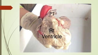
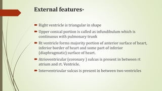
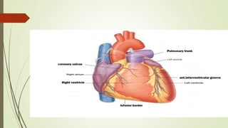
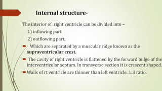
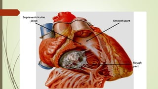
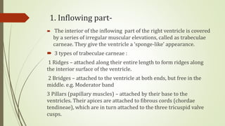
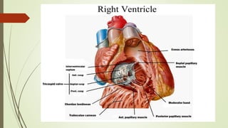
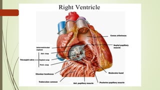
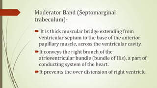
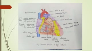
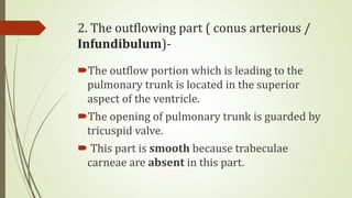
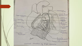
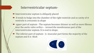
Right ventricle
Dr Karle Geetanjali
Professor, PMT’s Ayu. College, Shevgaon
Introduction-
The right ventricle receives deoxygenated
blood from the right atrium through
tricuspid valve, and pumps it to the lungs
through the pulmonary trunk
External features-
 Right ventricle is triangular in shape
 Upper conical portion is called as infundibulum which is
continuous with pulmonary trunk
 Rt ventricle forms majority portion of anterior surface of heart,
inferior border of heart and some part of inferior
(diaphragmatic) surface of heart.
 Atrioventricular (coronary ) sulcus is present in between rt
atrium and rt. Ventricle.
 Interventricular sulcus is present in between two ventricles
Internal structure-
The interior of right ventricle can be divided into –
1) inflowing part
2) outflowing part,
 Which are separated by a muscular ridge known as the
supraventricular crest.
 The cavity of right ventricle is flattened by the forward bulge of the
interventricular septum. In transverse section it is crescent shaped.
Walls of rt ventricle are thinner than left ventricle. 1:3 ratio.
1. Inflowing part-
 The interior of the inflowing part of the right ventricle is covered
by a series of irregular muscular elevations, called as trabeculae
carneae. They give the ventricle a ‘sponge-like’ appearance.
 3 types of trabeculae carneae :
1 Ridges – attached along their entire length to form ridges along
the interior surface of the ventricle.
2 Bridges – attached to the ventricle at both ends, but free in the
middle. e.g. Moderator band
3 Pillars (papillary muscles) – attached by their base to the
ventricles. Their apices are attached to fibrous cords (chordae
tendineae), which are in turn attached to the three tricuspid valve
cusps.
Papillary muscles of rt ventricle-
 These represent the pillars of trabeculae carneae. Their bases of
papillary muscles are connected to the ventricular wall and their apices
are connected by thread-like fibrous cords (the Chordae tendinae) to the
cusps of the tricuspid valve.
 There are 3 papillary muscles in the right ventricle: –
(a) Anterior - the largest one
(b) Posterior (inferior) – small
(c) Septal - it is generally split into 2 or 3 nipples.
 The papillary muscles of right ventricle are connected to the cusps of the
tricuspid valve
Moderator Band (Septomarginal
trabeculum)-
 It is thick muscular bridge extending from
ventricular septum to the base of the anterior
papillary muscle, across the ventricular cavity.
It conveys the right branch of the
atrioventricular bundle (bundle of His), a part of
conducting system of the heart.
It prevents the over distension of right ventricle.
2. The outflowing part ( conus arterious /
Infundibulum)-
The outflow portion which is leading to the
pulmonary trunk is located in the superior
aspect of the ventricle.
The opening of pulmonary trunk is guarded by
tricuspid valve.
 This part is smooth because trabeculae
carneae are absent in this part.
Interventricular septum-
 Interventricular septum is obliquely placed
 It tends to bulge into the chamber of the right ventricle and so cavity of rt
ventricle is crescentic in shape
 upper part of septum - The septum becomes thinner as well as more fibrous
near the aortic valve orifice – termed the membranous part of the
interventricular septum. It is oval in shape.
 The inferior part of septum- is muscular part forms the majority of the
septum and it is thick
Clinical anatomy-
Right ventricular hypertrophy – thickening of
walls of rt ventricle
due to pulmonary hypertension
Ventricular septal defect – congenital defect –
abnormal opening in septum
Thank you !!!
Special thanks to Neha Dethe for colored diagram of
internal structures and to Durva Pawar for pencil
diagram of internal structures.

Right ventricle

  • 1.
    Right ventricle Dr KarleGeetanjali Professor, PMT’s Ayu. College, Shevgaon
  • 2.
    Introduction- The right ventriclereceives deoxygenated blood from the right atrium through tricuspid valve, and pumps it to the lungs through the pulmonary trunk
  • 4.
    External features-  Rightventricle is triangular in shape  Upper conical portion is called as infundibulum which is continuous with pulmonary trunk  Rt ventricle forms majority portion of anterior surface of heart, inferior border of heart and some part of inferior (diaphragmatic) surface of heart.  Atrioventricular (coronary ) sulcus is present in between rt atrium and rt. Ventricle.  Interventricular sulcus is present in between two ventricles
  • 6.
    Internal structure- The interiorof right ventricle can be divided into – 1) inflowing part 2) outflowing part,  Which are separated by a muscular ridge known as the supraventricular crest.  The cavity of right ventricle is flattened by the forward bulge of the interventricular septum. In transverse section it is crescent shaped. Walls of rt ventricle are thinner than left ventricle. 1:3 ratio.
  • 8.
    1. Inflowing part- The interior of the inflowing part of the right ventricle is covered by a series of irregular muscular elevations, called as trabeculae carneae. They give the ventricle a ‘sponge-like’ appearance.  3 types of trabeculae carneae : 1 Ridges – attached along their entire length to form ridges along the interior surface of the ventricle. 2 Bridges – attached to the ventricle at both ends, but free in the middle. e.g. Moderator band 3 Pillars (papillary muscles) – attached by their base to the ventricles. Their apices are attached to fibrous cords (chordae tendineae), which are in turn attached to the three tricuspid valve cusps.
  • 10.
    Papillary muscles ofrt ventricle-  These represent the pillars of trabeculae carneae. Their bases of papillary muscles are connected to the ventricular wall and their apices are connected by thread-like fibrous cords (the Chordae tendinae) to the cusps of the tricuspid valve.  There are 3 papillary muscles in the right ventricle: – (a) Anterior - the largest one (b) Posterior (inferior) – small (c) Septal - it is generally split into 2 or 3 nipples.  The papillary muscles of right ventricle are connected to the cusps of the tricuspid valve
  • 12.
    Moderator Band (Septomarginal trabeculum)- It is thick muscular bridge extending from ventricular septum to the base of the anterior papillary muscle, across the ventricular cavity. It conveys the right branch of the atrioventricular bundle (bundle of His), a part of conducting system of the heart. It prevents the over distension of right ventricle.
  • 14.
    2. The outflowingpart ( conus arterious / Infundibulum)- The outflow portion which is leading to the pulmonary trunk is located in the superior aspect of the ventricle. The opening of pulmonary trunk is guarded by tricuspid valve.  This part is smooth because trabeculae carneae are absent in this part.
  • 16.
    Interventricular septum-  Interventricularseptum is obliquely placed  It tends to bulge into the chamber of the right ventricle and so cavity of rt ventricle is crescentic in shape  upper part of septum - The septum becomes thinner as well as more fibrous near the aortic valve orifice – termed the membranous part of the interventricular septum. It is oval in shape.  The inferior part of septum- is muscular part forms the majority of the septum and it is thick
  • 17.
    Clinical anatomy- Right ventricularhypertrophy – thickening of walls of rt ventricle due to pulmonary hypertension Ventricular septal defect – congenital defect – abnormal opening in septum
  • 18.
    Thank you !!! Specialthanks to Neha Dethe for colored diagram of internal structures and to Durva Pawar for pencil diagram of internal structures.