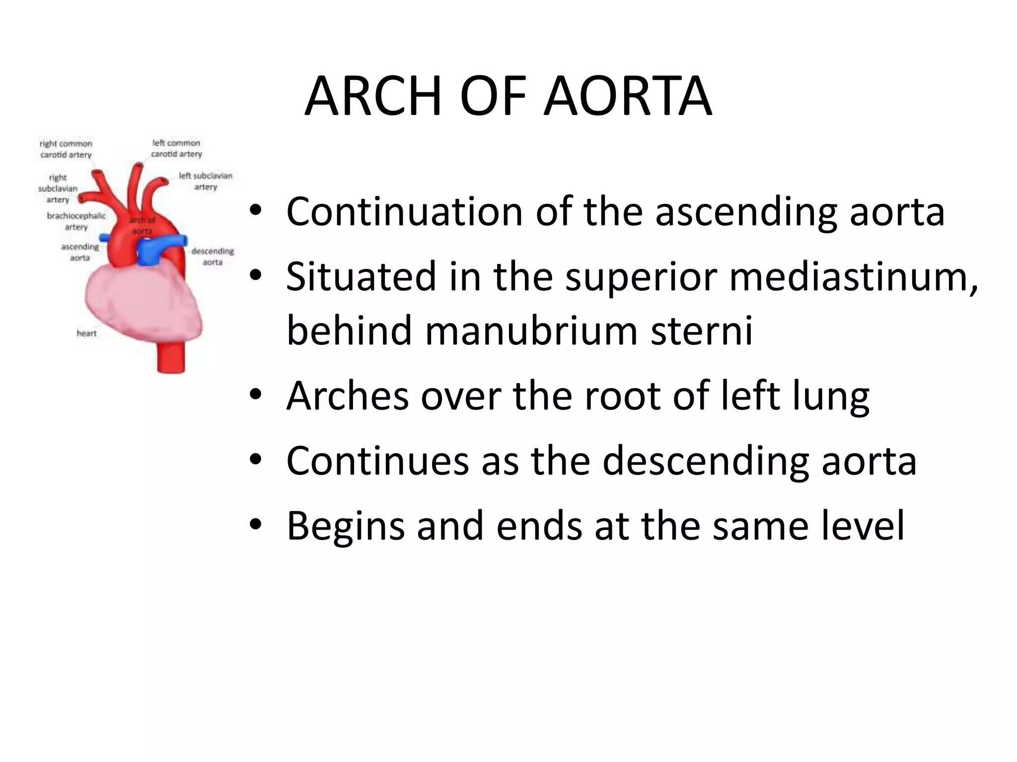
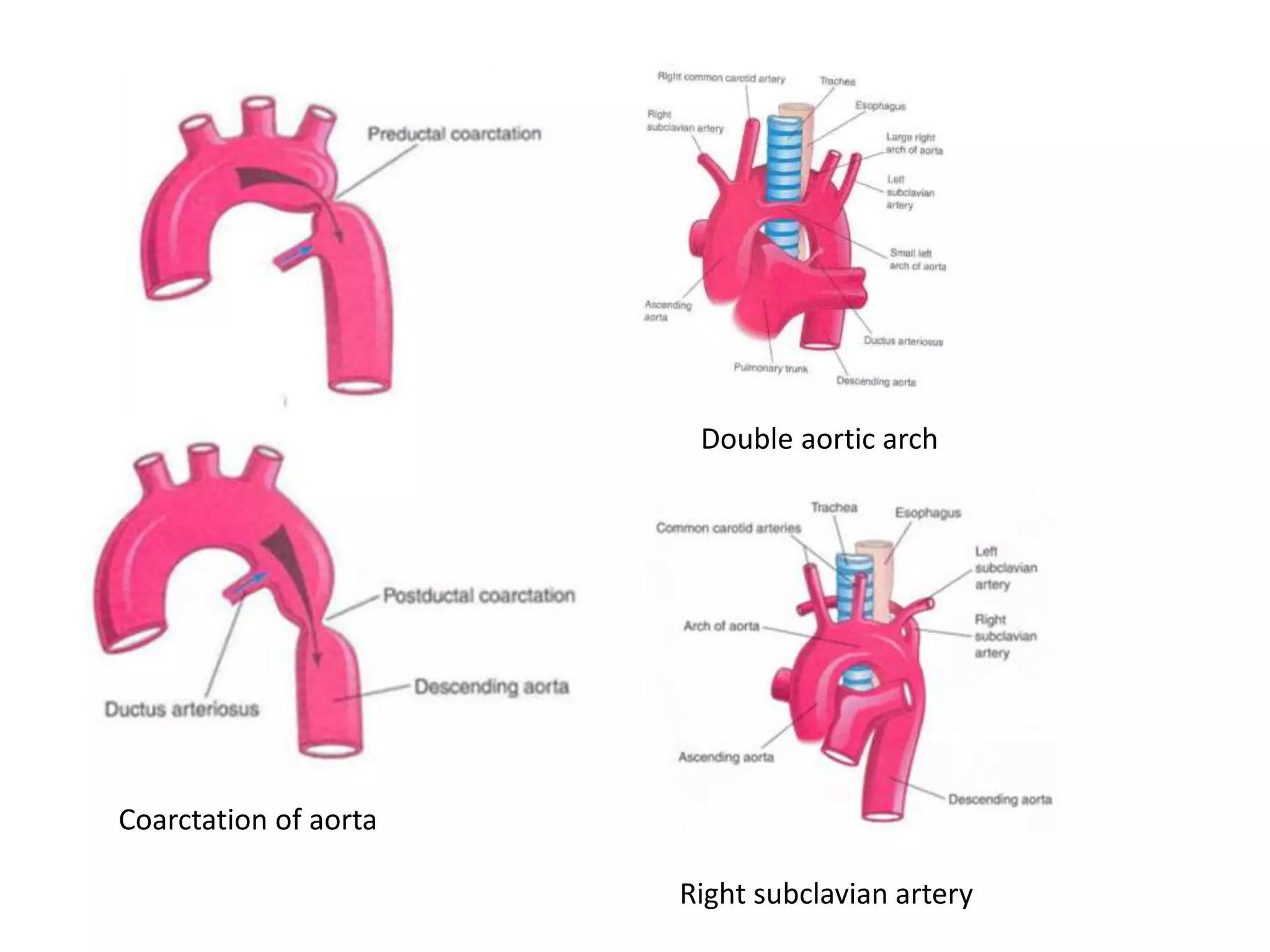
2. ARCH OF AORTA
• Continuation of the ascending aorta
• Situated in the superior mediastinum,
behind manubrium sterni
• Arches over the root of left lung
• Continues as the descending aorta
• Begins and ends at the same level
3. COURSE
• Begins behind the upper border of 2nd Right
sternochondral joint
• Runs upwards, backwards and to the left across
the left side of the bifurcation of trachea
• Passes downwards behind the left bronchus and
on the left side of the body of 4th thoracic
vertebra
• Arches the root of the left lung
• Ends at the lower border of 4th thoracic vertebra
4. RELATIONS
A. Anteriorly and to the left
1. Four nerves (from before backwards)
• Left phrenic
• Lower cervical cardiac branch of left vagus
• Superior cervical cardiac branch of sympathetic chain
• Left vagus
2. Left superior intercostal vein, deep to phenic
nerve and superficial to vagus
3. Left lung and its pleura
4. Remains of thymus
5. B. Posteriorly and to the right
Trachea with the deep cardiac plexus and the
tracheobronchial lymph nodes
Oesophagus
Left recurrent laryngeal nerve
Thoracic duct
Vertebral column
6. C. Superior
Branches of arch of aorta
• Brachiocephalic trunk
• Left common carotid
• Left subclavian
All these are crossed by the left brachiocephalic
vein, close to their origin
7. D. Inferior
Bifuraction of pulmonary trunk
Left bronchus
Ligamentum arteriosum with superficial cardiac
plexus on it
Left recurrent laryngeal nerve
8. BRANCHES
MAIN BRANCHES
• Brachiocephalic trunk
• Left common caritid
• Left subclavian
OCCASIONAL BRANCHES
• Left vertebral artery
• Arteria thyroidea ima
• Inferior thyroid artery
• Internal thoracic artery
• Left bronchial artery
• Right subclavian artery
9. DEVELOPMENT
• Three major sources of development
– Left limb of the aortic sac (truncus arteriosus)
• Forms the part between the brachiocephalic trunk and
the left common carotid artery
– Left fourth aortic arch
• Forms the part between left common carotid and left
subclavian artery
– Left dorsal aortic arch
• Forms the rest of the arch of aorta up to the
descending aorta
10. DEVELOPMENTAL ANOMALIES
1. Right sided aortic arch
– Due to persistence of right dorsal aorta below
the 7th intersegmental artery
– Disappearance of left dorsal aorta
– Common in birds
2. Double aortic arch
– Due to persistence of both the aortic arches
11. 3. Origin of Right subclavian artery from the
junction of aortic arch and descending aorta
– Due to persistance of right dorsal aorta below 7th
intersegmental artery and
– Disappearance of fourth aortic arch
4. Coarctation of aorta
– Due to stenosis or atresia of arch of aorta distal
to the origin of left subclavian artery
– May be of
• Preductal – ductus arteriosus remains patent
• Postductal – ductus arteriosus is obliterated